Umbilical Cord
Historical document, translated for reference. It reflects medical knowledge of the 1920s–30s and is not medical advice.
Summary
The umbilical cord is a cord connecting the navel of the fetus to the placenta, containing vessels that serve the purposes of nutrition and respiration of the fetus. It develops from the abdominal stalk and contains remnants of the allantois and yolk duct.
Encyclopedia article (1928–1936)
UMBILICAL CORD (funiculus umbilicalis), synonym pupochtnyy kanatik, represents a cord connecting the navel of the fetus with the placenta and containing vessels which serve the purposes of nutrition and respiration of the fetus (umbilical vessels-2 arteries and 1 vein), as well as remnants of the allantois and yolk duct. Embryology of the umbilical cord. The origin of the umbilical cord in the embryo begins at the time when, due to the development of the amniotic cavity, the embryo separates from the egg membranes, remaining in connection with them only by means of the abdominal stalk (see Embryo); in the latter are placed the yolk duct (see Intestine, development of) and the developing allantois (see), enclosed in a mass of gelatinous embryonic tissue. The amniotic membrane begins from the skin of the fetus on its abdominal surface in the place where the lateral parts of the abdominal wall, bending outward, transform into a tube for the formation of the abdominal stalk; here the water membrane, stretched by the fluid accumulating in it, covers the organs emerging from the abdominal cavity, the yolk duct and allantois, compresses them together with the gelatinous embryonic tissue into a common cord and passes to the inner surface of the chorionic sac. The formation of the cord proceeds progressively in both directions-toward the fetus, where with the completion of the development of the abdominal wall it ends with the formation of the navel, and toward the villous membrane, where the amnion accompanies the umbilical cord to the place where the placenta subsequently forms. The allantois as a hollow organ (urinary bladder) begins to disappear already from the 2nd month of embryonic life; remain its vessels-2 arteries and 2 veins; the latter subsequently merge into one, while the arteries are limited only by an anastomosis which they form before entering the placenta. With the establishment of placental circulation, the yolk duct with the yolk sac and yolk vessels (vasa omphalomesenterica) atrophies. Thus, in the umbilical cord only the former vessels of the allantois remain functioning, now greatly developing and receiving the name umbilical vessels. In the development of the latter, peculiar features are noted: first, a tendency to spiral growth is observed, sharply manifested in the arteries and less noticeable in the vein, where it is expressed by the appearance of grooves on the outer surface of this vessel and folds or valves (valvulae Hobokeni) on the inner; second, a more rapid growth of the arteries compared to the vein; as a result of these two properties, the arteries spiral around the vein, most often from left to right (counting from the embryo); spirals of the arteries begin to become noticeable from the 2nd month of embryonic life; their number can reach 40 or more (95-according to Meckel). The rapid growth of the arteries is also expressed by the development of coils (Fig. 1) and corkscrew twists, due to which nodular-like thickenings appear on the umbilical cord, bearing the name of false nodes (nodi arteriosi). Other false nodes of the umbilical cord are formed by varicose dilatations of the umbilical vein (nodi varicosi), as well as local accumulations of Wharton's jelly (nodi gelatinosi), which develops unevenly along the umbilical cord. False nodes should not be confused with true nodes, which form as a result of tying the umbilical cord (see below). In the further development of the umbilical cord, it is necessary to note the appearance of spiral twists of the entire cord, occurring due to the rotational movements of the fetus; they are observed in greater or lesser numbers, directed in one direction or another; they may sometimes be completely absent; they apparently arise at a time when the fetus is small and possesses considerable mobility. Structure of the umbilical cord. In its fully developed state, as we see it in the newborn, the umbilical cord represents a grayish-white cord with blood vessels visible through it in places; its length varies greatly in different cases, on average equaling 50-60 cm; the thickness of the umbilical cord also fluctuates, on average equaling the thickness of a finger (11-13 mm); it varies in different fetuses depending on the amount of Wharton's jelly (in this respect, thin umbilical cords are distinguished-about 14% of all cases, medium-75% and fat, or juicy-10-11%). The umbilical cords also differ in strength; they withstand a load of 3 to 12 kg, on average-up to 6 kg; the vessels possess the greatest strength, then the amnion and the end of Wharton's jelly, which easily tears when the umbilical cord is stretched. Starting from the navel of the fetus with a sharp transition of subcutaneous tissue into Wharton's jelly and the skin into the amnion, the umbilical cord ends on the placenta, attaching to it most often in the region of the center (central attachment-insertio centralis); the vessels of the umbilical cord immediately after attachment diverge over the placenta, penetrating it with their branches. Eccentric attachment of the umbilical cord (ins. excentrica) is also quite often observed; much less frequently the umbilical cord attaches at the very edge of the placenta (marginal attachment-ins. marginalis); in individual cases it does not reach the placenta, attaching at some distance from its edge to the membranes of the fetal sac (membranous, or membranous attachment, see Insertio velamentosa); sometimes the umbilical cord attaches to the placenta already being divided into 2 branches-ins. furcata. The amnion, passing from the placenta to the umbilical cord, forms on it the so-called caruncles-multiple formations arising due to the proliferation of epithelium with the oral

Figure 1. False nodes of the umbilical cord: a-nodus arteriosus; b-large varicose node of the umbilical vein. <
surface of the umbilical cord. The umbilical cord consists of: 1) blood vessels-2 arteries and 1 vein; 2) the remains of the allantois in the form of Wharton's jelly; 3) the amniotic membrane covering the cord. The arteries have a thicker wall than the vein; their lumen is narrower. The vein is wider and has a thinner wall. The arteries are twisted spirally around the vein. The umbilical cord is covered by the amniotic membrane, which forms caruncles on its surface. Wharton's jelly fills the space between the vessels and the amniotic membrane. It is a gelatinous substance containing mucin, fibroblasts, and collagen fibers. The umbilical cord varies in length and thickness depending on the amount of Wharton's jelly. The strength of the umbilical cord also varies, with the vessels being the strongest part. The attachment of the umbilical cord to the placenta can be central, eccentric, marginal, membranous, or bifid. The umbilical cord is a vital connection between the fetus and the placenta, providing oxygen and nutrients to the fetus and removing waste products.

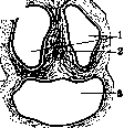
Figure 3. Cross section of the umbilical cord of mature fetuses: 1-v. umbilicalis; 2-aa. umbilicales.
Figure 4. Cross-section of the umbilical cord of mature fetuses: 1-arteries; 2-remnant of the allantois; 3-vein. The umbilical cord is covered by an amniotic sheath, tightly fused with the underlying tissue. The main tissue of it is Wharton's jelly (fig. 2). This tissue becomes denser toward the center of the cord, forming here a kind of core, sending processes to the umbilical vessels around them (chordae funiculi umbilicalis). 7& The vessels, besides the thickening of connective tissue around them, are also provided with a well-developed muscular membrane. Generally, the vessels of the umbilical cord have thicker walls than other vessels of the fetus's body. On a cross-section of the umbilical cord (fig. 3 and 4), with the help of a magnifying glass, one can find remnants of the allantois in the form of a cluster of cells, located in the middle between two arteries; closer to the navel this remnant lies more peripherally, then it shifts to the center and finally at the placental end approaches the vein; in places (not always and far from on all sections) remnants of the yolk duct are also found in the umbilical cord in the form of clumps of epithelial cells. The umbilical cord apparently does not possess its own vessels and nerves; there are also no true lymphatic vessels in it, but only juice canals, through which the nutrition of its tissues occurs. The function of the umbilical cord is the connection of the fetus with the sources of its nutrition and respiration-after the formation of the placenta, it falls exclusively on the umbilical vessels; along the arteries, the fetus's heart pumps the used blood into the placenta, and from the latter, oxidized, purified and enriched with nutrients blood returns to the fetus through the vein. The umbilical cord is an organ of exclusively intrauterine life and dies after the birth of the fetus. In the umbilical cord of a newborn child, the pulsation of the vessels is clearly felt, but after a short time the pulse in it begins to weaken and after 5-15 min. stops altogether. The weakening of the pulse wave comes from the placenta, and in the segment closest to the navel, the pulsation lasts the longest. The cessation of the activity of the umbilical vessels depends on two reasons: first, the respiratory movements of the child and the sucking force of his chest, in connection with the increase in the blood vessel area in the lungs, cause a decrease in blood pressure in the distant umbilical vessels; secondly, the umbilical vessels, very sensitive to cooling and stretching, strongly contract, and their strong musculature pushes the inner vascular membrane into the lumen of the vessel in scallops until the lumen is completely closed (Stravinsky). The umbilical cord has found technical application as a resorbable ligature material for replacing catgut. Bykov (1924) split the little-coiled umbilical cords lengthwise, threw out the vessels and prepared threads, which he successfully tested on animals and humans. Pathology of the umbilical cord. A. Irregularities of development. 1. Anomaly of length. Excessive length of the umbilical cord-up to 11/2-2 m and more-can be the cause of incorrect positions of it (entanglements, true knots, prolapses-see below), which in some cases can be the cause of death of the fetus. (Cases of L. up to 3 m-Schneider are described.) Excessive shortness of the umbilical cord-30 cm and less-usually does not cause complications until the expulsion of the fetus during labor; when the progressive movements of the fetus begin (in the second stage of labor), it can lead to their delay, to premature separation of the placenta, and with a close connection of the placenta with the uterus-to its inversion and finally to the rupture of the umbilical cord with the risk of fatal bleeding for the fetus. Such excessive shortness of the umbilical cord can be suspected during labor if the expulsion of the fetus is delayed without other visible apparent causes, under seemingly completely normal conditions; at this time signs of asphyxia of the fetus appear due to tension and compression of the umbilical vessels. Another sign of it may be the appearance of a cup-shaped depression on the wall of the uterus, in the place corresponding to the position of the placenta and the attachment of the umbilical cord (beginning inversion of the uterus). In all cases where shortness of the umbilical cord is suspected, it is recommended to accelerate the delivery (expulsion of the fetus by Kristeller's method, Verbov's bandage, forceps, etc.); a direct indication for intervention here is asphyxia of the fetus; one must keep in mind the possibility of intrauterine rupture of the umbilical cord and the need in such a case for its speedy ligation (see below). The shortness of the umbilical cord can reach its complete absence. In such cases we have to deal with the eversion of the fetus's internal organs, accompanied by other developmental defects; the fetus in this case turns out to be non-viable. 2. Umbilical hernia (hernia funiculi umbilicalis, syn. eventratio, exomphalos, omphalocele), sometimes incorrectly called umbilical hernia, represents underdevelopment of the anterior abdominal wall, the umbilical ring and the umbilical cord, arising in the early period of embryonic life (3rd-5th week) (fig. 5). In essence
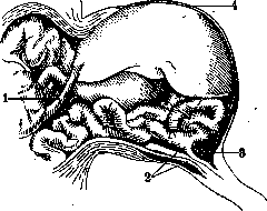
Figure 5. Umbilical hernia: 1-v. coeliaca; 2-umbilical arteries; 3-ductus mesentericus; 4-liver.
this is a malformation—not a hernia, since here there are no hernial gates (the matter did not go as far as the formation of the navel), nor the protrusion of the internal organs into these gates, since they do not protrude, but on the contrary, are retained in the cavity of the abdominal stalk, from which the umbilical cord is formed. Several forms of umbilical cord hernia are distinguished depending on the degree of development of the malformation: a) stalk-like hernias with a narrower neck, formed as if by an elongated navel, and a round tumor covered by the amnion and usually containing only the intestines; b) hernias with a broad base, where the underdevelopment of the abdominal wall is already clearly visible; the tumor has a dome-shaped form, with its broad side communicating with the abdominal cavity, and on the apex of its amniotal covering it bears the attachment of the umbilical cord; such hernias often contain the liver, more or less deformed, and other organs (stomach, spleen); these two forms bear the predominant name of umbilical cord hernia; c) the third form is given the predominant name 'eventration'; the form of the tumor here is irregular, the walls of the abdomen are clearly defective, sometimes passing into the amnion already on the lateral parts of the abdomen; due to the underdevelopment of the diaphragm, such hernias may contain the heart; they are accompanied by numerous malformations in other parts of the body. The coverings of the hernial sac in umbilical cord hernia consist of the amnion and peritoneum, and in severe degrees of malformations—of only an avascular amnion, since the peritoneum, like the abdominal wall, also turns out to be underdeveloped; sometimes in the lateral parts of the sac it is possible to find embryonic rudiments of the abdominal muscles. The membranes in a healthy (non-inflamed) and fresh state are transparent, allowing the contents of the hernia to be seen. In 60% of cases there are only loops of intestines, in 25%—the liver, one or together with intestines, more rarely the stomach, heart, kidneys, uterus with ovaries are found. Among the accompanying malformations, the following should be noted: lordosis, curvatures and underdevelopment of the limbs, spina bifida, labium leporinum, extrophy of the bladder and other underdevelopments of the urogenital organs, amniotic adhesions, etc. Umbilical cord hernias occur in approximately 0.02% (1:5,000) of births, in boys almost one and a half times more often than in girls. Many theories have been put forward to explain the occurrence of umbilical cord hernias. Until the 19th century, they were explained by the mechanical squeezing out of the internal organs (Sandifort, 1799). Oken (1810) was the first to point to developmental delay, namely the insufficient retraction of intestinal loops into the abdominal cavity, and Meckel (1812)—to the underdevelopment of the abdominal coverings; Neugebauer (1850) introduced the element of underdevelopment of the intestinal ligaments; in Panum and Dareste (1892) the idea is first expressed of embryonic inflammation as the cause of many malformations, and Preobrazhensky (1900) to explain the genesis of umbilical cord hernias puts forward the idea of focal embryonic inflammation caused by toxic causes. The theories cited in their totality create a coherent doctrine of the genesis of this malformation, whereas other explanations (Scarpa, 1822—pulling out the internal organs by a too short or twisted umbilical cord; Simpson, 1839—fetal peritonitis and adhesions of the internal organs with the vaginal process of the umbilical cord; St. Hilaire, 1832—amniotic adhesions, Alfeld—persistence of the ductus omphalo-entericus, pulling the intestine into the umbilical cord; Aschoff—lordosis of the spine, etc.) proved to be either incorrect or applicable only to individual cases. Recognition of umbilical cord hernia before birth is difficult; it can be palpated during internal examination only when it is presenting, i.e., in the transverse position of the fetus; it has happened that the eviscerated intestine of the fetus (in case of intrauterine rupture of the hernial sac) was confused with the umbilical cord and even with the intestine of the mother herself, which had penetrated into the uterine cavity in what was apparently a rupture of the latter. Sometimes umbilical cord hernia can mechanically slow down the birth of the fetus by itself, due to its size or due to the associated shortness of the umbilical cord. At birth, the hernia threatens the fetus with rupture of the membranes with subsequent death from peritonitis or from internal bleeding on the basis of damage to the umbilical vessels, liver vessels, etc. Peritonitis occurs due to necrosis of the amniotal covering. From peritonitis, those newborns who cannot be helped surgically due to the extent of the defect or for other reasons usually die. Treatment. Only very few, small hernias can heal spontaneously, by scarring of the defect in the abdominal wall after the umbilical cord falls off. Other hernias, of small and medium size, can be cured by surgical means, with the reconstruction of the skin-muscular abdominal wall. In the literature, only about 200 cases of operation for umbilical cord hernia have been published with 67% recoveries. In these cases, mainly the radical operation was used—a circular incision through the intact skin, opening of the abdominal cavity, ligation of the umbilical vessels, freeing the organs from adhesions, reducing them, suturing the abdominal wall, as in ordinary laparotomy. In this way, hernias up to the size of two fists or the head of a newborn were operated on (Colley, Lindfors, etc.) with resection of the liver, Meckel's diverticulum, intestines, etc.; among the complications, asphyxia and colic should be noted when reducing the internal organs into a narrow abdominal cavity, due to compression of the diaphragm and pressure on the heart. Umbilical cord hernias in newborns should be operated on as soon as possible, before the onset of necrosis of the membranes. Among other surgical aids, the method of Olshausen, which does without opening the peritoneum, and the so-called subcutaneous ligature of Breus can be mentioned. The operation can be performed without anesthesia (newborns do not react to incisions with pain), chloroform was found necessary only for the reduction of the internal organs, to suppress the reflexes.
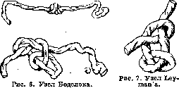
3. Velamentous attachment (see Insertio velamentosa) is observed in 0.5-1.0% of all births. In most cases, this anomaly proceeds without complications and is recognized only after birth. It acquires clinical significance in those cases when the vessels passing between the membranes are found in the lower pole of the egg, near the uterine os; then when the membranes rupture they can also easily rupture and cause fatal bleeding for the fetus; the fetus can be saved only by its rapid extraction. In velamentous attachment, sudden intrauterine ruptures of vessels have also been observed, apparently due to the shortness of the umbilical cord and movements of the fetus. B. Incorrect position of the umbilical cord. 1. Twists of the umbilical cord around the body, limbs, and neck of the fetus occur very often (in 25% of all births). In the vast majority of cases, due to the slipperiness of the umbilical cord and the protective role of Wharton's jelly, they remain harmless; however, where the twists are tight or multiple, the following can occur: a) tension and compression of the umbilical cord vessels, and during auscultation of the pregnant woman's abdomen, blowing sounds synchronous with the fetal heartbeat (umbilical cord noises) may be heard; b) difficulty in blood circulation in the constricted limbs, their retardation in development, formation of grooves and even complete separation of them (spontaneous amputation); c) difficulties in the expulsion of the fetus (especially in cases of twisting around the neck), similar to those observed with a short umbilical cord (see above) and with the same consequences; if difficulties occur when the head is not yet born, the threatening asphyxia of the fetus forces the use of forceps with the risk of rupturing the umbilical cord or turning the uterus inside out; when the head has already been born with the neck twisted by the umbilical cord and with difficulty in further expulsion of the fetus, it is recommended to slip the loop of the umbilical cord off the head or to cut it on the neck, clamping both ends with torsion forceps. 2. True knots (tying the umbilical cord in a knot, fig. 6 and 7) also occur due to the movements of the fetus, when it slips through a loop of the umbilical cord and then tightens it; for this to happen, the umbilical cord must be very long or the fetus must be small, which is why this complication occurs most often in early periods of pregnancy. The tightening knot gradually causes atrophy of Wharton's jelly at the points of pressure, obstruction of the umbilical vessels and death of the fetus. In later periods, knots can occur during childbirth itself, during the passage of the fetus through the loop of the umbilical cord and the birth canal; in these cases they usually do not lead to harmful consequences. An extremely rare phenomenon is knots in twins, when the umbilical cord of one fetus is wound or tied around that of the other (for this it is necessary that both fetuses have a common amniotic cavity, which occurs once in 30-35,000 births); in this case both fetuses usually die due to disruption of the placental circulation. 3. Presentations and prolapses of the umbilical cord. Normally, the umbilical cord, folded in a loop, is placed with its main mass on the abdominal side of the fetus, between its small parts. Being a long movable organ, it is easily displaced and if the lower segment of the uterus
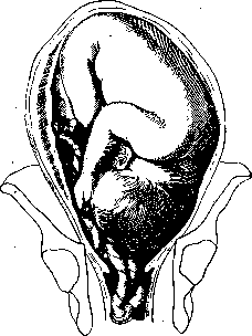
Fig. 8. Prolapse of the umbilical cord in cephalic presentation.
If, upon examination, we feel it in this position with the waters not yet discharged, then there is present a presentation of the umbilical cord; but if the sac is already gone, then we speak of prolapse of the umbilical cord (fig. 8). Not always does presentation of the umbilical cord, upon discharge of the waters, turn into its prolapse—the presenting part, inserting itself, may push the cord upward; sometimes, on the contrary, the displacement and prolapse of the loop of the cord occurs only at the moment of rupture of the sac, when suddenly flowing waters carry along with them the movable cord, before the large presenting part has had time to insert itself. Among the factors favorable to presentation and prolapse of the cord should be mentioned excessive length of the cord, large amount of amniotic fluid, distensibility of the lower segment of the uterus, multiple pregnancy, pendulous abdomen, narrow pelvis, asymmetrical pelvis, small volume of the presenting part (small head, breech, legs), transverse position, incorrect insertion of the head, and finally, low attachment of the placenta. Prolapse of the cord is observed in 0.6-1.0% of births; it is least common in cephalic presentations, more common in breech (10%), footling and transverse (20%), and occurs more easily in multiparas than in primiparas; it is relatively frequently observed in premature births. Completely indifferent for the mother, prolapse of the cord is extremely dangerous for the fetus, which during expulsion compresses the cord and dies from asphyxia. In spontaneous births, fetal mortality from this anomaly reaches 90%; often the fetus dies despite the assistance provided, and here the position and presentation of the fetus play an important role: in cephalic presentations 44% of fetuses die, in breech 33%, in transverse position 32%, in footling presentations 22% (Gusev). This abnormality is diagnosed only by internal examination, very easily when the fetal sac is absent and with some difficulties when the waters have not yet discharged; a valuable sign in this case is the palpable pulsation of the umbilical vessels. Before internal examination, suspicion of compression of the prolapsed cord may be aroused by the sudden onset of signs of fetal asphyxia during the entry of the presenting part into the pelvis in the presence of the aforementioned predisposing conditions. If such suspicion arises, one must immediately resort to internal examination, in which the presenting cord is most often found in the sacroiliac articulation area; generally the cord prolapses more easily through the lateral parts of the pelvis. The therapy of this anomaly has in view exclusively the interests of the fetus. If the latter is dead (absence of pulsation in the prolapsed cord between contractions, absence of fetal heartbeat), then labor is managed expectantly; but if the fetus is alive but is in immediate danger (the cord is compressed during contractions, there are present signs of fetal asphyxia), then prompt delivery is indicated, the form of which is determined by the conditions existing in this particular case. Specifically, with full dilation of the cervix, the fetus is immediately extracted by the method that is most suitable for the given position and given presenting part—with the head that has entered the pelvis, extraction is performed with forceps; with a still mobile head or transverse position—version to a footling extraction; with breech presentation—lowering of a foot with extraction or extraction by the breech; finally, in some cases the question of cesarean section may arise, if conditions are suitable and the mother absolutely desires to have a living child. With incomplete dilation of the cervical os, conditions are forcibly created for extraction of the fetus (by digital or instrumental dilation of the cervix or, under suitable conditions, vaginal section) and further management proceeds as indicated above. It goes without saying that for measures involving danger to the mother, her consent is required. In other cases, where the cord is not yet compressed, e.g., when the sac is still present and the fetus is not in immediate danger, one should, sparing the mother, adopt measures as gentle as possible toward her. Here the question first arises of replacing the prolapsed or presenting loop of the cord. This replacement, with the sac intact, sometimes succeeds by changing the position of the parturient (she assumes either a position with the pelvis elevated or the knee-chest position, or on the side opposite to that from which the cord prolapses); if this method does not succeed in replacing the cord, one can try to push it back by hand, and in order to avoid entrapment of the cord upon repeated prolapse, one should preserve the fetal sac as much as possible. A prolapsed umbilical cord is replaced with more difficulty than a presenting one. Its replacement is again performed directly by hand, preferably with the parturient's pelvis elevated, under anesthesia; the loop of the cord is pushed upward with the fingers behind the presenting part (head) and then one tries to fix the latter in the pelvis during a contraction or artificially, by pressing it into the pelvis, so that further prolapse does not occur. In former times, replacement of the prolapsed loop of the cord was recommended with the aid of instruments, so-called repositories—probes or sticks with a loop into which the cord was placed; fig. 9 shows such a Brown stick, with which the loop slips off upon reverse extraction of the instrument, freeing the cord, which remains in the uterine cavity; elastic repositories, e.g., Robertson's, were left in the uterus until the birth of the child; now, however, repositories are rarely used due to the comparative lack of success and danger of their application. It should be kept in mind that replacement of the cord very often fails (in three-quarters of cases); furthermore, frequently (in almost half of cases) the cord replaced by hand ceases to pulsate (spasm of the vessels due to cooling and irritation); finally, replacement with the hand introduced into the uterine cavity gives a considerable number of postpartum infections; therefore, if the conditions of labor are such that labor can be quickly completed without harm to the mother, it is completed without prior replacement of the cord. If replacement of the cord for some reason is undesirable or has failed, then the prolapsed cord is protected as long as possible from compression by the following methods: a) the parturient is placed in a suitable position (see above); b) external version to the breech is performed, which easily succeeds with a sufficient amount of fluid or in a premature fetus (this is often the case with cord prolapse); the resulting breech presentation provides more favorable conditions for the fetus.

Fig. 9. Robertson's Brown's repositorory.
and for further manipulations when significant dilation of the cervix occurs; c) with sufficient dilation of the latter (by two fingers or more), a combined rotation can be performed, during which the umbilical cord is reduced using the methods mentioned above; d) many authors recommend with minimal dilation of the cervix and after the waters have broken to insert a metrerinter, which, by pushing the presenting part upward, prevents it from pressing on the umbilical cord and at the same time promotes dilation of the cervical canal. With these operative measures undertaken in the interests of the fetus, it is not always possible to save its life, and the mortality rate of fetuses reaches 17-20%; therefore, in cases where the mother is willing to take the risk to save the child's life, the question of performing a cesarean section - vaginal (Doederlein, Seitz, etc.) or abdominal (in narrow pelvis - Lebedev, Shirshov, Serezhnikov-Pekarskaya, Couvelaire, Lepage, etc.) - may arise even before urgent indications for ending the labor appear. B. Diseases and other abnormalities of the umbilical cord. 1. Twisting of the umbilical cord and obstruction of its vessels. Spiral twists of the umbilical cord may sometimes be absent; in other cases, they can be very numerous (Hyrtl, for example, once counted up to 380 of them). With a small amount of Wharton's jelly, such twists can lead to compression of the umbilical vessels and death of the fetus. Obstruction of the umbilical vessels can occur, besides twisting, also in certain diseases, for example in syphilis, when obliterative processes are observed, which sometimes lead to complete closure of the vascular lumen. 2. Tumors of the umbilical cord. In the short time of the umbilical cord's life, only a few forms of tumors, mostly of embryonic origin, manage to develop in it. These include, for example, cysts of the allantois with greenish-yellow contents consisting of secretions from the intestine and kidneys of the fetus (Ahlfeld), and cysts of the bile duct (Ruge) - small formations (the size of a pinhead and larger), found on the periphery of the umbilical cord and filled with fatty-degenerated epithelial cells, detritus, and calcareous masses. Bueden (Büdin) described a single case of teratoma of the umbilical cord in a full-term fetus; in this case, the tumor consisted of several sections containing cavities lined with stratified squamous epithelium, blood clots, smooth muscle fibers, cartilage, bones, etc. Fava and Panciera described a cyst of the umbilical cord from a two-year-old child, lined inside with endothelium and containing transparent fluid. It is also necessary to mention false cysts of the umbilical cord, resulting from liquefaction of Wharton's jelly and appearing as small bubbles, which in exceptional cases reach the size of a chicken egg; inflammation of the umbilical vessels, edema of Wharton's jelly, and twisting of the umbilical cord play a role in their development. Most tumors of the umbilical cord were found in premature fetuses (4-7 months). False cysts of the umbilical cord may be of postmortem origin. Tumors of the umbilical cord have no clinical significance. 3. Varicose dilations of the umbilical cord are quite common. When expressed to a slight degree, they have no practical significance and do not cause any harm. In more pronounced cases, they can be the cause of intrauterine death of the fetus (due to rupture of a varicose node during pregnancy or labor). Extremely rarely occurring complications include hematoma of the umbilical cord. 4. Inflammatory diseases of the umbilical cord are rare. They are most often observed in syphilis. They can lead to thrombosis of the umbilical vessels, which is often found in the umbilical cord of dead and macerated fetuses. 5. Ruptures of the umbilical cord. The umbilical cord can usually withstand the weight of the fetus, but with jolts and in certain pathological conditions, it can rupture relatively easily; for example, in the experiments of some researchers, it tore when a weight of 500g, suspended from its end, fell from a height of one meter. Most often, the umbilical cord ruptures during labor in the standing position of the mother, when the falling fetus breaks it with its weight; however, ruptures are also often observed in the lying position of the parturient, during rapid labor (in 20-30% of cases), when the fetus is thrown some distance from the mother's genital organs by the last contraction. Furthermore, ruptures of the umbilical cord have been observed in normal labor in cases of excessive shortness of the cord, during the release of entanglements, when the pelvis of the parturient is accidentally raised with the fetus not yet separated and the placenta not yet expelled, in cases of velamentous attachment of the umbilical cord, at insertio velata, during operations-rotations, forceps; finally, in individual cases, inexplicable intrauterine ruptures of the umbilical cord have been described. Predisposing to ruptures are pathological conditions of the umbilical cord, inflammatory infiltration, weak development of Wharton's jelly and its loose structure, varicose dilations of the umbilical vessels, their excessive tortuosity and thinness. When the umbilical cord ruptures, it stretches, the amnion tears; the vessels withstand the longest. The edge of the rupture is most often oblique, uneven, jagged, which distinguishes it from a cut. In most cases, the umbilical cord ruptures in its fetal segment, less often in the placental segment, and even less often in the middle, sometimes (about 20% of cases) the umbilical cord is torn off right at the navel; there have been cases of rupture of the umbilical vessels even in the abdominal cavity of the fetus. In most cases, ruptures of the umbilical cord bleed almost not at all, since the strong tension preceding the rupture causes the vessels to contract, and the rolling of the torn intima further contributes to the closure of their lumen; nevertheless, significant, even fatal bleeding can sometimes occur here, especially when the rupture of the vessels occurs in the area of the navel itself or even inside the abdominal cavity. Cases of the latter kind, if the rupture is recognized in time, require immediate transverse incision for ligation of the vessels; if the umbilical cord has torn off right at the skin of the navel and the latter is bleeding, then the umbilical vessels are grasped and ligated individually; if it is impossible to grasp them, the navel has to be infiltrated; in other cases, the umbilical cord is handled in the usual way, ligating it to prevent subsequent bleeding. Ruptures of the umbilical cord in cases of fetal death may have forensic-medical significance (infanticide, leaving the newborn without help). Separation of the fetus and ligation of the umbilical cord. Mammals separate the fetus after the birth of the placenta, biting through the umbilical cord with their teeth; predators eat the placenta, starting with the afterbirth; eating of the placenta has also been observed in non-predatory animals, for example horses; thus, separation of the fetus occurs in animals some time after birth. Primitive peoples bite off, tear, rub between stones, or burn the umbilical cord, leaving a rather large umbilical stump; separation of the fetus is also performed by them after the birth of the placenta - by some peoples only on the next day after birth. All these methods, unconscious or empirical, aim to ensure for the newborn the cessation of bleeding from the umbilical vessels. But waiting too long contributes to contamination of the umbilical cord and infection of the fetus, while too early separation can cause bleeding. Therefore, in most civilized peoples, early separation of the fetus (before the expulsion of the placenta) is practiced with hemostasis ensured by ligation of the umbilical cord. At the same time, despite the simplicity of the mentioned operation, there are still different opinions regarding the method of separating the fetus and handling the umbilical stump, for which reason various methods are also practiced. The differences in these methods mainly come down to handling the umbilical stump and are expressed in the techniques that can be divided into three main groups: 1) ligation of the umbilical cord, 2) crushing it with instruments, 3) leaving it unligated. Common moments in all methods are: a) waiting with manipulations until complete cessation (or at least significant weakening) of pulsation in the umbilical cord, and b) the final act of cutting with scissors. Waiting for the cessation of pulsation in the umbilical cord aims not only for better hemostasis in the umbilical vessels but also for establishing equilibrium in the amount of blood between the fetus and the placenta. The newborn, which has undergone significant compression in the birth canal, is often anemic, and during the continuing extrauterine pulsation of the umbilical cord, it receives from the placenta the so-called 'reserve blood' in an amount of 50 to 200 grams, which is by no means indifferent to the economy of the newborn's organism. In addition, according to existing observations, the bloodless placenta separates better from the uterine wall. For cutting the umbilical cord, the so-called 'umbilical cord scissors' (Fig. 10) are used; they are curved at the edge, with relatively long branches, with blunt, rounded ends so as not to injure the child during its accidental movements, and with not too sharp cutting edges: they should somewhat traumatize the umbilical vessels being cut, in order to cause the intima to roll up and better occlude the vascular lumen of the vessel.
The cutting of the umbilical cord is usually done at a distance of 1-2 cm from the place where a clamp, etc., has been applied to the cord; the cord stump is bandaged to the newborn's abdomen after preliminary treatment with drying or anti-putrefaction substances (a mixture of Runge-potato flour with boric acid, gypsum according to Sutugin-Fagonsky, dermatol, salicylic acid, alcohol according to Ahlfeld, collodion and bismuth according to Rogak, etc.). Ligation of the Umbilical Cord. The oldest and most common method of dealing with the cord is ligation with a strong linen tape, about 1/2 cm wide; it is preferred to silk ligature, which easily cuts through the tissues of the cord. The tape is applied at a distance of 1 1/2-2 cm from the navel, i.e., as close to the latter as the thickness of the cord allows, so as not to cause irritation of the navel with the ligature. The knot of the tape must be tightened tightly, as it easily loosens on the pliable mucous tissue of the cord. For this reason, it is recommended to pull the cord with two knots, namely, after tying the tape with a surgical knot on one side, to transfer its ends to the other side and there again tighten it with a double knot. It goes without saying that the ligation should be done with sterile material; the tape is boiled and before use is stored in a mercuric chloride solution. Despite careful ligation, cases of successive bleeding from the umbilical vessels are still sometimes observed; bleeding is all the more unpleasant because it is difficult to monitor it; to prevent loss of heat, newborns are usually well wrapped, and
Figure 11. Reliable ligation of the umbilical cord remnant: 1 - skin knot; 2 - place of cord cutting; 3 - first knot; 4 - second knot,
v
it is often discovered only after the child has already lost a significant amount of blood. True, fatal bleeding from the umbilical remnant is very rare (in the material of maternity hospitals, Kakushkin found 3 cases out of 220,000 births), but even less significant blood loss cannot be considered indifferent to newborns. The causes of these bleedings, apart from cases of poor ligation (done by the mother's own hands or unskilled bystanders, or deliberately performed for the purpose of infanticide, with weak material), are mostly abnormalities in the structure of the cord, changes in its vessels on the basis of inflammation, syphilis, as well as hemophilia, etc.; bleeding occurs more easily in poorly breathing and asphyxiated children. The fear of successive bleeding led to the use of double or two-moment ligation of the cord (Alfeld, Jaschke, Pfaundler, Bumm, Pinard (fig. 11)), with the latter first ligated at a palm's distance from the navel and cut off, and then after bathing, the ligated end of the cord is bent to the navel and the tape is tied again with a knot near the skin navel; thus the cord remnant remains hanging in the form of a "loop". Hemostasis with this method is excellent, but the long cord remnant, representing a considerable mass of dead tissue, is unfavorable in terms of facilitating infection. Generally, in the tape method of ligation, in the tape itself, which easily becomes moist and therefore hinders drying (mummification) of the cord remnant, some authors saw a certain chance for infection. Therefore, proposals were made to ligate the cord with an elastic rubber cord (Büdden, 1880), or to apply a tight rubber ring to the cord remnant (Lebedev, Kuzmin, 1898); in the latter method, the cord is first cut off, and then a rubber ring is applied to it with the help of a special instrument, which lies almost at the skin navel (fig. 12). Omphalotripsy. With the aim of possibly reducing the cord remnant and at the same time achieving good hemostasis, crushing of the cord with an angiotribe (e.g., Tuffier's instrument) or another strong instrument with strong clamping surfaces (omphalotribe) was proposed. After grasping the cord, the instrument is fixed by an assistant, and the opera
Figure 13. Vag's pincer for omphalotripsy: 1 and 2 - place where the cord is clamped.
Figure 14: Dubinchik's clamp in open, b - in closed view. tor closes its branches with the help of a lever or other device; when crushing, the traumatized intima turns and blocks the vessel lumen under the compressed place, which turns into a thin plate. Later, heavy instruments were replaced by lighter ones, like pincers, which could be left on the cord for 3-36 hours after clamping. Kakushkin (1905) proposed using an ordinary Kocher's pincer with the teeth cut off for omphalotripsy, applying it so that the rings are turned towards the child's chest, and Bar (Vag, 1907) constructed a special instrument (fig. 13), now in sufficient use. In the vast majority of cases, it is sufficient to leave the instrument for 3 hours; however, cases of successive bleeding 30-40 hours after the birth of the child have sometimes been observed. Recently: Dubinchik proposed a new clamp for the cord. Dubinchik's clamp has an ellipsoidal shape and consists of 2 arched blades connected to each other by a hinge and clamped with a toggle screw (fig. 14). On the inner surface of each blade there is a narrow crossbar; on one crossbar there is a recess, and on the other, corresponding to this recess, a protrusion. When both blades are closed, the umbilical cord is compressed until the lumen of the umbilical vessels is completely closed. The clamp is light, portable, does not traumatize the delicate skin of the newborn, and does not injure the amniotic leaf of the cord. The clamp is removed after 3-4 hours, after which the cord segment remains completely dry under a light, changeable sterile dressing until it falls off. Clinical observations (Gok-Smrcek and Sarne) have shown the advisability of using this clamp in practice. Separation of the fetus without ligation of the cord. The negative aspects of the ligature method of caring for the cord - secondary bleeding due to weakening of the ligature, irritation of the navel caused by a foreign body, inflammation of the umbilical vessels, delay in mummification of the cord remnant - led to the proposal to do without ligation of the cord altogether. The basis for this proposal was empirical observations indicating the rarity of bleeding in cases of cord rupture, slipping of the ligature, etc., further - examples of animals and primitive peoples who do without ligation, and finally, acquaintance with the physiological properties of the cord, namely the spontaneous cessation of blood circulation in it after the birth of the fetus. Non-ligation of the cord. was first proposed in the early 18th century. In this method, the cord is not cut until pulsation in it has completely ceased (12-18 min) at a distance of 4-4 1/2 cm from the navel (so that it can be ligated in case of immediate or successive bleeding); if drops of blood appear after cutting, it is recommended to massage the cord remnant with pressing movements from the navel to the periphery; the infant remains under close supervision for 2-3 hours. Out of 16,000 births conducted in this way (Rakhmanov), it was necessary to ligate the cord in 17% of cases, mostly pathological (immaturity of the fetus, asphyxia, bleeding in the mother, preventing waiting, etc.). This method, inconvenient due to the long waiting and not giving confidence that everything has been done for hemostasis, at one time went out of use, then reappeared; at present, it is hardly applied anywhere. Among other methods of caring for the cord, one can mention cauterization of the cord remnant with red-hot pincers, burning the cord with Pakelen (Martin) (both methods require great caution due to the danger of burns to the fetus) and excision of the navel with subsequent suturing.






Related articles
Mentioned in
Cite this page
“Umbilical Cord.” Soviet Medical Encyclopedia. English translation of Bolshaya Meditsinskaya Entsiklopediya, 1st ed. (Moscow, 1928–1936), ed. N. A. Semashko. https://sovietmedicalencyclopedia.pages.dev/article/umbilical-cord/