Ventrofixation
Historical document, translated for reference. It reflects medical knowledge of the 1920s–30s and is not medical advice.
Summary
Ventrofixation is a surgical procedure used to fix the uterus to the abdominal wall to treat prolapse or retroversion. This 1930s article details the historical evolution of the technique, its associated complications during pregnancy and childbirth, and various modifications designed to preserve uterine mobility.
Encyclopedia article (1928–1936)
VENTROFIXATION (ventrofixatio), an operation in which the uterus is fixed to the abdominal wall by one method or another. Ventrofixation is used for prolapse or descent of the uterus, and for its deviation and bending backward; ventrofixation is also recommended after operations to remove uterine appendages, ectopic pregnancy, etc., to avoid the development of retroflexio uteri fix. In cases of uterine descent and prolapse, ventrofixation is combined with plastic surgery on the pelvic floor. The ventrofixation operation arose by chance, and it appears that Olshausen was the first to propose ventrofixation as an independent method for correcting the position of the uterus. Initially, Olshausen's method consisted of passing a silkworm gut suture through the base of the round ligament, then through the peritoneum and aponeurosis and back; the suture was tied from the side of the abdominal cavity. In 1887, Leopold modified the ventrofixation operation by passing sutures in the lower corner of the abdominal wound through the aponeurosis, muscles, and peritoneum, grasping the fundus of the uterus; in order to achieve firm fixation, a portion of the serous covering at the fundus of the uterus was previously removed with a scalpel. Later, Czerny proposed, in addition, to place two or three sutures passing through the aponeurosis, muscle, and peritoneum, and onto the anterior surface of the uterus. As early as 1888, in order to obtain a more sharply defined anteverted position of the uterus, Kelly proposed placing sutures on the posterior surface of the uterus at its fundus. Over time, Olshausen changed the original technique: after opening the abdominal cavity, the subcutaneous
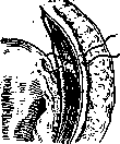
fatty tissue was pushed away from the aponeurosis, and, retreating 2-3 cm from the midline, a silkworm suture was passed through the aponeurosis and peritoneum, the base of the round ligament was pierced, and the suture, passed in the opposite direction, was tied over the aponeurosis (see Figure 1). These methods do not always yield lasting success in cases of uterine descent and prolapse. Furthermore, the Leopold-Czerny operation can cause a whole series of complications during pregnancy, labor, and the postpartum period. These complications arise more easily the wider the area of uterine fixation. It is not always possible to truly predict the extent of the fixation, and often the latter turns out to be more significant than intended. The Olshausen operation preserves the mobility of the uterus; however, even with [Fig. 1. The suture is passed through the aponeurosis, peritoneum, and the site of origin of the round ligament; it is tied over the aponeurosis]
it, broad adhesions at the fundus are possible if the uterus is injured during its extraction from the abdominal cavity. Relatively often after ventrofixation of the uterus, pregnancy is interrupted. This is explained by the fact that the placenta, developing at the site of uterine fixation, which does not participate in the general hypertrophy, is not attached firmly enough. With broad fixation of the anterior surface of the uterus, the posterior wall of the uterus participates mainly in the formation of the gestational sac during pregnancy, as a result of which the cervix is displaced toward the sacral promontory and higher. In connection with such a configuration of the uterus, the fetus often assumes an incorrect position (transverse, oblique). Fixation of the fundus and anterior surface of the uterus also contributes to insufficient labor activity, and dilation of the cervix is often delayed. If, nevertheless, dilation of the cervix has occurred, the fetus often receives an incorrect direction—not toward the pelvic outlet, but toward its posterior part. Sometimes, despite prolonged contractions, dilation of the cervix does not occur, and the uterine os remains closed. The posterior part of the uterus is thereby stretched to the extreme, and thus, a danger of rupture arises, for the prevention of which a classic cesarean section is indicated. Performing a version on the foot in suitable cases may encounter significant difficulties, for the elimination of which one has to incise the anterior wall of the cervix. At the same time, the high position of the cervix, however, may not always allow this operation to be performed quite clearly. Cases have been described where the placental site, being located at the site of fixation, caused severe blood loss in the postpartum period due to weak contraction. The site of fixation can stretch during pregnancy, causing pain, and form a cord (the so-called ligamentum medianum tertius uteri), which sometimes contributes to the occurrence of ileus. In order to avoid various complications during pregnancy and labor, various methods of shortening the round ligaments were proposed for ventrofixation in women of childbearing age in order to preserve the mobility of the uterus. These include: the Bum method, according to which loops of the round ligament are passed through the peritoneum and attached to its inner surface; the Doléris method, according to which the loop of the round ligament is passed, in addition, through the rectus muscle and strengthened over it; the Gilliam method, according to which the round ligament is, in addition, passed through the aponeurosis and strengthened there with several sutures (see Figure 2). The methods of Brodescu and Gosset are analogous. Kiparsky, for the purpose of more firm strengthening of the uterus, sutures the distal end of the formed loop of the round ligament at the site of the ligament's origin, then fixing the loop in the usual way to the aponeurosis. In all these methods, the passage

[Figure 2. Loops of the round ligaments, pulled]
through openings made in the peritoneum, muscle, and aponeurosis and attached to the latter with interrupted sutures (Gilliam's operation). of the loop of the round ligament is performed approximately 2-2.5 cm away from the midline and at the level of the internal inguinal ring. The success of these operations was marred by cases of bowel strangulation at the site where the round ligament passes through the abdominal wall. To eliminate this, Amann proposed a special modification, consisting of the fact that after a suprapubic incision of the abdominal wall, one passes with a curved forceps through the rectus muscle and then further to the internal opening of the inguinal canal, where the peritoneum is opened and the round ligament is grasped; the formed loop of the latter is pulled out and strengthened to the anterior surface of the rectus muscle and to the aponeurosis. The modifications of Caballero, Heymann, Heinsius, and others are analogous. In cases of significantly pronounced uterine prolapse in the climacteric period or in cases where the possibility of conception is excluded by resection of the tubes, ventrofixation is performed according to the method of Kocher, who proposed attaching the parietal peritoneum around the fundus of the uterus (exohysteropexia uteri) (see Figure 3). Shirshov, in addition, fixes the uterus to the aponeurosis during this operation. Tieder, using a transverse incision of the abdominal wall, modified the Kocher operation, fixing the parietal peritoneum to the anterior wall of the uterus and passing silk ligatures through the aponeurosis, muscle, and anterior wall of the uterus. The advantages of the Kocher operation, in addition to firm fixation, consist in the high position of the uterus, as a result of which the weakened connective tissue structures (retinaculi uteri) are tightened, which favorably affects the position of the bladder and vagina. This is expressed to an even greater extent in the modification

[Figure 3. The parietal peritoneum is attached around the fundus of the uterus, which is thus located extraperitoneally (Kocher's operation).]
Müller's (P. Müller), who after supravaginal amputation of the uterus sutures the stump to the abdominal wall, or in Küstner's method, which removes the uterus while fixing the vagina to the abdominal wall. A particularly firm fixation of the uterus is achieved by Murphy's method, which after excision of the broad ligaments amputates the body of the uterus, preserving only the two lateral flaps, which are then attached to the aponeurosis. The weak point of Murphy's operation is the possibility of infection, bleeding, and pain from excessive tension. With the aim, besides firm and high fixation, to preserve the ability to bear children, Bumm proposed fixing the cervix to the abdominal wall (collifixura): a transverse incision in the abdominal wall as close to the pubis as possible, incision of the plicae vesico-uterinae, the bladder is pushed downward; a longitudinal incision of the cervix without opening the canal of the cervix; the edges of the cervical incision are attached to the rectus muscles; the peritoneum of the bladder is sutured to the parietal peritoneum, the round ligaments are shortened according to Doleri's method. Halban added to this operation reinforcement of the uterus with strips cut from the aponeurosis of the rectus muscles. With the aim of improving results, V. Shirshov, besides the Leopold-Cherny operation, sutured the anterior fornix of the vagina from both sides to the Poupart's ligament—a modification of the anterior colpopexy of V. O. Snegirev. Similarly, A. P. Gubarev proposed using Snegirev's operation through laparotomy. Flatau (Flatau) after resection of the tubes freshens the surface of the round ligaments, and the latter are sutured to the parietal peritoneum, starting from the internal inguinal canal to the edge of the abdominal wound, where the uterus is fixed. Thus, the vesico-uterine fossa is excluded from the influence of intra-abdominal pressure. Something similar, with the aim of preventing the formation of ileus, was proposed by Werth, who sutured the bladder to the fixed uterus (vesico-ventrofixatio uteri). A particularly complex method for uterine prolapse was proposed by Lichtenstein: suturing the edges of the sacro-uterine ligaments (collifixura), suturing the round ligaments to the abdominal wall; the parietal peritoneum is closed in such a way that the uterus lies completely extraperitoneally and the influence of intra-abdominal pressure on the uterus and bladder is eliminated (see Prolapse of the uterus, Retroflexio uteri).
Related articles
Mentioned in
Cite this page
“Ventrofixation.” Soviet Medical Encyclopedia. English translation of Bolshaya Meditsinskaya Entsiklopediya, 1st ed. (Moscow, 1928–1936), ed. N. A. Semashko. https://sovietmedicalencyclopedia.pages.dev/article/ventrofixation/