Uterine Prolapse
Historical document, translated for reference. It reflects medical knowledge of the 1920s–30s and is not medical advice.
Summary
Uterine prolapse is a common condition where the uterus descends from its normal position, either partially or completely through the vaginal opening. This article discusses the various types of uterine and vaginal prolapse, their causes, and the anatomical mechanisms involved, particularly focusing on the role of childbirth and pelvic floor damage.
Encyclopedia article (1928–1936)
UTERINE PROLAPSE, VAGINAL PROLAPSE, a very frequently occurring disease. Descent of the uterus (descensus uteri) is distinguished, when the latter is located below its normal point of attachment without exiting the vaginal opening, and prolapse of the uterus (prolapsus uteri), if it is located outside the vaginal opening either partially (prolapsus partialis, see Figure 1) or completely (prolapsus totalis, see Figure 2). With downward displacement of the uterus, there is always a descent of the uterus and vagina (descensus vaginae) or their prolapse (prolapsus vaginae). Vaginal prolapse can also be partial (prol. vaginae incompletus) or complete (prol. vaginae completus). In some cases, only one wall, the anterior or posterior, descends or prolapses (descensus s. prolapsus ant. s. posterior). When the integrity of the perineum is compromised, a defect may form in the rectovaginal septum, through which the anterior wall of the rectum protrudes along with the posterior vaginal wall (rectocele, see Figure 3). With perineal tears and damage to the integrity of the urogenital diaphragm, the anterior vaginal wall descends, and with it the base of the urinary bladder also shifts (cystocele, see Figure 4). Relatively rarely, displacement of the upper part of the anterior
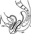
Figure 1. Partial prolapse of the uterus and vagina, elongatlo colli uteri: VU-urinary bladder; R-rectum; SD-Douglas space (according to Weibel).
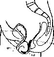
Figure 2. Complete prolapse of the uterus and vagina: VU-urinary bladder; U-uterus; R-rectum; SD-Douglas space (according to Weibel).
and posterior vaginal walls occurs, in which loops of intestine may descend into the resulting protrusion—so-called vaginal hernia (enterocele vaginalis anterior, posterior). During lactation, in the climacteric period, with atrophic processes, etc., vaginal displacements may be observed as an independent phenomenon; in the overwhelming majority of cases, however, vaginal displacements occur together with descent or prolapse of the uterus. In nulliparous women and in girls, uterine prolapse is rare. Thus, childbirth is one of the important etiological factors in prolapse. Already during pregnancy, the vagina becomes relaxed, longer, wider; the fetal head, exerting pressure on the anterior vaginal wall, stretches and traumatizes the cellular tissue surrounding the cervix and vaginal vaults, which is particularly evident during the act of delivery; injuries, hemorrhages, tears of the pelvic floor muscles with operative delivery have long been considered the most frequent causes of uterine and vaginal prolapse. In this regard, tears and damage to the perineum have always been given special importance. The role of the vagina and urinary bladder, which must displace the uterus downward when prolapsed, was also noted. The elongation of the prolapsed vagina was used to explain changes in the cervix—its enlargement and elongation. According to Schroder, with prolapse of the anterior wall, the middle part of the cervical canal usually elongates, with prolapse of the entire vagina or only the posterior wall—the supravaginal part of the cervix. Halban and Tandler attribute almost exclusive importance in the origin of uterine prolapse precisely to the disruption of the integrity of the pelvic floor and perineum (the "supporting apparatus") and intra-abdominal pressure. M. levator ani, according to Halban-Tandler, has a slit, in the anterior part of which (hiatus genitalis) the vagina and urethra pass. With increased intra-abdominal pressure, the m. levator ani contracts, and this slit shortens in the longitudinal and transverse directions. In addition, the hiatus genitalis is closed by the urogenital diaphragm located below it
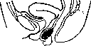
Figure 3. Prolapse of the posterior vaginal wall and rectocele (according to E. Martin).
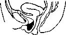
Figure 4. Prolapse of the anterior vaginal wall and cystocele (according to E. Martin). (diaphragma urogenitalis), the posterior part of which is in close connection with the external muscles of the perineum. With increased intra-abdominal pressure, the uterus, located in anteversio, is pressed against the urinary bladder and symphysis. The intra-abdominal pressure acting on the urinary bladder is transmitted to the anterior vaginal wall and the urogenital diaphragm, and the anterior vaginal wall is pressed against the posterior one, which has support from the perineum (see Figure 5). The conditions for preventing displacement of pelvic organs would be less favorable if intra-abdominal pressure acted downward (defecation, straining); ultimately, expansion of the hiatus genitalis occurs. Under such conditions, a certain part of the urinary bladder and the upper part of the anterior vaginal wall lose support from the m. levator ani and are therefore held only by the urogenital diaphragm and perineum. According to Halban-Tandler, such expansion of the hiatus genitalis has no significance for the uterus if it is in the position of anteversio, since its axis then passes behind the mentioned opening. With tears of the perineum and urogenital diaphragm, the anterior vaginal wall, along with the urinary bladder, loses its support, forming a cystocele. The latter can exist separately or together with uterine prolapse, which, in the case that is often observed with perineal tears, involves damage to the m. levator ani, with the dimensions of the hiatus genitalis increasing from 4 to 7½ cm in the longitudinal and from 2½ to 6½ cm in the transverse direction. For uterine prolapse, according to Halban-Tandler, preliminary descent of the uterus and its retroverted position are also necessary, whereby the cervix, and sometimes part of the body of the uterus, enters the expanded hiatus genitalis and is expelled through these so-called hernial gates by intra-abdominal pressure. Subsequently, due to the descent of the uterus, the upper part of the posterior vaginal wall everts and also descends into the hiatus genitalis. The part of the uterus located outside the latter is pressed and fixed by intra-abdominal pressure to the underlying tissues. Uterine prolapse progresses as the hiatus genitalis enlarges. In rarer cases, uterine prolapse occurs, according to Halban-Tandler, in an anteverted position. The uterus, located in anteversio, descending deeper, enters the area of the hernial gates. Intra-abdominal pressure acts in these
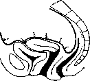
Figure 5. Diagram of the effect of increased intra-abdominal pressure on the uterus in anteversio. The black line (Figures 5, 6, 8) indicates the sphere of action of intra-abdominal pressure, arrows indicate the direction of the driving force (according to Halban-Tandler).
under conditions on the posterior wall of the uterus, pushing it toward the pubic symphysis and urogenital diaphragm. As a result, the posterior fornix and the intestinal loops located there are pushed out and protrude into the vagina (enterocele vaginalis), with the cervix, falling into the area of the genital hiatus, prolapses and elongates. The doctrine of Halban-Tandler can serve to explain uterine prolapse only in some special cases of severe trauma to the pelvic floor muscles (for example, uterine prolapse after gebostetomy, after application of high forceps). Kustner, pointing to the insufficiency of the pelvic floor as a cause favorable to uterine prolapse, attaches great importance to the stability of connective tissue and, especially, to the deviation of the uterus backward. Emphasizing the importance of connective tissue formations, Kustner in this case follows Schulze, who considered uterine prolapse as the final stage of insufficiency of parametrial, paravaginal, and especially perivesicular fat, as well as connective, elastic, fascial and fatty tissue, considering retroversio-flexio uteri as the initial stage of such prolapse. Some authors (Rosthorn, Freund and others) had earlier pointed to the important significance of connective tissue in the broad sense of the word (the "suspending" apparatus) for holding the uterus and vagina in normal position. This "suspending" apparatus is described in detail by E. Martin under the name retinaculum uteri (the mesh apparatus of the uterus). In this strengthening connective tissue apparatus of the uterus, according to Martin, three parts can be distinguished: anterior (pars anterior), middle (pars media) and posterior (pars posterior). The anterior part includes connective tissue bundles that make up the vesicocervical and pubovesical ligaments. The middle part is formed from similar connective tissue bundles, with an admixture of smooth muscle fibers, located at the base of the broad ligaments of the uterus (lig. cardinalia). Finally, the posterior part of the connective tissue apparatus consists of fibers forming the retractors (lig. sacro-uterina). Based on anatomical research and clinical data, Martin and Bumm, giving due importance to the muscular apparatus of the pelvic floor and the role of intra-abdominal pressure, come to the conclusion that the cause of prolapse and uterine prolapse lies in the insufficiency of connective tissue formations. In some cases, this insufficiency is primary, in others it is of secondary origin, after the supporting apparatus has become inadequate. Disagreements about the predominant significance of individual tissues in the etiology of uterine prolapse and vaginal prolapse have subsequently been considerably smoothed out. Halban himself now recognizes the role not only of the pelvic floor muscles, but also of connective tissue formations and especially emphasizes the important significance which the fascial plate of the anterior vaginal wall has, which together with the pelvic fascia closes the genital hiatus and supports the bladder. Halban also explains the origin of retroversio uteri and the subsequent displacement of the uterus downward with well-developed m. levator ani by the relaxation of connective tissue formations. Martin, proposing to suture the fasciae in all cases of uterine displacement, thus also extends the significance of the muscular diaphragm of the pelvis, since, undoubtedly, strengthening the fasciae contributes to improving the functional capacity of the muscle itself. Thus, there should be no complete functional demarcation between the pelvic floor muscles and connective tissue formations. The pelvic floor represents a complex formation consisting of muscular and various connective tissue formations and functions as a single structural whole (Scipiades). Agreeing with this, some (Menge, Jaschke, Sellheim) still assign a greater role to the pelvic floor muscles. Sellheim calls it the "obligatory" apparatus holding the pelvic organs, and connective tissue formations the "facultative" apparatus. The very mechanism of the occurrence of uterine prolapse and vaginal prolapse is also interpreted differently. Halban-Tandler and Kustner recognize only primary forms of prolapse; Grusdev, R. Schroeder, E. Martin admit the participation of neighboring organs (vagina, bladder) in the origin of uterine prolapse - secondary prolapse. It is understandable that one can also speak of a mixed type of uterine prolapse. Many point out that retroversio, retroflexio uteri, the "saddle" position of the uterus especially favor the occurrence of prolapse (see below). Nevertheless, it must be admitted that in details the mechanism of prolapse is little known to us. It must be emphasized that any displacement of the uterus and vagina downward (excluding complete prolapse) is in relatively rare cases a stationary phenomenon; usually it progresses and moreover with various individual deviations which cannot always be explained by existing schemes. Thus, in some cases, with seemingly equal conditions in terms of tissue damage, displacements and prolapse of the vagina are insignificant, while in others, on the contrary, the prolapse is quite fully expressed and moreover progresses rapidly. In cases of perineal ruptures, for example, with destruction of the perineal center (the point where the main perineal muscles, all pelvic fasciae converge and part of the fibers of m. levator ani are interwoven), when, therefore, all conditions exist for uterine prolapse and vaginal prolapse - in some cases there is only gaping of the genital cleft, in others - prolapse of the vagina, and finally in third cases - uterine prolapse. Here, much can seemingly be explained by the state of intra-abdominal pressure. Indeed, as observations show, in women engaged in heavy physical labor, uterine prolapse occurs especially frequently. The influence of straining in persistent constipation, coughing, etc., has also been noted. This, however, does not completely resolve the question. For many it has long become obvious that in elucidating the etiology of uterine prolapse and vaginal prolapse it is necessary to recognize the significance of such a major factor as the functional state of tissues. According to the data of Hoeven, for example, it is seen that uterine prolapse most often develops after the first birth, then after the second and third, and then cases of prolapse, as the number of births increases, significantly decrease. This can be explained by the fact that birth trauma serves as a test for the stability of tissues, which sometimes, especially after the first birth, is quickly lost, while in others it retains its properties. The study of constitutional features has fully confirmed this assumption. Jaschke in 490 cases of uterine and vaginal prolapse noted constitutional anomalies in 477 cases. A special tendency to displacement of the uterus is noted in asthenic women, whose tissues, having reduced tone, easily undergo overstretching. It is quite understandable that with such a state of tissues in women suffering from uterine prolapse, displacement of other abdominal organs (gastroptosis, wandering kidney, etc.) and the presence of hernia can often be observed. Inadequacy of tissues and consequently uterine prolapse and vaginal prolapse may also manifest in other diseases (tuberculosis, diabetes, chlorosis, alcoholism, rickets, etc.). Uterine prolapse and vaginal prolapse is often discovered only with the onset of the preclimacteric and climacteric periods, when the elasticity and stability of tissues decrease and their atrophic state develops. The large number of cases of uterine prolapse and vaginal prolapse during wartime, when the population was in conditions of poor nutrition, also indicates the role of tissue inadequacy in the etiology of this condition. All this can explain the frequent cases of prolapse, uterine prolapse and vaginal prolapse in persons engaged in heavy labor under conditions of poor nutrition. Early onset of work soon after childbirth has long been noted as a substantially important etiological factor in the origin of uterine prolapse, explained not only by increased intra-abdominal pressure but also by reduced functional state of tissues. There are indications that prolonged work in a sitting position leads to uterine prolapse, namely, depending on the developing inadequacy of pelvic floor tissues. With greater justification here should be included weakenings of pelvic tissues on the basis of disturbed innervation in spina bifida occulta in girls and nulliparous women. In such cases, hypoplasia of tissues constituting the pelvic floor - congenitally inadequate development of muscles and connective tissue formations - can also be assumed. With complete paralysis of the third and fourth sacral nerves supplying the pelvic muscles, the formation of uterine prolapse in spina bifida is observed in the first days of the child's life. Certain forms of infantilism, which are characterized by rigidity of tissues, their small stretchability, due to which they undergo significant trauma during the act of childbirth, can also play a significant role in the origin of uterine prolapse. In general, here the same relationships are created,
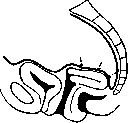
Figure 6. Diagram of the action of increased intra-abdominal pressure in retro-versio-flexio (according to Halban-Tandler).
Ban-Tandler. In elderly primiparas, who often subsequently exhibit uterine prolapse and vaginal prolapse (Fetzer). Speaking of the role of abnormal constitutional factors, it is necessary to point out the significance in the etiology of prolapses of the pelvic inclination (Flatau). Special mention in the etiology of uterine prolapse is deserved by retroversio and retroversio-flexio uteri. According to many authors, such a position has enormous significance, because in it the intra-abdominal pressure easily displaces the uterus downward, along the axis of the vagina. However, the 'median' position of the uterus, retroversio, is for the most part only temporary and therefore, in essence, it cannot have the significance that Schulze, Küstner, and Halban attribute to it. According to Halban-Tandler, the uterus, being pressed by the action of intra-abdominal pressure against the plate of m. levat. ani, descends only to a certain limit; being in the sphere of hiatus genitalis, only the cervix uteri prolapses and elongates (see Figure 6). However, far from always does retroflexio uteri, even when existing for a long time, accompany such changes and lead to uterine prolapse. Retroflexio does not always accompany uterine descent, and this phenomenon is often only apparent (see Figure 7) (Winter, Meuge, Schroder). Uterine descent can occur only with weakening of the pelvic floor tissues; according to Halban, descent becomes possible, besides this, only in the case that the uterus assumes the position of retroversio (see Figure 8). In fact, this is not so: uterine descent is observed also with anterversio uteri, as is evident from the clinical data of Küstler, Jaschke. If the uterus subsequently prolapses and assumes the median position, retroversion, retroflexio, this happens ONLY due to the insufficiency of the pelvic tissues that preceded this position of the uterus. Observations show that operations that only correct the position of the uterus, in retroflexio uteri and its descent, often do not succeed. On the other hand, it has been noted that in many cases it is sufficient to restore the pelvic floor for the uterus subsequently to assume the position of anteversio. Therefore, 'preventive' correction of the position of the uterus to prevent its prolapse (Küstner) is in many ways inferior to the proposal of Selheim to perform only plastic surgery of the pelvic floor in such cases.
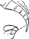
Figure 7. With a thick line is shown the typical normal position of the uterus; with a dotted line is shown the apparent low position of the vaginal part in retroflexio uteri; with a thin line is shown the actual prolapse of the uterus. Observations show that operations, only correcting the position of the uterus, in retroflexio uteri and its descent often do not give success. On the other hand, it has been noted that in many cases it is sufficient to restore the pelvic floor for the uterus subsequently to assume the position of anteversio. Therefore, 'preventive' correction of the position of the uterus to prevent its prolapse (Küstner) is in many ways inferior to the proposal of Selheim to perform only plastic surgery of the pelvic floor in such cases.
In the initial stages, uterine prolapse is detected only with increased intra-abdominal pressure; subsequently the uterus begins to prolapse without any strain, reducing itself when in the recumbent position; with progressive weakening of tissue tone, uterine prolapse and vaginal prolapse become stationary, accompanied by changes depending on congestive phenomena. In rare cases, an acute onset of uterine prolapse is noted—with severe straining, falling from a great height onto the feet, etc. Sometimes uterine prolapse and vaginal prolapse are associated with the presence of a large tumor in the abdominal cavity, ascites, etc. The changes observed in the sexual organs in uterine prolapse and vaginal prolapse are various. With slight degrees of descent, the genital gape is open; during straining, the walls of the vagina bulge out—more significantly and more often only the anterior wall. During straining, when the uterus is significantly displaced, usually first the anterior wall bulges out, then the cervix uteri appears from the genital gape, and finally the posterior wall of the vagina. With significantly expressed hysterocele, the relationships may be reversed. With complete prolapse from the genital gape, the hypertrophied cervix uteri and the walls of the vagina protrude. The cervix is edematous, cyanotic; the external os is often gaping. On the cervix there are often ulcers. With significant elongation of the cervix, retention of discharges from the uterine cavity is possible, which gives rise to the formation of pyometra. With large prolapses, the vaginal mucosa loses its character, its folds disappear, it becomes dry and epidermized. On the basis of pressure and trauma, ulcers form, which become infected and turn into deep ulcers. In the uterus, congestive phenomena are noted; its cavity is significantly enlarged (up to 18-20 cm), mainly due to the elongated cervix. The shape of the urinary bladder is significantly altered, which is divided into supra- and infra-symphysar parts; in the latter urine is retained, which favors infection and the formation of cystitis; with bends of the ureters, urine retention and development of pyelonephritis are possible. In uterine prolapse and vaginal prolapse, from the genital gape in severe cases a tumor protrudes, reaching the size of a child's head; the presence of an opening on its lower pole serves as an important sign in differential diagnosis. Grasping this tumor with the hand, through the vaginal walls it is clearly possible to palpate
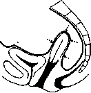
Figure 8. Diagram of the action of increased intra-abdominal pressure on the uterus in retroversio (according to Halban-Tandler).
an elongated cervix, and sometimes the entire uterus. In complete uterine prolapse, hypertrophy of the cervix is usually absent. Palpation determines to what extent the vagina participates in the prolapse. By introducing a catheter, which is directed downward, it is established that the urinary bladder is located in front of the vaginal wall; rectocele is additionally diagnosed by examination per rectum. Due to severe swelling, the volume of the prolapsed part can become so significant that repositioning becomes difficult, sometimes directly impossible; tissue necrosis develops, and without timely assistance, patients die from septicemia. Painful manifestations are not always dependent on the degree of prolapse. In nervous women, even slight downward displacement of the uterus often causes a feeling of pressure, pulling pains in the groin and lower back; in other cases, even with significantly pronounced prolapse, the pains do not bother the woman at all. Pains in the lower back and lower abdomen often appear after heavy work, decreasing and sometimes disappearing completely when sitting or lying down. Leukorrhea and bloody discharge are often noted. Symptoms from the urinary bladder are detected early - frequent urges, difficult urination. With significantly pronounced cystocele, patients are often forced to reposition the prolapsed uterus before urination. With significantly pronounced rectocele, defecation is sometimes delayed (part of the feces does not enter the anal opening but goes into the rectocele). Due to stagnation in the pelvis, menstruation often becomes more profuse. Conception is possible, but with severe forms of prolapse it is rarely observed; during pregnancy, the uterus gradually repositions itself spontaneously. With acute uterine prolapse, severe pains are noted with signs of vagus nerve irritation - weakness, fainting, dizziness, vomiting. Prevention. It is necessary to pay great attention to prevention. Reasonably applied sports and physical exercises lead to strengthening of the tissues. It is necessary, as much as possible, to limit operative interventions during childbirth. The use of forceps is especially harmful when the head is high (significant trauma to the levator muscle). In primiparas, delivery should be carried out with special care. It is desirable to use perineotomy more widely, as this prevents excessive trauma to the pubo-rectal part of the levator muscle and overstretching of the pelvic fasciae. Any tears of the perineum after childbirth should be carefully sutured. Great attention should be paid to the rational management of the postpartum period. Early ambulation promotes better involution, contributes to the proper function of the bladder and intestines. To strengthen the abdominal wall and pelvic floor, it is recommended to perform light gymnastics systematically and cautiously from the 2nd-3rd day after childbirth. Lifting from a horizontal position causes contraction of the abdominal and pelvic muscles. Drawing in of the anus, as is done to hold back defecation, coughing - are accompanied by contraction of the levator ani muscle. Along with this, heavy work requiring strain and increased intra-abdominal pressure should be prohibited. Treatment of uterine prolapse is mainly operative; in case of the patient's unwillingness to undergo surgery or contraindications to it, orthopedic treatment (pessary) and massage are still used to this day. Many different methods have been proposed for the operative treatment of prolapses. The choice of method depends largely on the degree of prolapse and the age of the patient, since the most radical methods are associated with disruption of the ability to conceive. In some cases, indications for such operations can be extended to women of childbearing age, as the results of plastic operations often become null and void after childbirth. Before the operation, the patient should be put to bed for several days, the uterus should be repositioned, which reduces hyperemia and swelling of the tissues; ulcers are treated in the usual way (cauterization, ointments with ichthyol, etc.). For small displacements of the vagina and uterus, plastic operations on the perineum and vagina are used, which achieve not only narrowing of the genital cleft and vagina, but mainly restoration of the pelvic floor, elimination of displacements of the vagina, as well as neighboring organs - the urinary bladder and rectum. Indications for these operations should be expanded: the earlier they are performed, the greater the chances of success. Usually, in the presence of a retroflexed uterus, the Alexander-Adaja operation (see) or ventrofixation (see) is added, although some authors consider it quite sufficient to limit oneself to plastic operations alone (D. O. Ott, Heidenhain, Gudim-Levkovich, etc.). The use of ventrofixation alone in cases of a tendency of the uterus to prolapse should be considered a mistake. For slight uterine prolapse and vaginal prolapse in elderly women, vaginofixation (see) with perineoplasty is appropriate. For significantly pronounced and complete uterine prolapse, various operative methods are used. Some authors, in addition to plastic operations on the perineum and vagina, add a firm and high ventrofixation of the uterus in order not to burden the pelvic floor, and also to pull up the weakened connective tissue formations of the pelvis, which facilitates the maintenance of the position of the urinary bladder. To this type of ventrofixation belong the so-called Promontorifixur Küstner-Schmidt, in which the uterus is attached to the periosteum between the first sacral and the last lumbar vertebra, and symphysiofixation (Symphisofixur nach Liepmann) - attachment of the anterior surface of the uterus to the periosteum of the posterior surface of the pubic bone. In the presence of a hypertrophied cervix, amputation of the cervix is desirable, which should, however, be avoided in women of childbearing age. For the purpose of possibly more reliable strengthening of the pelvic floor, instead of the usual plastic surgery, isolated suturing of the levator ani muscle is recommended. G. Freund, Menge propose transplantation of the fascia of the thigh, and Halban-Tandler - transplantation of muscle from the gluteus maximus. Many authors in patients in the climacteric or close to it age, especially with significantly pronounced cystocele, readily apply the Wertheim-Shaute operation (see Wertheim's operation). With significantly pronounced changes in the prolapsed organs, it is often necessary to remove the uterus per vaginam. Frequent recurrences - vaginal prolapse - have forced to abandon the wider application of this method. Individual authors (Parsamov), using careful plastic surgery of the pelvic floor, however, report good results. Fritsch, in order to avoid recurrences, proposed to remove not only the uterus but also the vagina. In old age, when coitus is absent, median colporrhaphy (colporrhaphia mediana Neugebauer-Le Fort) can be of great service in prolapse, which consists in cutting out two flaps 2-3 cm wide from the anterior and posterior walls of the vagina; the denuded surface is sutured, which prevents uterine and vaginal prolapse. In general, one should refrain from using too complex operations, as this creates a danger to the patient's life; with significantly pronounced prolapses, with weak tissues, one can never be sure of obtaining a durable success. After the operation, it is desirable to apply measures aimed at improving the general condition, limitation of heavy work, etc. Orthopedic treatment (pessaries). For uterine prolapse, the most commonly used are Hodge pessaries, eight-shaped Schultze pessaries, soft rubber Mayer rings, etc. A pessary cannot cure prolapse. By exerting pressure on the surrounding tissues, the ring ultimately leads to worsening of the pathological condition, as a result of which, after a certain time, the inserted pessary falls out and has to be replaced with a larger pessary. When wearing a pessary, the patient should be under medical supervision, perform daily douching and remove the pessary from time to time. If these rules are not observed, bedsores form. The pessary, sinking into the underlying tissues, becomes surrounded by granulation tissue, and later by scar tissue; in this case, operative removal of the pessary sometimes presents great difficulties, and injury to neighboring organs is possible. The hysterophore is a pear-shaped or other-shaped pelotte, which is inserted into the vagina and held by tapes attached to a belt worn around the waist (see the figure in the article Hysterophore). Massage and special gymnastics, proposed by Ture Brandt for the purpose of strengthening the pelvic musculature, are unsuitable for curing prolapse; these measures are appropriate only with prolonged use as an auxiliary means for slight downward displacements of the uterus.
Related articles
Mentioned in
Cite this page
“Uterine Prolapse.” Soviet Medical Encyclopedia. English translation of Bolshaya Meditsinskaya Entsiklopediya, 1st ed. (Moscow, 1928–1936), ed. N. A. Semashko. https://sovietmedicalencyclopedia.pages.dev/article/uterine-prolapse/