Uterine Inversion
Historical document, translated for reference. It reflects medical knowledge of the 1920s–30s and is not medical advice.
Summary
This article from the 1928-1936 Great Medical Encyclopedia examines the causes, symptoms, diagnosis, and treatment of uterine inversion, distinguishing between postpartum and non-puerperal cases.
Encyclopedia article (1928–1936)
UTERINE INVERSION is such a displacement of its body where the internal surface of the body, covered by the mucous membrane, is fully or partially indented into the uterine cavity and passes through the dilated os into the vagina, during which the body surface covered by the peritoneum descends into the funnel formed by the lateral parts, into which the tubes and ovaries are naturally partially drawn. If a part of the uterine body is indented into the cavity, this condition is called incomplete inversion; if the entire body is inverted and passes through the os into the vagina, complete inversion is obtained (see figure 1). In complete inversion, the body of the uterus may be located in the vagina, and in other cases even emerge outside through the vulvar ring together with the walls of the vagina. There is no consensus on the causes of uterine inversion development. While some (Martin, Eulenburg, and others) believe that uterine inversion develops as a result of unskilled or
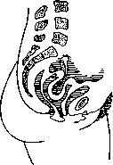
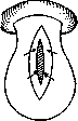
Figure 1. Scheme of complete inversion of the uterus.
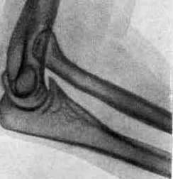
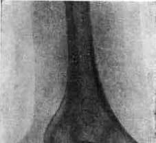
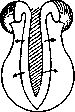
Figure 2. Scheme of Küstner's method.

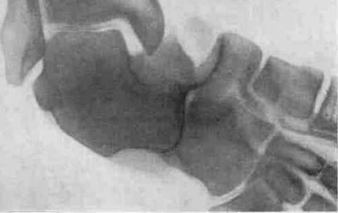
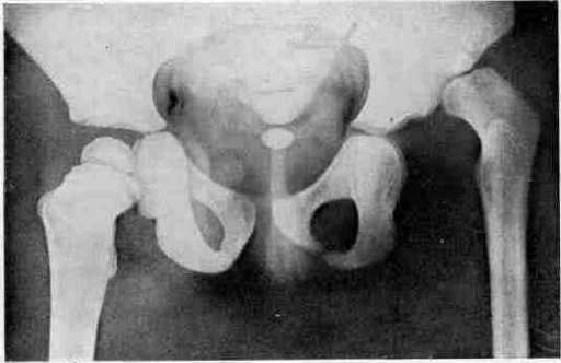
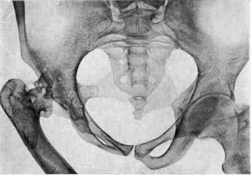
irrational obstetric care (Credé's method with a insufficiently well-contracted uterus, pulling on the umbilical cord), others (Spiegelberg, Thorn, Kroner, Jones, Beckman) attach great importance to the change in pressure within the uterine cavity during its rapid emptying. Beckman and Thorn, based on their materials, conclude that the number of spontaneous uterine inversions prevails over violent ones. Non-puerperal (oncogenetic) inversions are those that occur outside the pregnant state of a woman and depend in most cases on the drawing of the uterine walls by tumors sitting on it, indented by uterine contractions towards the os and through the os. One of the first places among these uterine tumors is occupied by submucous fibromyomas sitting at the fundus of the uterus; less often by sarcomas and carcinomas. Stephan collected 17 cases of uterine inversion together with its complete prolapse, where inversion also occurred in the absence of a tumor and not in the postpartum period. He associates such an inversion with prolapse and calls it "prolapsinversio." If postpartum inversion is not corrected in time, the gradually contracting os compresses the inverted body, and stasis with edema develops in it, and in severe cases even necrosis. Symptoms of acute uterine inversion include shock and hemorrhage from the placental site. Shock is explained (Schauta, Geiseler) by nervous influence. Along with these main symptoms, there are such phenomena as a feeling of pressure in the vagina, tenesmus, and the urge to urinate. Symptoms of chronic uterine inversion are pains in the sacrum, abdomen, and a feeling of distension in the pelvis. Bloody serous discharges or hemorrhages are also observed, which are sustained due to the kinking and compression of veins in the narrowed cervix. Snegirev notes the unusual pallor of women having an inversion, which, in his opinion, depends not on blood loss (sometimes not so great), but on spasm in the cervical membrane and some pinching of the tubes. Pallor may depend, although not on strong, but on prolonged bloody discharge. Diagnosis in cases of complete postpartum inversion does not present difficulties if phenomena of severe pinching and necrosis are not joined, but even in such cases, the absence of the uterine body on bimanual examination, the definition above the tumor located in the vagina, of a ring-shaped formation encompassing the tumor (os) facilitate diagnosis (it can be confused with a nascent fibroid or cancer of the vaginal portion). In uncomplicated cases, the uneven surface of the tumor, the presence of the tube openings right there, the absence of the uterine body in its usual place, and the presence of a funnel make it possible to quickly orient oneself. Examination under anesthesia, as well as per rectum, facilitates diagnosis. Prophylaxis. According to statistics, inversion occurs in the postpartum period mostly spontaneously; a certain percentage is nevertheless caused by irrational management of the postpartum period. In view of this, it is necessary to strictly observe all the rules of modern obstetrics when managing the placental period and especially carefully perform Credé's method, which is far from indifferent and unsafe in unskilled hands. Therapy in fresh cases of uterine inversion reduces to reduction by manual maneuvers. Under anesthesia, trying to press on the corners or lateral parts of the uterus, they carefully advance the uterus through the os. One can proceed to reduction only under the condition of a sufficiently good condition of the puerperal woman. In case of unsuccessful reduction, there is a danger, if time is lost, of losing the patient from hemorrhage and shock, in view of which reduction using a colpeurynter or tamponade can be resorted to only in the absence of the indicated complications. In complete inversions, in the presence of continuing hemorrhages, temporary ligation according to Kocks is recommended. Ligation should not be very tight; it should only restrain bleeding. In the therapy of chronic cases of inversion of puerperal origin, there are many methods; most gynecologists use Küstner's method: after opening the pouch of Douglas with a transverse incision, an index finger is inserted penetrating into the funnel, and the posterior wall of the uterus is dissected enough to accomplish reduction; then the wall is sutured, the uterus is reduced, and the posterior fornix is restored (see figure 2). In the method of Piccoli, Westermark, Borelius, Duret, the dissection of the uterus is continued onto the cervix (see figure 3). Of other methods, one can point to the method of Spinelli, Thorn with anterior colpotomy. The abdominal wall route (recommended by Thomas, Munde, Werke, and others) consists in dissecting the ring from the outside after detaching the bladder; one can also try to produce reduction by the path of careful pulling on the appendages and ligaments of the body, during which in case of failure the body is gradually dissected. In cases where the tumor was the cause of the inversion, it is possible, by enucleating the latter, to reduce the uterus; in other cases, with significant damage to the uterine body by the tumor and with its malignancy, complete removal of the uterus is necessary (in the latter case, with appendages). Statistics: out of 627 cases collected by Zangemeister, 531 inversions occurred after childbirth, premature births, and abortions; of them, normal births account for 521 cases; with tumors, inversion was observed 83 times, of which in 79 cases with myomas; idiopathic inversions were noted 13. Here also belong 12 cases of inversion after death, during childbirth in dead women. The frequency of postpartum uterine inversions is estimated at 1:10,000 (Zangemeister). Among Russian obstetricians, Beckman (on the material of the St. Petersburg Lying-in
institution for the period from 1840 to 1912) notes 2 cases of inversion per 270,000 births; Krivsky for 10 years (based on the material of St. Petersburg city maternity shelters) does not indicate a single case. As for inversions of non-puerperal origin, they represent an even greater rarity, constituting approximately 9-11% of the total number of uterine inversions.
F. Solovyov
Related articles
Mentioned in
Cite this page
“Uterine Inversion.” Soviet Medical Encyclopedia. English translation of Bolshaya Meditsinskaya Entsiklopediya, 1st ed. (Moscow, 1928–1936), ed. N. A. Semashko. https://sovietmedicalencyclopedia.pages.dev/article/uterine-inversion/