Ultraviolet Rays
Historical document, translated for reference. It reflects medical knowledge of the 1920s–30s and is not medical advice.
Summary
Ultraviolet rays are invisible radiation with wavelengths from approximately 4,000 to 500 Angstroms. They were discovered relatively late in 1801 by Ritter and have significant biological effects including bactericidal action, biochemical changes, and erythema production.
Encyclopedia article (1928–1936)
ULTRAVIOLET RAYS, invisible radiation occupying the wavelength range from approximately 4,000 to 500 Å (Angstrom = 0.0001 mm). Since instruments with glass optics do not transmit rays shorter than 3,500 Å, ultraviolet rays were discovered relatively late: Ritter in 1801 detected them by their action on silver chloride. The ultraviolet region is conventionally divided into the following groups: near ultraviolet - from 4,000 to 3,500 Å, far ultraviolet - 3,500-2,500 Å (Fig. 1), extreme ultraviolet - 2,500-1,850 Å.
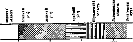
Figure 1
Regions of ultraviolet rays (wavelength in Å). 1. Near ultraviolet - 4,000-3,500 Å. 2. Far ultraviolet - 3,500-2,500 Å. 3. Extreme ultraviolet - 2,500-1,850 Å. 4. Schumann region - 1,850-1,200 Å. 5. Lyman region - 1,200-500 Å; directly adjacent to the Lyman region is the region of soft X-rays. Working with ultraviolet radiation presents considerable experimental difficulties. The following table shows the transparency limits for some of the most commonly used substances: Substance | Limit Substance | Limit Ordinary glass | 3,500 Å Fluorite (CaF2) | 1,850 Å Quartz crystal | 2,850 Å Gelatin | 1,800 Å Fused quartz | 2,500 Å Air | 1,200 Å. To work with rays shorter than 1,850 Å, it is necessary to evacuate the air from the instruments. In the Lyman region, there are no solid or liquid transparent substances. For practical purposes, only the region up to 1,850 Å is significant. Ultraviolet rays exhibit noticeable biological action only starting from 3,200 Å and shorter (Dorno rays). Rays from 2,800 to 1,850 Å are called mitogenetic rays (see).-Of the biological actions of ultraviolet rays, we can note: 1) strong bactericidal action (see below); 2) various biochemical actions, for example, the formation of vitamin B in milk and other irradiated preparations (see Irradiated preparations); 3) the appearance of erythema on areas of skin that have been irradiated. In this case, wavelengths of 3,100-2,900 Å are particularly active. The depth of penetration of ultraviolet rays through tissues depends on their intensity (see Phototherapy). The most accessible source of ultraviolet rays is the sun, whose ultraviolet radiation in middle latitudes during summer reaches down to 3,000 Å. Artificial light sources provide much more intense radiation, of which the principal ones are the mercury-quartz lamp, the arc between metal and carbon electrodes, and the high-voltage spark discharge. The mercury arc gives a series of intense lines extending to 2,500 Å; the maximum intensity is at 3,000 Å. Arcs between carbon and metal electrodes are an extremely powerful source of near ultraviolet radiation, though inferior to the mercury arc in the far ultraviolet. Finally, the spark discharge is used mainly for work in the extreme ultraviolet, where it gives a series of exceptionally intense lines depending on the electrode material (Cd, Zn, Al).-Of the applications of ultraviolet rays, we can mention the ultraviolet microscope, in which the increased resolving power of the optical system that appears with decreasing wavelength is utilized. Since quartz and fluorite optics cannot be well achromatized, it is necessary to use monochromatic light through a special quartz monochromator for illumination. Fig. 2 (a and b) shows the diagram of a monochromatic ultraviolet microscope. Rays from source F after spectral decomposition in prisms P1 and P2 pass through a diaphragm to the total internal reflection prism P and are reflected onto the object. The path of the rays after prism P is shown in part b of Fig. 2. Here V is a plate of fluorescent uranium glass used for instrument alignment, K is the condenser, O is the slide and cover glass. Both dry and immersion systems exist; glycerin is usually used for immersion. The resolving power of such an ultraviolet microscope is several times higher than that of ordinary ones. For the therapeutic significance of ultraviolet rays, see Phototherapy, Heliotherapy.
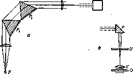
Figure 2.
a) Diagram of monochromatic ultraviolet microscope. b) Path of rays after prism P. F-source, P1 and P2-prisms, P-prism of total internal reflection, V-uranium glass plate, K-condenser, O-object slide and cover glass.
n. ultraviolet rays. Ultraviolet rays in microbiology. The first experiments, in which the bactericidal action of light was established, date back to 1877 and 1878 (see Light). Geissler in St. Petersburg (1891-1892) first showed that short-wave U. rays have the greatest bactericidal action, which was confirmed by Ward (1893) and Kruse (1895). At the end of the 19th century, interest in the bactericidal action of light increased due to the work of Finsen on phototherapy. A new field of knowledge emerged - Lichtmikrobiologie, systematically studied by Bie, Bang, Hertel, Thiele, Wolf, and Wiesner. In the following years, interest in the action of U. rays on bacteria declined. And only from the 1920s, with the refinement of methodology, a number of works devoted to this question reappeared. Already the first works showed that individual parts of the ultraviolet spectrum have a particularly strong bactericidal action. Thus, Bie observed that rays between 295 and 200 mμ are 12 times more effective than the entire spectrum of rays with a wavelength above 295 mμ. Bang established two maxima of intensity in the spectrum of a carbon arc lamp: a lower one - 360-340 mμ - and a higher one - 300-200 mμ. Coblentz and Fulton (1924) showed that to kill Bact. coli at a wavelength of 280-220 mμ, 7 times more energy is needed than when irradiated with waves of 220-170 mμ. Gates (1929), studying the action of U. rays on staphylococci, also showed that to obtain the same bactericidal effect, more energy must be used, the longer the waves being tested. Apparently, as shown by the work of Browning and Russ (1917, 1918), different parts of the spectrum are bactericidal for different bacteria: for Bact. coli - 296-220 mμ, for Bac. typhi abdom. - 300-210 mμ, for Staphyl. aureus - 295-238 mμ. Ehrismann and Noethling established that the smallest amount of energy causing the death of bacteria Staph. aureus, Bac. pyocyan., V. Finkler - at 265 mμ, Bact. coli - at 254 mμ, Bac. prodigiosus - at 281 mμ. It is even possible that different strains of the same bacterial species have different sensitivity to the same parts of the ultraviolet spectrum. If bacteria distributed in a thin layer on the surface of an agar dish (immediately after seeding) are irradiated, the rays of a mercury-quartz lamp at a distance of 50 cm kill the seeded bacteria in a few seconds; when irradiated with a carbon arc lamp, minutes are required. Different types of bacteria show different sensitivity in this case. With sufficient time, even the most resistant spore-forming types perish. In some works, data are given on the duration of irradiation necessary to kill various types of bacteria under these conditions. However, these figures are very relative, since the time required for killing by U. rays depends on a multitude of factors: the state of the irradiated bacteria (their age, density of seeding), temperature of the environment, pH of the medium, intensity of radiation, which will vary depending on the voltage of the supply network, on the age and individual qualities of the burner if a mercury-quartz lamp is used, on the type and quality of the carbons if the source of radiation is a carbon arc, etc. Bacteria can also be killed by U. rays when irradiated in a suspended state in a liquid. Suspensions must be in a quartz glass vessel. Here it is even more difficult to speak of the time required for irradiation, since it will depend, in addition to the factors mentioned, on the thickness of the irradiated layer, on the properties of the liquid, on the density of the suspension. The liquid must be colorless, since a colored liquid (e.g., broth) absorbs U. rays and thereby protects the suspended bacteria from the harmful action of the rays. Coblentz and Fulton attempted to calculate the amount of energy required to kill one bacterium. According to the authors' calculations, when irradiated with the most active rays, this energy amounts to 19.10-12 watt, or 4.5. 10-10 erg/cal. It is possible that the method of delivering radiant energy also has significance in this regard. Wiesner (1907) showed that the bactericidal action is enhanced when intermittent light actions are summed up. More recently, Coblentz and Fulton, comparing the action of continuous and intermittent irradiation, came to the conclusion that no difference is found in this case. The mechanism of the bactericidal action of U. rays remains unexplained to this day. Older authors explained this action by the formation of hydrogen peroxide or ozone in the medium in which the bacteria are located, which causes the death of the bacteria. But already Wiesner and a number of other authors explained the bactericidal action of U. rays by their direct action on bacterial cells. Bacteria absorb U. rays. According to some authors, changes in the electrical charge of bacterial bodies occur during this process, leading to their death. In small doses, U. rays have an irritating effect on bacteria. The old experiments of Hertel showed that during irradiation, activation of bacterial mobility first occurs, expressed in the bacteria fleeing from the irradiated area. After this, with further irradiation, a slowing of movement occurs, followed by death. Nadson and Philippov established that very small doses of U. rays accelerate the growth of yeast and mold fungi. The same was shown for bacteria by Trotsky and Sviridova. U. rays have a destructive effect on bacterial toxins. The question has been most studied in relation to diphtheria toxin. Hartoch, Schürmann, and Stiner showed that when irradiated with U. rays for 1 hour, a 1% solution of the toxin was detoxified to such an extent that 20 doses could be tolerated by guinea pigs without reaction. 3-hour irradiation of a 5% solution gave less clear results. Sometimes guinea pigs that received irradiated toxin died late without the phenomena characteristic of acute diphtheric intoxication. Thus, irradiation completely destroyed the toxic components acting locally, while the nervous and vascular toxins remained. These same authors established that irradiated diphtheria toxin lacks immunizing properties and also lost the ability to bind antitoxin. Later, Welch and Megrail (1930) came to the same conclusions, studying the action of a carbon arc on purified diphtheria toxin containing very little protein. However, these works do not answer the question of which parts of U. rays have the greatest destructive effect on the toxin, and what doses of U. rays are necessary to destroy the toxin. Ultraviolet rays have an effect on antibodies. The work of a number of researchers (Abelin, Stiner, Baroni, Jonesco-Mihaiesti, Doerr, Moldovan) has established the destructive effect of U. rays on serum antibodies. The most resistant are agglutinins and antitoxins, hemolysins, bacteriolysins, and substances that bind complement have less resistance, and finally complement is particularly sensitive. The time of irradiation required to destroy antibodies depends on the degree of dilution of the serum. The more diluted the serum, the faster the destruction of the antibodies contained in it occurs. For example, if complement under certain irradiation conditions in a 1/100 dilution is destroyed in 1 min., then under the same irradiation conditions, a 1/10 dilution is destroyed only in 5 min. When irradiating undiluted sera, just as when irradiating undiluted toxin, no effect is obtained.
Related articles
Mentioned in
Cite this page
“Ultraviolet Rays.” Soviet Medical Encyclopedia. English translation of Bolshaya Meditsinskaya Entsiklopediya, 1st ed. (Moscow, 1928–1936), ed. N. A. Semashko. https://sovietmedicalencyclopedia.pages.dev/article/ultraviolet-rays/