Cathode Rays
Historical document, translated for reference. It reflects medical knowledge of the 1920s–30s and is not medical advice.
Summary
Cathode rays are a straight beam of electrons emanating from the cathode when high voltage current passes through metal electrodes in a vacuum. These rays possess negative electrical charge, travel at high speeds, and can produce X-rays when striking solid surfaces.
Encyclopedia article (1928–1936)
Cathode rays, a straight beam of electrons (see), emanating from the cathode and arising when high voltage current is passed through metal electrodes placed in an extremely
high vacuum, also when
illuminating the sur-
A' face of met-
als,
placed in a vacuum, with ultraviolet rays and under certain other conditions, for example from the surface of an incandescent cathode. When cathode rays strike the surface of a solid body, X-rays (see) are produced. If a cylindrical sealed tube R, having the shape shown in Figure 1, with electrodes- cathode K and anode A sealed into it- is connected to the corresponding poles of a high voltage battery, an induction coil, or a high voltage transformer and then the gas is gradually pumped out of this tube, then at a pressure of about several tenths of a mm mercury column, a faint blue glow S emanating from cathode K becomes noticeable, causing
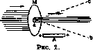
bright green fluorescence on the opposite wall L. The bluish glow of the residual gas and the green fluorescence of the glass are caused by cathode rays. Cathode rays exhibit the following properties. They pass through solid bodies, and if a screen with an opening M, made of a very thin metal sheet or glass, is placed in their path, then beyond this opening the cathode rays spread in a straight line having the dimensions of the opening (Figure 2). If a positively charged body A is brought near the beam K. l., it will bend toward this body, as
s shown in the form of ___. £j> a dashed line on Fig. 2. k m-_ -t ё Figure 3 If the surface A is charged negatively, then the cathode ray is repelled (see the dashed curve ac). By placing a magnetic field such that the positive magnetic pole N is in front and the negative is behind (Figure 3), deviation of the cathode beam KK (dashed MT) is caused in the plane perpendicular to the direction of the cathode beam and to the direction of the magnetic field lines (in the plane of the paper), and this deviation is greater the stronger the magnetic field. (The cathode ray* goes in the direction of the arrow.) By changing the direction of the magnetic field, the direction of deviation is also changed. The cathode ray K, falling on a metal cylinder C (Figure 4), protected from electrostatic action by an insulated metal shell A, connected to ground (insulator T), and located in the cathode tube, charges the cylinder negatively, which shows that K. l. carries a negative charge. If K. ray falls on an instrument with which one can measure t° (a thermometer or thermocouple), then the instrument shows an increase in temperature. Finally, if light objects that can be deflected are suspended in the path of K. l., then when K. l. strikes them, deflection occurs in the direction of movement of K. rays from the cathode to the opposite wall. All these phenomena allow us to recognize that K. rays are a stream of the smallest particles having a negative electric charge and equivalent to an electric current, which is deviated by a mag- A Figure 4.

netic field. The stream of negative particles is deviated by an electrostatic field, being attracted by a positively charged body and repelled by a negatively charged one. The high speed of the K. l. particles is the cause of the heating they produce: upon absorption they give up all their kinetic energy. K. l. can be released outside through an opening a in the wall S (Figure 5), covered with a thin aluminum plate having a thickness of a hundredth of a millimeter. Under these conditions, K. l. emerge outside as a glow. Recently, by this method, Coolidge has obtained K. rays of enormous intensity. These rays have a strong physiological effect: pro-
Fig. 5. duce severe burns
and can serve as a therapeutic agent. -The study of deviations of K. l. in magnetic and electric fields makes it possible to determine the ratio of charge to mass of the particles constituting K. l., and their speed, which turns out to be on the order of a tenth of the speed of light (about 30,000 km per sec.) and depends on the electric field. The charge of the particles of cathode rays is the same in magnitude as the charge of monovalent ions, and the mass is equal to -1/1837- the mass of a hydrogen atom. When using electrodes made of various metals and when introducing various gases into the tube R (Figure 1), one always obtains the same mass and the same charge for the particles of K. l. Thus, cathode rays are indeed a stream of primary negatively charged particles-electrons. At high speeds approaching the speed of light, electrons change their mass, and the study of these mass changes shows that the mass of an electron should be considered as a mass of electromagnetic ORIGIN.
P. Lazarev. Biological action of C. rays. Thanks to the work of Pauli and Coolidge, a special electron tube was constructed for obtaining C. rays of high intensity. This tube resembles an X-ray tube of the Coolidge type, i.e., it has a heated cathode; in the anticathode appendage of the tube, directly opposite the cathode, is placed a metal tube open in the direction of the cathode. At the opposite end of this tube is placed a metal window for the exit of electrons, i.e., cathode rays, into the surrounding air. This window is made of a nickel plate 0.0127 mm thick. To avoid damage to this plate, it is soldered onto a thick molybdenum grid; due to a special device, this entire window is cooled by running water. According to Pauli's calculations, the number of electrons obtained in such a tube is equal to the number of beta rays emitted by 1 mg of radium; their energy, however, is 4 million times greater than the energy of the X-rays formed by these same C. rays in the X-ray tube. Possessing such great energy, C. rays exhibit sharp biological action, which was studied by a number of authors. Pauli, Grober, and Hartmann established that plant cells, apparently due to the presence of a denser shell, react differently to illumination by C. rays than animal cells: in the latter, the shell is first damaged, and then due to the release of its contents, the cell itself perishes; in the plant cell, however, despite the integrity of its shell, its contents perish. Baensch and Finsterbusch illuminated the leaves of some plants with C. rays and observed the appearance of bubbles filled with milky sap. The aforementioned authors also studied the bactericidal action of C. rays. It turns out that C. rays kill any bacteria and spores within 10 seconds. This bactericidal action was used by Htihne together with Baensch and Finsterbusch for the sterilization of catgut. The action of C. rays on the animal organism was studied by many authors. The shaved abdominal skin of a guinea pig was subjected to illumination. Changes in the skin depend on the dose of C. rays applied. With a small dose, already after a few hours, redness of the skin, painful to the touch, is observed at the illuminated site, i.e., signs of inflammation. With somewhat larger doses, a stronger redness appears, which lasts up to 14 days; then the exfoliation of the epidermis and its replacement with new tissue begins. With a further increase in dose, blisters and finally ulcers appear. Thus, here the same phenomena are observed as with illumination by X-rays; the difference lies only in the fact that X-ray ulcers have a weak tendency to heal, while ulcers from C. rays heal in 8-15 days, leaving behind a delicate, shiny scar; keloids do not form. In general, the action of C. rays is limited only to the skin, as they do not penetrate deeper. Of animal tissues, epithelial tissue is most sensitive to them, less so is muscular tissue, and even less is nervous tissue, i.e., the sensitivity of tissues to them runs parallel to X-ray sensitivity. The eye is very sensitive to C. rays: with small doses, conjunctivitis appears, with larger ones-clouding of the cornea and ulcers on it. C. rays also have a general action: not only after intensified general illumination of the experimental animal, but also after intensified local illumination, the animal dies after a few days. Initially, appetite decreases, the animal becomes restless, its hair bristles, and it dies without any particular characteristic symptoms. In mass autopsies of such animals, no particular typical changes in the internal organs could be found; in only one animal was a picture of liver cirrhosis found. Thus, the cause of death of the animals has not yet been established. The question of whether this biological action is explained exclusively by the action of C. rays or whether X-rays act here, arising when C. rays hit the window, causes much controversy. Baensch and Finsterbusch showed that if the illuminated place is covered with a postcard, then no changes are noticed. This proves that the changes occur under the influence of C. rays, since X-rays would pass through the postcard. They proved the predominant predominance of C. rays in this case by a whole series of physical measurements. On the other hand, the same authors agree that when C. rays hit the skin, soft X-rays arise in it, which also act biologically, adding to the action of the cathode rays. To reduce the amount of X-rays produced, the illuminated place must be covered with protective rubber. Therapeutic application. Baensch and Finsterbusch in 1927 in the clinic of Pyrre first used C. rays for therapeutic purposes, but since C. rays are already absorbed by the superficial layers of the skin, they began to use them only for certain skin diseases (skin cancer, lupus, psoriasis, chronic eczema, and poorly healing infected wounds). They already have observations on 52 patients with various forms of skin cancer. In all cases, a favorable result was obtained: even in patients whom X-rays and radium did not help, and after surgery, constant recurrences occurred, healing was achieved from C. rays. In some cases, with extensive decay, it decreased under the influence of C. rays to such an extent that surgical intervention became possible. In Fig. 1 and 2 (separate table) is shown a case of skin cancer before and after treatment with C. rays. Good results are observed in the treatment of lupus exulcerans and lupus verrucosus. Old cases that did not respond to other treatment improved after several illuminations. In Fig. 3 and 4 (separate table) is presented a case of lupus verr. before and after treatment with C. rays. In local psoriasis and local chronic eczema, a good therapeutic effect was obtained; the therapeutic action of C. rays in poorly healing infected wounds is doubtful. Dosimetry of C. rays is carried out experimentally: on the skin of a guinea pig, the dose that does not cause an ulcer is determined, and the patient is illuminated with a correspondingly smaller dose. Usually illumination is carried out at a distance of 5 cm from the painful focus to the window, at a voltage of 95 kV and a current strength of 3 tA for 10-30 seconds. Illumination can be repeated after the reactive phenomena disappear several times with intervals of 8-14 days. The therapeutic effect is observed only 10-14 days after illumination. The observations of authors who have used C. rays as a therapeutic factor need to be checked and further developed. The same applies to dosimetry.
M. Mapikov.
Related articles
Mentioned in
Cite this page
“Cathode Rays.” Soviet Medical Encyclopedia. English translation of Bolshaya Meditsinskaya Entsiklopediya, 1st ed. (Moscow, 1928–1936), ed. N. A. Semashko. https://sovietmedicalencyclopedia.pages.dev/article/cathode-rays/