Light Therapy
Historical document, translated for reference. It reflects medical knowledge of the 1920s–30s and is not medical advice.
Summary
Light therapy (phototherapy) is a medical treatment based on the chemical effects of light, particularly ultraviolet rays, on bacteria and human tissues. The article details the historical development of light therapy from its beginnings with natural sunlight to the use of electric arc lamps and mercury-quartz lamps, explaining how different wavelengths penetrate tissues and cause biological effects like erythema.
Encyclopedia article (1928–1936)
Light Therapy (phototherapy, from Greek phos, photos—light and therapeia—care, treatment). Modern light therapy is based on knowledge of the so-called chemical action of light. First and foremost, the action of light on bacteria was studied. In 1877, Downes and Blunt established that light (short-wave rays) is capable of killing bacteria. Based on this, the Danish scientist Niels Finsen (1860-1904) developed his method of actinic therapy (in the 1890s). Starting with sunlight, he then moved to electric arc light. Subsequently, heliotherapy (see) at mountain altitudes appeared (Bernhard, 1902, and Rollier, 1904), which gained particular recognition in surgical tuberculosis. With the progress of technology, especially in the application of electricity, light therapy naturally turned toward the use of electric light. In 1894, the American physician Kellogg proposed his electric light bath. In 1891, the Russian physician Minin proposed the blue incandescent lamp with reflector, which became so widespread. Due to their significant content of ultraviolet (chemically active) rays, mercury-quartz lamps play a particularly important role in modern light therapy, introduced by Kromayer (1906) and then successfully modified by Bach (1911). In recent times, data has been obtained indicating an indirect action of light in its short-wave portion and its ability to activate food substances (A. Hess) (see Irradiated preparations). On the physical nature of radiant energy and biological action of light—see Radiant energy, Radiation, Light. Passage of light through tissues. Various rays penetrate the human body differently. The most common opinion that the human body generally poorly transmits rays is not entirely accurate. Thus, with sufficient intensity, long-wave rays can penetrate the body to considerable depth, but short rays also penetrate much deeper than was initially assumed. According to Godnev's experiments, glass tubes filled with chlorosilver paper and placed under the skin of dogs, cats, and under the human prepuce, blackened under the influence of light. Busk, by passing concentrated sunlight through a hand 2.8 cm thick, could note its effect on isochromatic plates. Similarly, an effect was obtained with concentrated arc light (70 amperes and 50 volts), requiring an exposure of 1 second, and through the wrist joint (3.7 cm thick)—4 minutes. Negative results were obtained when irradiating the forearm (thickness 5.6 cm), even with exposure duration up to 30 minutes. Comparative studies of the ability of various rays to penetrate animal tissues (blood-filled rabbit ear) showed that the curve, starting from the extreme ultraviolet rays where it is very low, gradually increases, reaching the infrared portion. In the final part of the infrared rays, the curve decreases again. On this question, Gasselbalkh conducted a series of interesting experiments. He took chest skin of adults and newborns. First, he established that there is no difference in the passage of ultraviolet rays through living or dead bloodless skin. As skin thickness increases, absorption of short-wave rays also increases, with a considerable portion of them reaching the vascular papillae of the skin, where they are captured by blood circulation. According to Keller, ultraviolet rays can penetrate tissues to a depth of 0.63 mm, i.e., to the capillary network of the papillae. However, there are also indications of deeper penetration of ultraviolet rays (Gassul, Remezov). Glitscher investigated the same question with respect to visible rays. He used pig bladder tissue, similar in structure to human skin. His data confirm previous observations about the increasing penetrating ability of light rays as their wavelength increases. The penetrating ability of infrared rays appears particularly great. Among the frequently observed phenomena, especially in spring and summer, is the inflammatory condition of human skin under the influence of sunlight (erythema solare, photoerythema). To determine which rays play a role in this, Finsen fixed a series of differently colored glasses, a plate of mountain crystal, drew letters with ink, and applied ointment on the flexor side of the arm. Exposing such an arm to the action of light from a strong carbon arc lamp, he could immediately after the experiment observe uniform reddening of the arm skin even on covered areas. After two hours from the end of irradiation, the redness somewhat decreased, but the skin remained uniformly colored. An hour later, the erythema intensified, but only on the uncovered areas. The next morning, the result became sharply defined. The skin covered with ink during the experiment was completely normal and sharply demarcated from the adjacent inflamed surface. At the site of the ointment and glasses, the skin was also unchanged. Where the skin was covered with a mountain crystal plate, it was intensely red, hot, and sensitive. The erythema lasted for several days and gradually began to weaken, being replaced by peeling and finally pigmentation. Finsen's experiments are conclusive in terms of the importance of ultraviolet rays for the appearance of photochemical reactions. On those places where there were various media not transmitting ultraviolet rays (glass, ointment, ink), photoerythema did not develop. On uncovered skin and at the site of the mountain crystal plate, which as is known transmits these rays, erythema appeared. Furthermore, Finsen noted the significance of pigment as a manifestation of the body's protective reaction. He showed that on all places where pigmentation already existed due to initial exposure, the skin no longer reacted to the irritating action of ultraviolet rays. Following these data, a series of experiments were conducted, in general confirming the role of ultraviolet rays as the cause of skin erythema. 75 v In the most recent time, this question was investigated by Hausser and Vahle. The authors found that shortly after irradiation, uniform redness appeared with wavelengths up to 313 μμ; the maximum was at wavelengths 313-280 μμ, rapidly appearing within a few hours and decreasing the next day after irradiation. In the redness developing after irradiation with shorter-wave rays, this process goes even faster. The authors establish that the ability of rays to cause erythema begins to manifest only from a wavelength of 320 μμ, after which (in the direction of decreasing λ) this ability rapidly reaches its maximum (300 μμ). Their later research showed that there is another, however less significant maximum at 250 μμ. The histology of the skin in light inflammation was studied by a number of authors. After 10-15 minutes, the first changes are found in the capillaries. The smallest capillaries dilate, blood circulation in them slows down and finally completely ceases, with thrombi forming. Gradually, diapedesis of numerous white and single red blood cells develops. After 24 hours, swelling usually increases, tissues become cloudy. Diapedesis occurs only in deeper, somewhat dilated, but not thrombosed vessels. After 48 hours, the edema already subsides considerably. At sites where there were thrombi, circulation is restored again. According to Dreyer and Jansen, small vessels remain dilated even when blood circulation has already been restored. These authors pointed out that this phenomenon completely coincides with the observation made by Finsen on humans that such dilation of vessels remains prolonged, and irradiated skin areas still after 5-6 months react more strongly to mechanical irritation (a more pronounced hyperemia appears) than unirradiated skin areas. The sensitivity of irradiated skin depends mainly on the following 3 factors: 1) the constitution of the individual, 2) the different reactivity of various skin areas to ultraviolet irradiation (regional light sensitivity) and 3) external additional irritations accompanying irradiation (Welliscn). Individual light sensitivity is one manifestation of constitution. Light sensitivity increases with increased blood filling of the skin. Increased light sensitivity is apparently characteristic of subjects with a labile vasomotor system, prone to urticaria, and asthmatics. Children show high light sensitivity, the elderly—lower. Women before menstruation are more sensitive to light than after it. In pregnant women, the abdominal skin is more sensitive to light than the chest skin. In the same individual, skin in different areas is differently sensitive to irradiation. The reason lies first in the different thickness of the horny layer, and then in the different adaptation of skin to light, covered and uncovered by clothing. External irritations can modify the photo-reaction. Rubbing the skin before or after irradiation enhances the reaction. On the other hand, pressure applied after irradiation suppresses the development of erythema.
When ultraviolet irradiation is applied simultaneously with a source emitting longer wavelengths or when preceded by exposure to a hot air bath, an intensification of erythema is observed. The same effect is obtained when using photodynamic substances such as eosin, methylene blue, chlorophyll, and hematoporphyrin. Longer wavelength rays (red, infrared) cause a reaction on the skin with certain peculiarities. In erythema from infrared rays, there is no latent period; with a larger irradiated surface, the redness is not uniform, but presents a distinct network pattern, which is explained by the deeper penetration of infrared rays to the subpapillary and deeper small arteries and veins and their dilation. The course of such erythema depends on the intensity of radiation: it may disappear after a very short time or persist for several days. The pigmentation fully corresponds to the network redness; it is darker than with ultraviolet rays (Wellisch). The influence of light, and in particular ultraviolet rays, on internal organs is undeniable. This is proven by numerous observations in the clinic and in experiments. Nevertheless, when it comes to the therapeutic application of light, it is hardly possible to speak of a direct action on internal organs. After all, the most active part of the phototherapeutic spectrum—ultraviolet rays—acts mainly as a skin irritant. Hence, the influence on internal organs is through the skin. The effect of irradiation on blood has been studied by many authors. However, the data from these studies are so contradictory that it is not yet possible to grasp any constancy in the changes obtained. Undoubtedly, both red blood cells and especially white blood cells undergo changes. Regarding the circulatory system, there are numerous observations concerning the state of blood pressure. Authors note a decrease in blood pressure as a long-lasting effect lasting for months. According to Kestner and Kimmerle, the decrease in blood pressure, where it occurs during irradiation, does not depend on the radiation but on the inhalation of gases developed during the burning of the arc lamp, and most likely on nitroxyl compounds. Plotnikov and Broderzon conducted experimental studies on the vessels of the rabbit's ear by the isolated organ method in Brushtein's laboratory on the same question. They obtained results indicating that radiation from a mercury-quartz lamp changes the lumen of the vessels, and in most cases their narrowing is observed, while in a minority of cases—expansion. This fact does not contradict clinical observations, since here the effect immediately following illumination is noted, whereas the expansion observed in the clinic refers to the time of erythema development. After illuminating a medium-sized area of skin with a carbon voltaic arc, the respiratory rate decreases, and the depth of respiratory movements increases. This phenomenon lasts longer than erythema (Hasselbalch). In the Alps (3,290 m above sea level), a similar state occurs even when only the skin of the face and hands is irradiated. The accelerated respiration due to altitude decreases when the skin is exposed to ultraviolet rays and erythema develops. The depth of breathing noticeably increases. When descending into the valley, the number of breaths increases, as well as to a lesser extent the depth of breathing (Hasselbalch and Lindhard). There are indications of the influence of light on gas exchange. In experiments on animals, an increase in oxygen absorption and carbon dioxide excretion is observed under the influence of light. The same has been noted in humans. Various tissues of the animal organism (blood, kidneys, liver, etc.) under the influence of light can absorb more oxygen than in darkness (Quincke). Ultraviolet irradiations increase the phosphorus content in the blood of rachitic patients and bring the calcium content to normal. Protein metabolism undergoes some intensification, which is apparently a reaction to the large intake into the blood of protein broken down due to irradiation. Residual nitrogen decreases under the influence of irradiation (Pincussen). The change in carbohydrate metabolism is manifested by a decrease in sugar content in the blood. Ultraviolet rays in small doses have an irritating effect on enzymes, while in large doses they have a suppressing effect. The body temperature during irradiation with a quartz lamp does not change (Bach), although after vigorous general and erythemal irradiations, a subfebrile temperature sometimes occurs, which according to Kellogg depends on the absorption of protein breakdown products. On the other hand, in febrile conditions, body temperature slightly decreases. Rays of the visible part of the spectrum can raise blood temperature to a greater extent than infrared rays. Experiments on the effect of general electro-light baths on the state of the combined motor reflex in humans showed that this reflex changes differently depending on the lighting used (colorless light, blue, red). Thus, the human nervous system responds to different irradiation perceived by the nervous apparatus of the skin with different reactions (Brushtein). In the literature, there are further indications regarding the influence of colored illuminations on the psyche. Immediately after irradiation, a certain hypalgesia appears, which, however, within an hour, therefore still during the latent period, passes into hyperalgesia, increasing with the development of erythema and decreasing with its disappearance. With the appearance of pigmentation, hypalgesia reappears. The state of the sensory nerves of the skin is of great importance for the appearance of erythema from ultraviolet irradiation. In the nerve endings of the skin, under the influence of irradiation, physicochemical changes develop that spread to the entire nerve and cause a decrease in its tone. Nerve endings in the skin under the influence of ultraviolet irradiation proliferate, nerve plexuses in the hair follicles increase. Changes in sensory nerve endings in the skin from the action of ultraviolet rays are similar to changes after the action of X-rays, differing from them only quantitatively. Light affects the sympathetic nervous system by acting on the endings of the sympathetic nerve. Ultraviolet rays lower the tone of the sympathetic nervous system. The tonic effect of light on the animal organism has been noted for a long time. This action ('photodermatic reflex') is also observed in animals deprived of vision (Graber). In lower animals, there is an antagonistic action of blue and red light (Finsen). Such antagonism is also manifested in the muscle tone of humans. Illumination of certain areas of the skin with red light from a Solux lamp affects outstretched horizontal arms, deviating them toward the illuminated side. When irradiated with blue light, deviation in the opposite direction is observed (photodermatic reflex of Ehrenwald). Blue light, acting on the skin, enhances, while red light weakens the combined motor reflex in humans (Brushtein). The reflex action of infrared rays on internal organs is of great importance for therapy. The redness they cause on the abdominal skin enhances the tone and peristalsis of the stomach musculature and reduces abdominal pain. In humans, higher and lower animals, pigmentation develops under the influence of light. However, pigmentation as a manifestation of a biological reaction can arise not only under the influence of light but also under the influence of a number of other irritants. In almost all animals, the coloration of body parts exposed to light is more intense (Finsen). The skin on the back of the human hand is colored more strongly than the palm. Pigmentation develops as a result of former photodermatitis. However, with gradual irradiation, skin pigmentation can develop without preliminary inflammation. As for which rays cause the development of pigmentation, short-wave rays must be placed in the first place (Unna, Finsen). However, the visible part of the spectrum and possibly its dark thermal part also participate in this. The question of the appearance of pigmentation from the so-called thermal rays has aroused very great practical interest in recent times. Kisch obtained intense darkening of the skin after exposure to devices that emitted almost no short-wave rays. According to him, short-wave rays alone cannot cause skin darkening in any way analogous to that obtained from sunlight. At the same time, he cites an observation that the skin of patients, heavily pigmented by the action of sunlight, gradually lost its pigmentation when treatment with a mercury-quartz lamp was continued. How does pigment arise? Is it a product of the coloring substance of the blood or does it arise in the epidermis of the skin? A series of studies has established that the pigment arises in the epidermis itself and is not brought there. Skin pigmentation should probably be attributed to the activity of the basal layers of the epidermis as a manifestation of a reaction to irritation.
At the same time, such a reaction—formation of pigment—is observed not only under the action of light, it also occurs with other irritations—chemical, thermal, and mechanical. What role does pigmentation play? On this score, various opinions exist. According to some (Unna, Finsen), the pigment is a kind of protector for the organism, shielding the underlying layers from the harmful action of rays. According to some authors, the pigment is characterized by an essential role in the regulation of heat in the organism. However, there are also opponents of the view on the positive role of the pigment. Rost, for example, even directly points to the need to avoid the formation of pigment as a phenomenon that interferes with the therapeutic effect. Thus, the significance of the pigment in the organism cannot yet be considered established with sufficient definiteness. In therapeutic practice, however, the question always arises of how one should relate to the pigment? Is it necessary to seek its appearance? We believe that in light therapy, the pigment is above all a manifestation of the organism's reaction to the action of short-wave rays on the skin. Without considering the various views on its significance sufficiently well-founded, we, on the basis of our experience, come to the conclusion that a therapeutic effect can rather be expected in cases where the skin reacts with pronounced erythema and where a more or less significant pigmentation develops. In most cases, however, we consider the appearance of pigmentation as a manifestation of the organism's ability to react positively. What is the mechanism of action of light? How can one imagine its therapeutic influence? The main site of application of light irritation is undoubtedly the skin, which perceives it directly through nerve endings (including sympathetic ones), which are very numerous in it. Through them, the irritation is transmitted via the central nervous system to the internal organs, causing in them various reactive phenomena (reflex theory). This same action can also be carried out in another way. In the skin, under the influence of irradiation, special substances are produced, some of which penetrate into the bloodstream, while others directly irritate the nerve endings and thus indirectly affect the nervous system through the blood. Light causes the breakdown of numerous epidermal cells (according to Keller, irradiation of the entire body with a quartz lamp only to the degree of slight redness results in up to 12 million damaged cells). Due to such cellular damage, products causing inflammation arise from cellular breakdown. At the same time, from the light-damaged cells, a special vasodilating substance of a histamine-like nature diffuses into the tissue spaces and lymphatic pathways. This histamine substance causes photoerythema and is the cause of vasodilation after mechanical, thermal, and other skin irritations. The products of protein breakdown also partly enter the bloodstream. The beneficial effect of various skin irritations on disease processes is explained by the protective ability inherent in the skin, which Hoffmann calls 'esophylaxis' and which consists in the formation of skin hormones, which he calls 'dermolexins' (see also Heliotherapy, Irradiated preparations). Treatment with light. The therapeutic action of light is based on its exciting, tissue-tonic effect, on its influence on the growth and development of the organism. Furthermore, light influences the nervous system, pulse, blood pressure, metabolism, and blood composition. Light has an unquestionable analgesic effect. One must also take into account the bactericidal action of light. Finally, it should be noted its diaphoretic, resorptive action and its ability to cause a special inflammatory condition in the skin, accompanied by persistent vasodilation. In particular, the application of infrared radiation is indicated in cases where the therapeutic effect of hyperemia is intended. It is contraindicated in acute inflammatory processes, as a result of the application of infrared rays, an intensification of pain may occur; then—in purulent processes when spread of infection to the entire organism can be expected; and finally in disease processes accompanied by a tendency to bleeding (Wellisch). In the treatment of light, attention was naturally first directed to the sun as a source distinguished by accessibility and possessing rays of different wavelengths. However, sunlight is not constant. Its brightness changes depending on the weather, cloudiness, dust, etc. In passing through the atmosphere, sunlight loses more or less a significant part of its rays depending on the state of the atmosphere, air purity, and height of the sun (see Heliotherapy). Light therapy uses light both solar and artificial. Both are applied either in an undecomposed state into their constituent parts—or in the form of individual parts of the spectrum—both in concentrated and non-concentrated form. Sunlight therapy (see Heliotherapy). In recent times, thanks to the work of Dorno, Hill, and in our country of Kalitin, special interest attaches to the use of scattered radiation of the blue sky for therapeutic purposes. In this case, a large biological effect is obtained and overheating of the body does not occur. Such application of radiation should have particular significance in pediatric practice. In addition to the use of solar radiation in toto, light therapy knows another form of sunlight therapy where not the entire spectrum but individual parts of it are used (chromotherapy). The following grounds exist for such therapy. Thus, Goethe already noted the influence of colored lighting on mood. In 1881, Moleschott and Fubini published their research on animals whose eyes had been completely destroyed by scissors and red-hot iron. In experiments with the influence of different colors (liquid filtrates), the authors obtained the following results. Blue-violet and red light increase the excretion of CO2 in birds and mammals, with red light being somewhat less. Red light has no effect on frogs. In blind mammals, violet and red light likewise increase the excretion of carbon dioxide, although to a lesser degree than in sighted ones. The amount of excreted carbon dioxide increases with the intensity of the light. This fact is observed equally in both blind and sighted animals. With regard to nitrogen metabolism, there are studies indicating that red light weakens the processes of assimilation and dissimilation, while yellow and violet light, especially the latter, most increase the life processes (Kogan). Under the influence of different lighting, the pulse filling changes. Violet light acts most depressingly on the pulse, red light least. Akopenko investigated the influence of colored rays on the speed of mental processes and came to the conclusion that colored lighting undoubtedly influences this phenomenon and moreover different rays have different effects depending on their position in the spectrum. Rays located closer to the thermal end of the spectrum act more invigoratingly. At the same time, mood also changes: it becomes more brisk and cheerful. Yellow color occupies a middle position. Its influence on mental processes is not perceptible. Green color already shows a slowing, depressing effect. This action increases as one approaches the violet part of the spectrum. Violet color extremely strongly influences both the speed of mental processes and mood, producing a sharply depressing effect. There are many indications regarding the influence of light and in particular its individual colors on the nervous system. Our research by the method of combined-motor reflexes (see above) give objective indications that the human nervous system reacts differently to different colors of the spectrum. Ponza found that under the influence of red light, the depressed mood of patients changed to a good disposition in a few hours. Patients who had previously refused food ate with appetite. Blue color had a calming effect on manic-excited patients. Schlager, on the basis of his observations, came to the conclusion that in most cases when light is used in the mentally ill, no influence can be detected. In some cases, however, in a state of strong excitement, calmness set in and that soon after the patient was placed in a blue room. Finsen, proceeding from the fact that light irritates the skin, mainly with rays of shorter wavelength, proposed treating smallpox patients with red light, placing them in wards whose windows were draped with red fabric. As a result, suppuration of the vesicles was very slight, as was the suppurative fever. There were also no pockmarks on the face. Finsen's observations were confirmed from various sides, although there are indications of the insufficient effectiveness of this method. Goldmann inoculated smallpox under red light and applied a red dressing to the site of inoculation. In this case, no swelling of the axillary glands or inflammatory reaction around the smallpox pustules was observed. In control observations, however, where inoculation was carried out under normal conditions, a fairly significant reaction was always observed.
At the same time, even with a weak reaction in the initial observations, complete immunization was achieved. Revaccinations after five weeks gave a negative result. In addition to smallpox1, the use of red light has also been proposed for measles, erysipelas, and scarlet fever. Furthermore, there are indications of the beneficial effect of red light in certain skin diseases. Winternitz covered the parts exposed to sunlight with red fabric in chronic eczema and observed a decrease in skin hyperemia and improvement of pathological conditions. Along with solar chromotherapy, electric incandescent light and arc light (Minin lamp, Solux, spotlight with color filters, etc.) are used to an even greater extent. The therapeutic value of concentrated light was first evaluated by Finsen. Initially, Finsen used sunlight, but then he switched to electric light. In phototherapy, electric light is used in the form of arc light as well as in the form of incandescent light.
Fig. 1. Diagram of a collecting apparatus device. This includes apparatus: 1) Finsen's large collecting apparatus for treating lupus [see separate table 2 (art. 711-712), fig. 4]. This apparatus aims to collect and concentrate rays on a specific diseased area. The apparatus itself is as follows: in two copper cylinders that slide into one another like a telescope (fig. 1), four lenses of quartz or rock crystal (only such a lens passes ultraviolet rays) are placed. These lenses have the task of making divergent rays convergent. Between the lenses is distilled water to absorb thermal rays. The source of light is a 60-80 ampere carbon arc, suspended from the ceiling in a special metal case. One arc serves four collecting apparatuses simultaneously. However, the radiation from such an apparatus still produces thermal irritation, which eventually becomes quite sensitive for the patient. Furthermore, research has established that tissues rich in blood, such as the skin, absorb ultraviolet rays. Therefore, Finsen introduced his compressors into practice. They consist of a wide metal ring on both sides of which two circular plates of rock crystal are placed. Cold water, constantly circulating, is passed through the cavity formed by the plates. Such a device is applied to the area being irradiated, thereby achieving, on one hand, cooling, and on the other, pressure and consequently local bloodlessness. The described apparatus - Finsen's original apparatus - is relatively expensive, quite complex, and consumes a large amount of electrical energy in operation. Therefore, the desire to simplify the apparatus itself appeared soon after its introduction into practice. A considerable number of devices were proposed, of which we will mention only some. Lortel and Genaud proposed a lamp with a current strength of 15-20 A, with the light source at a distance of 3-4 cm from the patient's skin, whereas in Finsen's apparatus it is 100 cm. However, this lamp did not achieve its purpose, as it did not provide sufficient concentration of rays with its relatively weak light source. More successful should be considered the proposal of Finsen's closest pupil, Axel Reyn. Reyn's apparatus also consists of a collecting apparatus and is designed to illuminate only one patient. The arc
Fig. 2. Finsen-Reyn apparatus. 7t>3 lamp (50 V and 20 A) is constructed such that the carbons in it are positioned at an angle to each other, which directs the radiation toward the concentrator, which is constructed on the same principle as in the large Finsen apparatus.
Fig. 3. General electric arc light bath.
The entire apparatus is mounted on a stand that allows it to move in various directions. Otherwise, the compressors are the same as with the large apparatus. The Finsen-Reyn lamp, emitting fairly strong concentrated radiation, is the most successful replacement for the original apparatus (fig. 2). 2) General electric arc light bath. The radiation of the arc (with carbon electrodes) is in its properties most similar to sunlight. At the same time, the spectrum of arc light is continuous, i.e., it lacks the absorption bands that are present in the solar spectrum, due to the latter losing certain rays when passing through the atmosphere (Fraunhofer lines). Arc light is used mainly for local treatment and only partly for general treatment. In addition, it is used in the form of non-concentrated radiation, in concentrated form, and finally after reflection from reflectors. For general use, carbon arcs suspended from the ceiling are used, with the radiation spreading throughout the room where unclothed patients are placed (fig. 3). With sufficient radiation intensity and with 10-15 minute application, erythema begins to develop on the skin, as happens with sunlight. For such a chemically strong light bath, the Finsen clinic uses an arc lamp of 150 A or two lamps of 75 A each at 50 V. The carbons are installed vertically. The lamps are equipped with regulation. The patient is placed o
Fig. 4. Jesionek's lamp.
lo the lamp at as close a distance as the high temperature developed by it during combustion allows. The patient's face and genitals, as well as eyes, are protected. Since the heat from such a lamp reaches a significant degree, it became necessary to devise some device that would eliminate this drawback. This goal is very successfully achieved with the help of a cooler proposed by Mahler. The design of such a cooler is that between the lamp and the patient, cold water is poured in the form of a water wall from a specially designed flat funnel throughout the session, thereby absorbing the heat developed by the arc lamp. The duration of the bath depends on whether a strong or weak erythema is desired. Usually sessions start with a duration of 5 minutes. Then one waits 2 days to determine the reaction. After that, gradually the session duration is increased to 30 minutes, prescribing the bath every 3-4 days. As a rule, it is necessary for erythema to appear after each bath. 3) Spotlight. This apparatus consists of an arc lamp of 10-30 A, enclosed in a nickel-plated metal cylinder. Behind the arc is a movable reflector, which allows the rays to be thrown onto the patient's body with sufficient completeness. The radiation from such a spotlight is quite considerable and moreover it contains both long and short wave rays. The spotlight is used in cases where an analgesic and resorptive effect is required (neuralgias, myositis, arthritis, exudates, etc.). 4) Of greatest importance in therapy are mercury-quartz lamps. As early as 1892, Aron Fig. 5. Cold (Arons) in Berlin established that mercury vapor in rarified air, when passing an electric current, gives off radiation richer in the ultraviolet part than carbon arc light. Kromeyer (Кго-тауег) in 1905, introducing certain technical modifications, proposed his mercury-quartz lamp for treating skin diseases according to Finsen's method. More perfect is Bach's lamp (see Bach's mercury-quartz lamp). For irradiating the entire body and simultaneous use by several people, Jesionek constructed a special apparatus consisting of a mercury-quartz burner of up to 3,000 candlepower and a large reflector (fig. 4). 5) Cold quartz lamp (Ultrakontaktlampe), differing in that the burner in it heats up so insignificantly that it can be brought into direct contact with the skin or mucous membrane (fig. 5). 6) Quartz-cadmium lamp (the poles consist of cadmium). The radiation spectrum is especially 76v
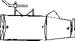
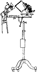
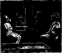
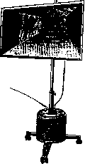

Fig. 6. Quartz-cadmium lamp. rich in lines between 330 mμ and 350 mμ, as well as between 280 mμ and 298 mμ (fig. 6). 7) In order to bring the radiation of artificial sources closer to the solar spectrum, a number of devices have been proposed: a) The Aureol lamp of Siemens (Siemens) is a glass ball in which a carbon arc is enclosed, b) The Spectro-sol lamp. The source of radiation is a metal filament placed in a glass tube. c) The Ultra-zone of Landeker (Landeker) with an arc, the carbons of which, due to metal impregnation, have the property of intensively producing ultraviolet rays. Due to the absence of irritating rays, the light of this lamp is especially suitable for irradiating mucous membranes (vagina, mouth, etc.). 8) Incandescent light. In addition to the already mentioned application of the blue bulb (Minin), general and local electric light baths are widely used. The general electric light bath was proposed by the American doctor Kellogg. A bath of the ordinary type is in the form of a cabinet having the shape of an octagonal prism, its height being such that the head of an adult person sitting in it on a chair is outside. For this purpose, an opening of appropriate size is cut in the cover. One can enter such a bath only through a special door. Inside the bath, in the middle, is placed a swivel chair, rotating around its axis and also giving the possibility, if necessary, to raise or lower it in height. Along the walls of such a box are evenly placed incandescent bulbs of 16-25 candlepower, one or more colors, in the amount of 50-90, and in some baths, on the sides, 3-4 arcs of 800-1000 candlepower each are installed. Both the incandescent bulbs and the arcs allow gradual and separate switching on. In addition, for the purpose of regulating the received radiation, a rheostat is also introduced into the circuit. Through special windows one can observe the patient's body as well as examine the state of his pulse. A thermometer (the mercury ball is inside) is embedded in the cover of the bath, showing the air temperature in
Fig. Solux lamp. it. The walls of such a light box are lined with mirrors, milk glass, painted with white enamel paint, etc.;-Physiotherapy also uses so-called intensive light baths, to which belong the «Radiotherm» and «Polysol». These baths, while maintaining the same device as ordinary ones, differ from them in the shape of the bulbs and the equipment of each bulb with a special reflector. The bulb of such a bath is in the form of a long tube with a vertically standing incandescent filament, and not in the form of a coil, as is the case in ordinary pear-shaped bulbs. Behind the lamp is placed a special rotating reflector, which is set up so that the filament of the bulb is exactly in its focus. In an ordinary electric light bath, most of the rays, despite reflection from the walls, do not reach the patient. Only about 1/10 of the radiation reaches him. At the same time, due to the heating of the walls of the bath and the air in it, the temperature around the patient is extremely high - on average up to 60-70°. In an intensive light bath, the air surrounding the patient heats up
Fig. 8 Small lamp directly applied to the body surface
relatively little and significantly more rays reach the patient. To achieve the same radiation that the patient receives in an ordinary electric light bath, it would be sufficient in an intensive light bath to install 5 tubular lamps with reflectors. However, the air temperature in the bath would then be too low. For this purpose, in intensive light baths, 16-20 tubular incandescent lamps with the same number of reflectors are installed. Thus, the radiation in such a bath will be 3 or more times stronger than in an ordinary one. The air temperature in the bath is then 25-40°. The general electric light bath is an agent causing more or less significant changes in the physiological functions of the body. These consist in an increase in body temperature, an increase in pulmonary gas exchange and general metabolism, increased sweating, accelerated respiration, changes in cardiac activity and blood pressure, changes in blood and functions of the nervous system. All these data fully justify the therapeutic use of general electric light baths, although here, as in many other cases, therapeutic practice has far outstripped and left behind the study of physiological influence. The extremely widespread use of electric light baths has contributed to their being applied outside medical supervision, under the guidance of persons who cannot realize the seriousness of this means. Such baths require strict individualization, and therefore the first 2-3 sessions should be conducted in the presence of a physician, who must determine the temperature and duration of the bath. Cases have been observed where the improper use of such baths has harmed the patient.-Local electric light baths, intended, as their very name indicates, for application to one or another part of the body, are constructed accordingly. The action of local electric light baths is the same as that of general ones. They have, compared to the latter, certain features related to the possibility of affecting not the entire body of the patient, but one or another part of it. Therefore, with a local bath, a significantly higher temperature (up to 100° and higher) is also tolerated. Unpleasant side effects (flushes to the head, states of weakness, excitement, etc.) are usually not observed. Duration
O O O: O O
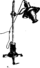


Figure 10. Device for determining individual light sensitivity. Figure 9. Profundus lamp. The duration of a session is on average 20-30 minutes. The indications are mainly gouty and rheumatic diseases of bones, joints, muscles, tendons, transudates and exudates, etc. 9) The solux lamp consists of a glass cap in which a luminous body [incandescent lamp with a metallic tungsten filament (fig. 7 and 8)] is enclosed. 10) The profundus lamp consists of a wire rheostat that heats up when current passes through it, glowing to the point of incandescence. It emits only infrared rays (fig. 9). Dosimetry. In practice, the dosage of ultraviolet rays is of particular importance. However, this problem remains to this day unresolved to any satisfactory degree. Among the methods proposed for this purpose, we note the following: 1) chemical method - the Bering and Meyer method, improved by Keller. It is based on the release of free iodine under the influence of ultraviolet rays from an acidified solution of potassium iodide; 2) photographic method: a) Keller's erythemadosimeter (most common) is based on the blackening of photographic paper under the influence of rays, placed under glass filters; b) Zalkind-son's photoquantimeter is based on the property of chloro-silver paper to change its color under the influence of violet and ultraviolet rays. As a unit (1 UFE), the degree of darkening of aristotypic paper is taken, which is obtained from irradiation of a new alternating current burner at 110 V at a distance of 1 m and an exposure of 1/2 min. In this way, the magnitude of the incident radiation in the ultraviolet part can be measured with greater or lesser accuracy. As for the individual sensitivity of a particular patient, it is determined by a very simple method. For this, 5 small circles with a diameter of 2 cm are cut out of a piece of paper (fig. 10). After covering the rest of the skin, all circles are irradiated sequentially for 1-2, etc., minutes each. The next day, from 5 different degrees of erythema, the desired one can be chosen and used for the planned session. With regard to dosage in various diseases, a scheme has been proposed covering 5 groups (Zalkind-son) (see table). Photopathology. Irradiation of excessive intensity in individuals with normal light sensitivity, on the one hand, and irradiation of medium intensity in subjects particularly sensitive to light, on the other hand, can cause a pathological condition. Under ordinary light regime conditions (climatic and others), people who are normal in terms of light sensitivity do not get sick from the effects of light. A certain predisposition - increased sensitivity to light - therefore plays the main role in the pathogenesis of light-induced diseases. They are divided into endogenous and exogenous. Exogenous include diseases after prolonged use of eosin and diseases in workers dealing with certain coal-tar dyes. From endogenous ones, hydroa, some forms of Character of irradiation Dose Skin manifestations Dose of 1st session in UFE Gradual increase in dose in UFE Dose in UFE Number of sessions per week I group Diseases of bones, joints and glands, bronchial asthma, metabolic diseases, rickets, furunculosis II group Ulcers and wounds, subacute eczema, pruritus, prurigo III group Peripheral tuberculosis, bronchial asthma, hypertension, lichen ruber, psoriasis vulgaris, eczema chronicum, perniones IV group Neuralgias, arthritis, myositis V group Alopecia areata, psoriasis vulgaris General Small Local Small Local Medium Local Large Local Very large Without erythema Without erythema Weak erythema Strong erythema Very strong erythema 2-3 2-3 5-30 30-100 100 and more 2-3 30-40 2-3 : 20-30 15-100 100 and more 100-200 200-300 up to 1,000 3-8 3-6 until disappearance of erythema urticaria, eczema solare, xeroderma pigmentosum and some others. By irradiating with mercury-quartz light, cases of latent malaria can be exacerbated and parasites can appear in the peripheral blood. Light therapy in individual diseases. Internal diseases. Among metabolic diseases, light therapy finds its application primarily in diabetes. Ultraviolet rays reduce the sugar content in the blood. The mercury-quartz lamp, ultrazone (for irradiating mucous membranes), solux (blue filter), and local electric light bath are used. For obesity, sun baths and general electric light baths are recommended. In view of the effect of short-wave rays on metabolism and in particular on purine metabolism, the general and local use of the mercury-quartz lamp is recommended. Further - sun baths and general electric light baths with incandescent lamps. In conditions of exhaustion due to physical or mental strain, as well as in the stage of convalescence, general ultraviolet irradiation is recommended. In secondary anemia, general mercury-quartz irradiation has a beneficial effect. With respect to diseases of the respiratory system, light therapy in pulmonary tuberculosis has special significance. Despite the large discrepancies existing in the literature on this issue, one thing is certain: when using light therapy in cases of pulmonary tuberculosis, it must be carried out with extreme caution, gradually increasing both the size of the irradiated surface and the duration of irradiation (Rollier). This should apply equally to heliotherapy and artificial sources (mercury-quartz lamp alone or in combination with solux). When clarifying the mechanism of light action in tuberculosis, it is necessary to proceed from the concept of tuberculosis as a disease of the whole organism. The treatment should aim to increase the immunobiological processes in the body. On the other hand, light therapy is one type of non-specific therapy that gives the desired effect with proper dosage. In this case, light therapy acts by causing inflammatory changes in the skin, accompanied by an increase in the vital activity of cellular elements, the formation of special substances irritating various tissues and organs (ezofilaksins), as well as the formation of vitamin D. Along with this, there is irritation of nerve endings in the skin, leading reflexively to a decrease in the tone of the sympathetic nerve with subsequent hyperemia. The influence of light on the psyche of patients (especially during insolation) is also of no small importance (Brushtein). Irradiation with a mercury-quartz lamp is indicated in the sequential treatment of acute pneumonias - croupous and influenza-like (resorption of infiltrates), as well as dry and exudative pleurisy. For long-standing limited exudates, a projector with a red filter, solux, and infrared light are indicated. Sometimes Minin's lamp can also be used (but only not blue, but red). Duration of irradiation 15-20 min. For chronic bronchitis - projector on the front and back of the chest, 10-15 min. For bronchial asthma - projector, general electric light baths (Striimpell), ultraviolet irradiation, infrared light. For diseases of the cardiovascular system, the use of ultraviolet therapy is based on reducing elevated blood pressure and is indicated in such conditions as arteriosclerosis, essential hypertension, renal hypertension. For local circulatory disorders - blue arc light or solux lamp, local electric light bath (Laqueur). For angina pectoris - ultraviolet irradiation. For diseases of abdominal organs, the use of a mercury-quartz lamp is indicated in tuberculous peritonitis. The best results are obtained in peritonitis with serous exudate, healing occurs in 50-70%. For scars and adhesions in the abdominal cavity - light from an arc lamp or solux (blue at first, with signs of significant irritability, and then - red). For ulcus ventriculi (duodeni), intensive local irradiation with a mercury-quartz lamp is recommended. For diseases of the stomach, intestines and gallbladder - infrared irradiation. For kidney diseases (chronic nephritis), sun baths and mercury-quartz irradiation are used. An attempt with satisfactory results was made in the initial forms of influenza, with ultraviolet irradiation with sub-erythemal doses applied to the trunk (in some cases together with solux). In cases of sharply expressed phenomena in the pharynx and on the nasal mucosa, these areas were also irradiated. Observations were made on workers of the 'Kauchuk', Lyubertsy and Kolomna plants. As a result - a rapid drop in temperature and disappearance of catarrhal phenomena. A beneficial effect of general irradiation of the whole body in case of carbon monoxide poisoning was noted. General ultraviolet irradiation is used for preventive purposes, e.g. in school-age children and during sports training. Ultraviolet irradiation increases work capacity. For rheumatic diseases (in the understanding of the unified classification adopted by the All-Union Committee for the Study of Rheumatism and the Fight Against It), light therapy in the form of electric light baths, mercury-quartz lamp, and solux is widely used in cases of chronic polyarthritis. For acute joint rheumatism, mercury-quartz irradiation relieves pain and has a beneficial effect on the course of the process.
Infrared rays are widely used both in chronic and acute rheumatism. In chronic joint rheumatism with a tendency to deformations, a combination of infrared baths followed by ultraviolet irradiation is recommended. For muscular rheumatism—sun baths, electric light baths, red projector, solux, spectrosol. For myalgias—quartz in combination with solux. For rheumatic neuralgias—quartz, Minin's blue light, solux, spectrosol. In nervous diseases, light therapy is used mainly in conditions accompanied by pain. The analgesic effect of solar irradiation has been known for a long time. In the doctrine of the local application of strong electric light, especially its so-called chemical rays, Russian physicians played a significant role. Stein and Gachkovsky were the first to note the analgesic effect of light from an incandescent lamp. As for the light of the voltaic arc, the first observations belong to Ewald, a physician at the Kolomna plant in the early 1890s. Ewald noted that since the introduction of iron smelting by the Benardos method (electric smelting) at the plant, the number of workers suffering from rheumatism, neuralgias, migraines, etc., had sharply decreased. Having found that the workers had found the source of their cure by exposing the affected part of the body to the light emitted during such smelting, Ewald set up a light therapy department in his factory hospital, where he began with great success to treat various nervous and rheumatic diseases. Subsequently, observations along the same principle were conducted by Kozlovsky and Griboyedov, and these authors obtained very encouraging results in treating various types of neuralgic diseases. Furthermore, in 1909, Brushtein for the first time proposed the use of the mercury-quartz lamp for neuralgias. Subsequently, Brushtein's observations were confirmed from various sides. Erythema doses of quartz often give good results in shooting pains in tabetics. There are observations indicating the beneficial effect of general ultraviolet irradiation in parkinsonism. Furthermore, sun baths and general ultraviolet irradiations can be used in neurasthenic conditions. Among mental diseases, in patients with manic-depressive psychosis and in schizophrenics, under the influence of ultraviolet irradiation, some improvement in appetite, weight gain, invigoration of muscular movements, and improvement in general well-being have been noted. The same applies to the use of colored lighting: blue—for states of excitement and red—for states of depression. Diseases of the skin. For the treatment of skin tuberculosis with light—see Tuberculous diseases of the skin. Diseases of the hair and the hairy part of the scalp are amenable to treatment with the mercury-quartz lamp (alopecia areata, alopecia seborrhoica). Subacute and chronic forms of eczema often respond well to quartz therapy. In lichen ruber, mercury-quartz irradiation gives exceptionally good results (Jesionek). The same is true for psoriasis. Children's diseases. There are numerous observations regarding the beneficial effect of heliotherapy on rickets. Recently, the question of treating rickets (see) with the mercury-quartz lamp has aroused great interest. It has been established that only a narrow band of the spectrum with a wavelength of 302-289 mμ (Ra-Strahlen) has this therapeutic effect in rickets. The anti-rachitic effect of ultraviolet irradiation is based on its effect on ergosterol, which is converted into vitamin D in the process. Preventive irradiation of children undoubtedly helps to protect them from rickets [see separate table (art. 711-712), fig. 5]. For the use of ultraviolet rays of the solar spectrum—see Lighting. Mercury-quartz irradiation is very effective in rachitogenic therapy and in childhood asthma. Ultraviolet irradiation is also recommended for scrofula, whooping cough, and erysipelas of the newborn. There are positive observations regarding ultraviolet irradiation of diphtheria bacillus carriers. Surgical diseases. Irradiation with mercury-quartz light is a very effective means for poorly healing wounds and for sluggish granulations. It can enhance the process of epithelialization. Ultraviolet rays are indicated in wound infections. Good results have been obtained in superficial wound infections with B. ruosa-pesh; the same is true for tetanus-infected wounds. When irradiating workers at the 'Kauchuk', Lyubertsy, and Kolomna plants who had suffered injuries with a mercury-quartz lamp, no cases of complications were noted, and a significant reduction in the number of abscesses, phlegmons, and lost working days due to them was achieved. In the question of treating surgical tuberculosis with irradiation, the school of Prof. Bier holds a special place. Based on observations conducted over many years in the clinical sanatorium Hohenlychen near Berlin, Bier's assistant Kisch published a special monograph in which the basic principles of the school on this question are set forth. Kisch criticizes existing views on the role of ultraviolet rays in pigment formation, on the physiological significance of pigment, and other questions of the theory and practice of phototherapy, and arrives at a number of conclusions that do not coincide with more or less established views. He asserts that pigmentation is caused not only by ultraviolet rays but also by rays of longer wavelength, and that perhaps the latter are capable of producing such strong pigmentation as is never achieved, for example, by a mercury-quartz lamp, especially, as is known, rich in ultraviolet rays. Based on measurements of the skin on artificially pigmented and unpigmented areas, he concludes that the main task of pigment is the regulation of heat in the body. The author identifies the essence of the action of heliotherapy with the action of congestive hyperemia. By combining both methods, he achieved a marked enhancement of the therapeutic effect. Regarding mountain heliotherapy, Kisch takes a decisive stand in the sense that it has no advantages over the same on plains, especially in summer; some—in spring and autumn; as for winter, the difference can be very significantly smoothed out by the hyperemizing power of Bier's bandage and the use of light apparatus proposed by Kisch, which abundantly emit so-called thermal rays. Proceeding from the view that the therapeutic effect of the sun is based on the power of its heat radiation, to which the body responds with hyperemia of the tissues as protection against burning, Kisch demands of the apparatus—to develop a high temperature and to cause a real brown coloration of the skin. Such an apparatus was constructed by the Zeiss firm for acetylene and electric light. In Hohenlychen, combined treatment is carried out, and its components are heliotherapy, treatment with artificial light, congestive hyperemia, and iodine. Success is achieved in general in 70-80%. In various forms of surgical tuberculosis, mercury-quartz irradiation is a very valuable method (in lymphadenitis, in lesions of bones and joints). The technique consists of general and local application. In osteomyelitis, heliotherapy is an excellent postoperative measure, i.e., after trepanation of an infected bone (Rollier). The same is true for quartz. Rollier's experience in organizing a clinic-factory that serves a double purpose: to treat patients with extrapulmonary tuberculosis with the sun and to serve as a workshop is interesting. The basis was the idea that 'in tuberculosis more than in any other disease, the outcome depends to a large extent on the patient's will to recover,' and that work should stimulate this 'will to recover' in chronic patients suffering from idleness and boredom. The wards and terraces of such a clinic (opened in 1930) are built like workshops and are equipped with electric motors as drives. Working patients make parts for small and precision mechanics instruments. More complex work is performed by convalescents in workshops specially located on the lower floor. All work is carried out under the best hygienic conditions, and working patients have the opportunity to use heliotherapy at the same time. The apparatus is constructed in such a way that its use is possible in any position required by orthopedic requirements. Women's diseases. For direct action on the genital organs, special instruments are proposed. These include: 1) Seitz's vaginal lamp, mainly of infrared radiation, 2) Wintz's vaginal lamp (in spectrum it approaches the lamp-spectrosol), 3) Landecker's ultrazone (see above) and 4) vaginal attachment for Bach and Croymer's lamps. Good results have been noted in inflammations of the vagina and external parts, in poorly healing perineal tears, and in pruritus vulvae. For endometritis and metritis, a combination of general irradiation with vaginal is recommended. Light therapy is also indicated for para- and perimetritis, as well as for retroflexio fixa caused by old inflammatory adhesions. Combined light therapy (superficial and vaginal) gives good results in adnexitis and oophoritis. Similarly, light therapy (quartz, ultrazone) has a beneficial effect on menstrual disorders, such as amenorrhea, excessive or too frequent bleeding, and dysmenorrheal disorders.
During pregnancy in weak, anemic women, general ultraviolet irradiations are indicated. General mercury-quartz irradiations have a beneficial effect on eclamptic seizures. Erythemal irradiations also give good results in cases of insufficient milk. La-ker attributes this to cases where the cause lies in the general condition, which is why he recommends giving general irradiations. Ear, nose, and throat diseases. The primary indication is for tuberculosis of the nose. - Eye diseases. Figure 1. Mercury-quartz
dental lamp-Dentaimodeii.
It is applied general irradiation with ultraviolet rays in kerato-conjunctivitis eczematosa (phlyctenulosa) with good results, also in hemeralopia idiopathica and in keratomalacia. Local irradiations using the Kromayer lamp are used in trachoma, in tbc. - Diseases of the teeth and oral cavity. For dental purposes, there is a special apparatus - Dentaimodeii (mercury-quartz lamp). Very good results are obtained in the treatment of scar formations (Fig. 11) in the oral cavity with quartz light, in paradentosis, in severe gingivitis, and in alveolar pyorrhea. Pain in periodontitis and periostitis in not too acute cases is relieved by the application of solux.
Related articles
Mentioned in
Cite this page
“Light Therapy.” Soviet Medical Encyclopedia. English translation of Bolshaya Meditsinskaya Entsiklopediya, 1st ed. (Moscow, 1928–1936), ed. N. A. Semashko. https://sovietmedicalencyclopedia.pages.dev/article/light-therapy/