Allantois
Historical document, translated for reference. It reflects medical knowledge of the 1920s–30s and is not medical advice.
Summary
An article from the 1928–1936 Soviet Great Medical Encyclopedia detailing the allantois, an embryonic organ originating as a diverticulum of the hindgut. It describes the anatomical development and differences of the allantois in reptiles, birds, and mammals, highlighting its roles in respiration, excretion, and placentation.
Encyclopedia article (1928–1936)
ALLANTOIS (from the Greek allas - sausage and eidos - form), the urinary sac, is formed (in reptiles, birds, and mammals) as a blind outpocketing of the anterior wall of the terminal gut. Its shape and subsequent role appear different in mammals and lower vertebrates. In reptiles and birds, the allantois appears very early (in the chicken on the third day) in the form of a small depression on the anterior wall of the terminal gut and rapidly grows into a blind sac extending between the amnion and the yolk sac into the extraembryonic part of the body cavity. Subsequently, its dimensions increase rapidly, and the end, in the shape of a mushroom cap, grows along the inner surface of the serous membrane, being connected to the site of its origin by a thin stalk. The allantois serves as a pathway for blood vessels that branch out onto its blind end and form a dense network here, serving for gas exchange both directly through the eggshell wall and especially through the membrane separating the air chamber of the egg. With the gradual reduction of the yolk sac, the allantois penetrates with its vessels also to the sharp end of the egg, where the thickened egg white accumulates; villous processes depart from the edges of the allantois, penetrating into the white mass and resorbing it (Duval's placenta). By the end of incubation, the fluid filling the allantois (kidney secretion) thickens and gradually disappears, so that only salt precipitates remain in the cavity of the sac. By the time the embryo hatches from the egg, the allantois atrophies. Thus, the allantois in birds and reptiles is a respiratory organ, as well as a reservoir for urine. In mammals, the allantois does not achieve such development as in lower vertebrates. It also develops as a blind outpocketing of the anterior wall of the terminal gut and extends as a thin cord into the extraembryonic section of the body cavity; in humans, it passes into the body stalk connecting the posterior end of the embryo with the chorion. Subsequently, together with the yolk sac, the allantois becomes part of the umbilical cord. The blood vessels formed on the stalk of the allantois form into two arteries and one vein and, entering into connection with the vessels of the chorion, form the vessels of the umbilical cord connecting the placenta with the fetus. The section of the allantois closest to the site of origin from the terminal gut retains its cavity and forms a spindle-shaped expansion, the primordium of the urinary bladder, from the apex of which a narrow passage departs, extending for some distance into the umbilical cord itself—the urachus, which subsequently becomes obliterated and turns into the median vesico-umbilical ligament. Thus, the allantois in mammals in the lower section, closest to the site of origin from the terminal gut, goes into the formation of the urinary bladder, and its vessels serve to connect the children's place (its embryonic part) formed from the chorion with the fetus.
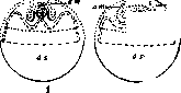
Development of the amnion and allantois in a chick in a longitudinal section through the embryo: 1 - on the third day of incubation, 2 - at the beginning of the fifth day, 3 - on the seventh day; am - amnion, al - allantois, s - serous membrane, ds - yolk sac.
Related articles
Mentioned in
Cite this page
“Allantois.” Soviet Medical Encyclopedia. English translation of Bolshaya Meditsinskaya Entsiklopediya, 1st ed. (Moscow, 1928–1936), ed. N. A. Semashko. https://sovietmedicalencyclopedia.pages.dev/article/allantois/