Childbirth
Historical document, translated for reference. It reflects medical knowledge of the 1920s–30s and is not medical advice.
Summary
This article from the 1928-1936 Soviet Medical Encyclopedia covers various aspects of childbirth, including obstetric shock, sudden maternal death, embolism, physiological effects on the female body, and premature artificial delivery.
Encyclopedia article (1928–1936)
257 XIX. Obstetric shock, sudden death of parturient women, puerperal women (embolism) . .
260 XXI. The effect of childbirth on the woman's body ....
271 XXIII. Artificial premature childbirth . .
272 1. Definition of the concept. Changes in the body during childbirth. Causes of the onset of labor. Childbirth is a physiological process as a result of which the entire egg detaches from the maternal organism, i.e., a mature fetus capable of extrauterine existence, together with all its appendages (secundina). As in every biological process, it is sometimes very difficult to draw a sharp, clear line between normal and pathological conditions, and therefore various authors, interpreting different variants of the mechanism of childbirth or generally any more or less significant deviations from their typical course, classify them—some as normal, others as pathological cases. Difficulty in predicting normal childbirth. If 'normal' is generally a very conditional and relative concept, then in its application to childbirth it turns out to be extremely elastic, depending on many reasons, and sometimes on completely accidental moments whose role cannot always be foreseen in advance. Even in the presence of certain obvious anatomical or functional anomalies which a priori should have affected both the normal course of the act of childbirth and its final outcome, childbirth can still fit within the framework of a purely physiological process and end successfully in all respects. On the other hand, even in the presence of anatomical-physiological prerequisites for a normal course, completely unexpected and unforeseen complications can occur during the act of childbirth, which can give a certain pathological deviation to one or another moment of this very complex physiological process. Childbirth is a general manifestation of life of the entire organism taken as a whole, and by no means a local process occurring only in the sexual sphere; they place heightened demands on all organ systems, on all active and hidden reserve forces of the female organism, and on all its life resources in general. Therefore, childbirth as a very multifaceted dynamic process requiring maximum, often extreme, tension of all the life forces of a woman, easily reveals any, often deeply hidden, defectiveness and inadequacy of her organism—both purely anatomical and functional (in particular biochemical). Static and dynamic-biological constitutional factors and their influence on the main components of childbirth. For the completely normal course and completion of the act of childbirth, of course, a normal state and action of all its main components (birth canal, birth object, and birth forces) are required, and above all, the correct harmonious ratio between them; however, the latter can be fully ensured only on the condition of a certain completeness of the entire organism as a whole. Therefore, various, even minor constitutionally pathological deviations, and all the more so already clearly expressed constitutional anomalies, can imprint a more or less sharp pathological mark on individual organ systems and their functions and thus affect all the main components of the act of childbirth, i.e., on the very structure of the birth canal, on the nature of the action of the birth forces, and directly or indirectly on the birth object, and consequently ultimately on the general outcome of childbirth (e.g., a narrowed pelvis in a woman with an infantile deviation or weak labor activity in a woman with an asthenic deviation can, due to spatial mismatch and because of the longer course of childbirth—and thus greater trauma to both mother and fetus—lead to various complications, some standing only on the border between normal and pathological, others already clearly pathological). Thus, the belonging of a woman to one or another constitutional type can to a certain degree influence the outcome of childbirth. III Significance of other, especially socio-domestic factors of motherhood. Determining significance are also racial, national, and age differences, and a whole series of other quite diverse individual peculiarities. However, dominant is the entire external environment in the broad sense of the word, surrounding the woman throughout her life path (starting from the first days of her life and ending with pregnancy and childbirth). The importance of all factors of a social order becomes obvious if one takes into account the conditions of growth and upbringing (especially the importance of rickets in the pathogenesis of pelvic anomalies), the general development of the organism, cultural-hygienic habits of the woman, physical culture, conditions of nutrition and housing, all professional and domestic moments, acquaintance with the sanitary minimum in the field of protection of motherhood and infancy, the woman's own attitude toward impending motherhood, greater or lesser rest before childbirth, provision of proper observation throughout the entire pregnancy, all preventive measures, and finally proper obstetric help at the right moment. Childbirth in woman from the point of view of comparative physiology and anthropology. Emptying the pregnant uterus of its contents, although representing a very complex physiological act, nevertheless has something in common with emptying the intestine and urinary bladder, at least to the extent that all these hollow organs are built from smooth muscle tissue (differing in their contractile activity by very characteristic features) and since their emptying takes a more or less active part of the abdominal press. This similarity is striking in lower mammals, where the expulsion of products of pregnancy occurs simply, easily, and quickly. In higher mammals this analogy gradually becomes less and less noticeable, as the mechanism of the act of childbirth gradually, as it approaches the human being, becomes more and more complicated. In connection with the changing ratio of parts of the fetus and with the modification of the shape of the pelvis as the birth canal and support for the entire trunk and lower extremities, obstacles gradually increase for the forward movement of the fetus and especially of its head end, which requires the participation in the act of childbirth of a number of auxiliary means and the involvement of the entire organism in the work of childbirth. In man, due to the relatively greatest predominance in the development of the brain, the head of the fetus is already so relatively voluminous that in its passage through the small pelvis it completely fills it, so closely touching everywhere against its walls that even a slight narrowing of it can significantly retard its forward movement as the most voluminous and densest part of the fetus. The birth canal as a whole (hard and soft birth passages). In order to understand the course of physiological childbirth and to penetrate into the very essence of their mechanism, one must first start from a clear concept of the individual components of this complex physiological act, of their essential properties and peculiarities, taken in the resting state, i.e., immediately before childbirth. The first component, pre-existing independently of pregnancy and childbirth—purely anatomical (predominantly static): this is the pelvic canal, the bony pelvic ring as a constant magnitude, almost not changing during the act of childbirth—the so-called hard birth passages. The supplement and as it were the direct continuation of the bony birth canal are the soft birth passages, which are formed only in the process of the act of childbirth, when under the pressure of expelling forces the forward-moving head stretches to the maximum limits all the musculature of the pelvic floor, its fasciae, the lower segment of the vagina, and the skin of the perineum, bulging all this together in the form of an 'additional outgoing cutaneous-muscular tube,' through which the entire fetus must pass as through the soft part of the birth canal. Normal spatial relations of the small pelvis. Taken in a skeletonized form the pelvis has the appearance of an almost solid bony ring (composed of the iliac, ischial, pubic bones and the sacrum together with the coccyx), closing a cavity, or canal, with a relatively voluminous entrance and a somewhat smaller exit. The spatial relations of this canal are not the same throughout its entire extent: since its side walls converge downward, it is somewhat narrowed in the transverse direction. A sagittal median section through the pelvis shows that its anterior wall, formed by the pubic bones, is much shorter than the posterior wall, formed by the sacral bone, and that due to the curvature of the anterior surface of the sacrum the entire pelvic canal is arched forward. The entrance to the pelvis has the shape of a transverse ellipse with a slight narrowing downward. It is bounded behind by the sacral promontory, on the sides by the linea terminalis, in front by the upper edge of the symphysis. The anteroposterior diameter of the entrance, i.e., the shortest distance from the sacral promontory to the nearest point on the posterior surface of the symphysis—the true or obstetrical conjugata (conjugata vera, s. obstetrica)—is 11 cm. The distance from the sacral promontory to the upper edge of the symphysis (1/2 cm more) is called the conjugata anatomica. (Measurement of pelvic dimensions—see Obstetrical examination). Its transverse diameter—the distance between the most distant points of the linea terminalis—is 13 cm. The right oblique diameter—from the right sacroiliac articulation to the left iliopectineal tubercle—is 12 cm. The left oblique—from the left sacroiliac articulation to the right iliopectineal tubercle—is also equal to 12 cm.
The pelvic outlet is bounded by the pubic arch, the ischial tuberosities, and the tip of the coccyx. Its anteroposterior dimension—from the lower border of the pubis to the tip of the coccyx—is 9.5 cm, but during childbirth, due to the backward displacement of the coccyx, it increases to 11.5 cm. The transverse dimension—the greatest distance between the ischial tuberosities—is 11 cm. By the pelvic cavity and specifically its middle part is meant that segment of the small pelvis which is located at the midpoint between the inlet and the outlet; it corresponds to the level below which the pelvic canal begins to change its direct direction downward and curves forward. Its anteroposterior dimension—from the point where the sacrum bends at the level of its third vertebra to the middle of the pubic symphysis, as well as its greatest transverse dimension—is 12 cm. Relative mobility of the pelvic joints. In addition to the above-mentioned increase in the anteroposterior dimension of the pelvic outlet (which is explained by the fact that the coccyx, due to the mobility of its joint with the sacrum, is pushed backward by the pressure of the fetal head), the most important obstetrical dimension of the pelvis, i.e., the anteroposterior dimension of the pelvic inlet—conjugata vera—also changes (though to a much lesser extent, approximately by 1/2 cm) in certain body positions, which Balandin demonstrated on cadavers, and Walcher by careful measurements on living subjects (see Walcher's hanging position). Finally, the concentric expansion of the entire pelvis as a whole, although very slight, has also been proven by the research of Loeschke. This is explained by the general loosening of tissues observed during pregnancy, affecting not only the soft tissues but the entire ligamentous apparatus and joints; it causes relatively greater mobility of both the sacroiliac and the pubic symphysis joints. All these data concerning the relative, although very slight, mobility of the pelvic joints should of course not be overestimated. Nevertheless, these data may play, although not visible to the eye, still a definite role. Geometric scales in application to the pelvis. The significance of the system of parallel planes according to Hodge. For the most vivid representation of the spatial relationships of the small pelvis (which is the only one that has significance from the mechanical point of view of childbirth), attempts have long been made to approach them with one or another purely geometric scale, using various mentally projected planes passing through different parts of the pelvis. Undoubtedly, all such planes are by no means such from a purely geometric point of view, and therefore such classics of obstetrics as, for example, Bumm, were skeptical of them, asserting that they by no means contribute to the theoretical understanding of the act of childbirth and cannot serve as a practical guide at the bedside of the parturient. Nevertheless, when speaking of the spatial relationships of the small pelvis, one cannot ignore the system of parallel planes proposed by Hodge, namely the following: the first parallel plane (terminal) corresponds to the plane of the pelvic inlet, the second corresponds to the level of the lower border of the symphysis, the third (spinal) corresponds to the level of the ischial spines, the fourth corresponds to the coccyx and the pelvic floor (Fig. 1). The use of these mentally projected planes (the distance between each two of them averages 3-4 cm) makes it easier to orient oneself at any given moment of childbirth in the spatial relationships between the pelvis and the presenting part of the fetus (usually the head) and to judge fairly accurately the height of its standing at one or another level of the pelvic (resp. birth) canal.
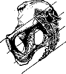
Figure 1. System of parallel planes according to Hodge.
A detailed study of the pelvic parallel planes of Godze showed that the pelvic cavity by no means represents a curved, forward and upwardly directed canal, but rather corresponds to a cylinder, obliquely cut downward and forward. Corresponding to this shape of the pelvic cavity, the fetal head passes through the pelvis not in the direction of a constantly forward-arching line, as was previously thought, but is pushed forward under the pressure of labor forces first directly downward, going in the direction of the axis of the pelvic inlet and gradually passing one after another all the above-mentioned 4 parallel planes down to the pelvic floor (and only here does the birth canal change its direction, see below). Axis of the pelvic inlet and pelvic axis (guideline of the pelvis). The axis of the pelvic inlet should be understood as a line mentally drawn through the middle of the pelvic inlet in a perpendicular direction (extended downward, it would reach the coccyx, and upward—approximately to the navel). This term, 'axis of the pelvic inlet,' should in no way be confused with another term 'pelvic axis,' which is a synonym for the concept 'guideline of the pelvis.' Under these last two equivalent terms should be understood a line mentally drawn through the midpoints of all the straight dimensions of the small pelvis. This line practically serves for orientation relative to the given position of the presenting part (i.e., forward or backward, left or right of the guideline). From the above it is clear that the guideline only in its beginning almost coincides with the direction of the axis of the pelvic inlet and precisely up to the middle of the pelvic cavity; here it begins to bend forward quite sharply and for a long time in an arc corresponding to the curvature of the pelvic canal.-Inclination of the pelvis. Since the promontory in the vertical position of a woman stands significantly higher than the upper edge of the symphysis, the plane of the pelvic inlet has an oblique direction relative to the horizontal: it is tilted forward and downward. The angle formed by the plane of the pelvic inlet with the horizontal plane is called the inclination of the pelvis; in the standing position of a woman it is 60°. Soft birth passages, pelvic floor or pelvic diaphragm, and urogenital diaphragm. The concept of the spatial relationships of the birth canal changes substantially if instead of a skeletonized pelvis, the pelvis is considered as a whole, i.e., together with all its soft parts. At once it becomes obvious that the upper segment of the pelvic cavity is spatially not limited at all by soft parts, whereas the pelvic outlet is closed by a group of tightly stretched and flatly arranged muscles—the pelvic floor, or pelvic diaphragm—through which only the exit openings of the intestine, sexual and urinary tracts pass. The main mass of this diaphragm is formed by muscular bundles of levatoris ani, which, starting from the tendinous line at the lateral and anterior walls of the pelvis, stretched out in a plane and divided on each side into two parts (portio pubica levatoris ani and r. iliaca), go to the coccyx and to the fibrous bridge in front of it. Here the bundles coming from both sides connect together, leaving however in the middle a free slit for the passage of the intestinal loop and vagina. To m. levat. ani on both sides behind, additional bundles m. ischio-coccygei are also attached, going fanwise from spina ischii to the coccyx. On their upper and lower surfaces, all these muscular bundles are lined with a dense fascia (fascia sup. and inf. of the pelvic diaphragm). Consideration of the pelvic floor from below shows that in front of the described diaphragm there is located another so-called additional, the so-called urogenital diaphragm, consisting of transversely directed bundles of m. compressoris ure-thrae and m. transversi perinaei profundi, as well as the musculature closing the vagina (i.e. m. bulbo-cavernosus = m. constrictor cunni and m. ischio-cavernosus) and closing the anal opening (i.e. m. sphincter ani externus). Only during childbirth under the pressure of the head, the muscles of the pelvic floor are smoothed out, stretched and pushed apart, thus forming an additional muscular tube that constitutes an extension of the bony birth canal (in length almost equal to it, and in width corresponding to the dimensions of the fetus). The stretching of the pelvic floor and the formation of this muscular, or rather skin-muscular tube from unyielding tissues of the perineum requires a very considerable expenditure of labor forces, especially in primiparas. Birth object. The birth object should be understood as all the contents of the uterus at the end of pregnancy and in this case one must first proceed from the correct concept of it as a whole taken in a resting state, i.e., immediately before childbirth. The individual parts of the ovum are not equivalent in their physical properties, since they are either liquid (amniotic fluid), or more or less soft (fetal membranes, all secundina), or hard (skeleton). All these parts of the ovum differ far from equally in 'deformability.' Only because the ovum as a whole, taken in its entirety, although not equally in its different parts, but in general still very significantly deformable, is it possible for the uniform transmission of intra-abdominal pressure during childbirth through the uterine walls to all the contents of the uterine cavity, and consequently the effect of this last (placed under conditions of increased pressure) in the direction of least resistance, i.e., directly on the exit opening of the uterus as the fetal container—on its internal os. Of course, the amniotic fluid, like any liquid medium, to a much greater degree than all other more or less dense components of the ovum, has the above-mentioned physical properties. The more amniotic fluid, the more easily the entire uterine content is deformed, and if one considers the fetal body itself, those parts of it that contain the largest amount of liquid are most easily deformed (a significant difference between its soft and bony parts). Therefore, during childbirth, the lower pole of the amniotic sac usually presents itself (comes forward) as the part that most readily yields to pressure and is therefore the first to be deformed. Fetal head. The main part of the birth object is the entire fetal body, but of course the head has the most significant importance as the most voluminous, dense and unyielding part and therefore relatively slowly deforming. On the fetal head, as on an adult's head, the skull (which is significantly the larger part) and the face must be distinguished. The skull consists of paired frontal and parietal bones, the occipital bone, the squama of the temporal bones and the wings of the sphenoid bone, which together form an ovoid-shaped bony capsule. Between these bones there exist thin fibrous connections, the so-called sutures, and namely the following: 1) sagittal (sutura sagittalis) between the parietal bones, 2) frontal (sut. frontalis) between the frontal bones, 3) coronal (sut. coronalis) on each side between the frontal and parietal, 4) lambdoid (s. lambdoidea) between the parietal and occipital bones. (The temporal and sphenoid, or mastoid sutures have no practical importance.)-Fontanelles. Where two or more of these sutures meet each other, more or less significant gaps remain, closed by fibrous membranes and called fontanelles: 1) large, or anterior (fonticulus frontalis), with rhomboid outlines, at the junction of the sagittal, frontal and both branches of the coronal suture; 2) small, or posterior (fonticulus occipitalis), at the junction of the sagittal and lambdoid sutures; this is not actually a free gap between the bones (at least in a mature fetus), but rather a small, triangular-shaped depression between the parietal and occipital bones, clearly palpable only during childbirth; 3) on the anterior and posterior parts of the squama of the temporal bone on each side there are also two lateral fontanelles, which however are of little practical importance. All these sutures and fontanelles significantly facilitate spatial orientation relative to the position of the head in one or another part of the pelvis and therefore serve as important reference points during childbirth. Dimensions and circumference of the head. For understanding the very essence of the mechanism of childbirth, it is also essential to know the following dimensions and circumferences of the head as average values in a full-term mature fetus: 1) vertical size, or diameter (diameter fronto-occipitalis)—from glabella to the most distant point of the occiput—12 cm, 2) large transverse (d. biparietalis)—distance between both parietal tuberosities—9½ cm, 3) small transverse (d. bitemporalis)—greatest distance between both branches of the coronal suture—8 cm, 4) large oblique (d. mento-occipitalis)—from chin to the most distant point of the occiput—13½ cm, 5) small oblique (d. suboccipito-bregmaticus)—from the suboccipital fossa to the anterior fontanelle—9½ cm.
Next, the following circles are distinguished: 1) the direct (circumferentia fronto-occipitalis), corresponding to the direct diameter and equal to 34 cm, 2) the large oblique (c. mento-occipitalis) - corresponding to the large oblique diameter, equal to 35 cm, and 3) the small oblique (c. suboccipito-bregmatica) - corresponding to the small oblique diameter, equal to 32 cm, and therefore the smallest and thus most important from the point of view of the mechanics of childbirth. Taken as a whole, the head represents a sort of biaxial or rotational ellipsoid (Sellheim) with a slight predominance of the long axis over the short one, and the long axis of this head ellipsoid forms an angle with the cervical vertebrae, the magnitude of which varies depending on the features of the engagement of the head in the pelvis during childbirth (A. Muller). Since under the influence of the forces of childbirth the head is compressed to a certain extent and in general therefore changes its shape, due to this (individually variable) plasticity, all the above-mentioned numerical data - diameters and circumferences (which can be accurately determined only on the head of a born fetus) - during childbirth in each individual case can vary considerably and therefore have the significance only of approximate average values. With considerable variability of the entire 'shape of the head, its total volume, on the contrary, remains relatively little changed; only with very strong compression is a certain outflow of fluid from the cerebral ventricles into the spinal canal possible and a corresponding slight decrease in total volume (Fehling). Much greater significance than the absolute values of all the mentioned diameters and circumferences of the head is its spatial relationship to the pelvis, and the most important, often decisive role is played by both its position and its property to mold itself, adapt to the dimensions and shape of a given pelvis, thanks to which, even in the presence of significant spatial discrepancy, its engagement in the pelvis by the most suitable, i.e., the smallest, diameter (resp. circumference) is possible. Configurational properties of the head. The considerable deformability of the head during childbirth or (using the generally accepted term here) its configurational ability is explained by the peculiar structure of the fetal skull from several relatively thin bone plates, which under the pressure of the forces of childbirth can to a certain extent bend, overlap one another and thus move considerably relative to each other. This is possible only thanks to the above-mentioned sutures as fibrous, relatively easily yielding, movable connections between the individual bones of the skull. When the head in its forward movement encounters significant obstacles from the hard or soft, but often still little yielding, rigid parts, after a more or less prolonged adaptation it can well configure itself, i.e., take the form needed for the given moment and place, more corresponding to the size and shape of one or another part of the birth canal; as a result of this, even a relatively large spatial discrepancy and, generally speaking, obstacle can be smoothed over and overcome. However, configuration of the head is possible only within certain limits - only on the condition of regular, very energetic and often very prolonged labor activity. The fetal body. Compared to the head, the fetal body as part of the birth object has much less significance. The greatest transverse size of the shoulders - 12 cm (the corresponding circumference - 35 cm). The greatest transverse size of the hips - 9 cm (the corresponding circumference - 27 cm). Since these, relatively soft parts of the body are easily compressed and displaced at the moment of passing through the pelvis, with rare exceptions they usually do not constitute a mechanical obstacle during childbirth. A mature full-term fetus, fully stretched in length, should be about 48-52 cm long and weigh on average about 2,800-3,200 g. However, one must always remember that, on the one hand, there are surprisingly light but undoubtedly full-term fetuses, and on the other hand, relatively heavy but clearly premature fetuses; hence it is clear that the concepts 'mature' and 'full-term fetus' are not always identical. Flexibility of the fetal spine and its significance in the mechanism of childbirth. From the point of view of the mechanics of childbirth, the greater or lesser flexibility of the entire fetus as a birth object, determined mainly by the flexibility of its spine, is of considerable interest. Only in 1904 Sellheim, in a whole series of (accompanied by all precautions and radiographically controlled) experiments on living newborns using a quadrant and dynamometer, comprehensively studied this question and arrived at the following important conclusions. The flexibility of the child's spine is comparatively very great (due to the greater content of cartilaginous tissue in it, greater elasticity of the ligamentous apparatus, etc.). The greatest flexibility is observed in the cervical part of the spine, which is explained here by the lower, less developed vertebrae and the very easy mobility of the joint with the head. In individual segments of the newborn's spine, flexibility in different directions turned out to be indeed unequal; the greatest flexibility (facillimum of it) of the cervical part - backward, of the thoracic region - to the sides, of the lumbar region - to the sides and backward, of the sacral-lumbar region - forward and backward. - Joints. For the course of childbirth, the very great mobility of the joints of the shoulder and pelvic girdles is very important. The mobility in the shoulder, in the acromioclavicular and in the sternoclavicular joints is so great that the shoulder bones can be freely brought into contact on the chest, with the shoulder blades strongly moving upward (whereas an attempt to push the fetus's hands to the back always encounters a strong obstacle). The fetus's thighs can easily be bent onto the abdomen and placed parallel to each other (as in breech presentation). Expulsive forces. The main source of the forces of childbirth is the uterus; as a motor hollow smooth-muscle contractile organ, extremely hypertrophied at the end of pregnancy and therefore fully suitable for very energetic mechanical work, it in the full sense of the word plays the role of the birth motor. Strictly speaking, this function is performed not by the entire uterus as a whole, but only by its upper part, i.e., its body (the actual fruit-container), the walls of which represent a thick dense network of closely interwoven in various directions strong muscle fibers, forming by their entire mass the so-called hollow muscle; it is precisely this part that is the main and very powerful expulsive organ. The isthmus (isthmus; according to Aschoff), i.e., the segment located between the body and the cervix, already involved in the composition of the fruit-container from the middle of pregnancy, and the cervix, remaining unchanged until the beginning of childbirth, have no direct relation to the birth motor. These parts with weak scanty muscle fibers play a secondary, much smaller role compared to the hollow muscle. Capable rather of passive maximum stretching than of active contractions, they only indirectly contribute to the forward movement of the fetus, being part of the 'outlet tube'. From a purely mechanical point of view, it is quite understandable that if all parts of the uterus contracted equally strongly during childbirth, the forward movement of the fetus in the direction of least resistance and its very expulsion would be impossible. - As auxiliary birth forces function: 1) the ligamentous (suspending) apparatus of the uterus, especially the strongly hypertrophied by the end of pregnancy, rich in smooth muscle fibers round uterine ligaments, and 2) powerful contractions of the striated musculature of the abdominal press. A secondary role is played, especially in difficult labors, by contractions of the muscles of the entire body (body press). And finally, when at the end of expulsion a significant part of the fetus has already passed through the vagina, the expulsion of the remaining part of it, as well as the delivery of the placenta, are indirectly helped by contractions of smooth muscle fibers located in the hypertrophied walls of the vagina. Experimental data on the physiology of the uterus. Experiments on the uterus both in vivo and on the isolated organ, carried out by a whole series of authors (over the last three decades with a significantly improved compared to previous times technique, guaranteeing the accuracy and objectivity of the results obtained), made it possible to thoroughly study the physiology of uterine contractions in general and in particular gave much that is interesting from the point of view of the physiology of the act of childbirth. It has been proven that the uterus, both outside the state of pregnancy, but especially during and at the end of it, possesses the ability to more or less regular and strong automatic contractions. This property is inherent in it, as in any other smooth-muscle organ, but to a much greater degree. The initial impulses for these automatic contractions arise (as is directly visible on the isolated organ) usually in the very upper parts of the genital canal. From here they spread peristaltically in a wave-like manner towards the exit from it.
Contractile waves are not limited to the uterus alone; usually they more or less simultaneously capture the entire contractile system of the sexual apparatus, i.e., not only the entire sexual canal, but also the fixing (suspending) ligamentous apparatus. The automatic contractile work of the round uterine ligaments and its significance during Childbirth (in experiment). In experiments on the isolated rabbit uterus (and especially on the pregnant one), Kurdinovsky often had to observe that the round uterine ligaments, actively participating in the contractions of the uterine horns, strongly pull and hold them, sometimes for */2 minutes, "in suspension" ("as on reins"); by fixing them in this way in the forward direction, they enable the uterus to better utilize its contractile work. This active role of the round ligaments is particularly striking when observing the act of childbirth on the isolated rabbit uterus; throughout the time of the forward movement of the fetus through the sexual canal, energetic contractions of the round ligaments are observed, facilitating the act of childbirth; strongly contracting, they raise the uterine horns, giving them a firm point of support (in vivo this point of support is even more secureg since the uterus is thus fixed directly in the direction of the anterior wall of the pelvis, i.e., to the place of attachment of these ligaments).-Automatic contractions of the broad uterine ligaments and their significance during Childbirth (in experiment). By fixing the entire uterus and bringing it closer to the exit from the sexual canal, the round ligaments facilitate the uterus in expelling its contents. The broad uterine ligaments also take an active part in the contractile work of the uterus. Only with superficial observation does it seem that they move purely passively due to the contracting uterine horns involving them in movement. With more attentive observation, however, one can undoubtedly ascertain that, in addition to these passive movements, they also possess the ability to completely independent active contractions, automatically occurring independently of the contractions of the uterine horns; in such cases, somewhere in one place of the broad ligament or in several simultaneously, the initial impulse to movement arises in the form of a peculiar play of fibers, which then quickly captures the entire ligament. The broad ligaments reveal even greater activity in the pregnant uterus, especially during the act of childbirth observed on the isolated organ. Direct observation here easily convinces us that they, by their contractions, contribute to the expulsion of the fetus. This happens as follows: when the contractions of the uterine horns displace the fetus into the lower part (into the upper part of the vagina), the broad ligament, which had been at rest before, begins to contract in a direction transverse to the axis of the sexual canal; with these contractions, it is given a forward movement in the direction of least resistance, i.e., toward the exit from the sexual canal; thus, the broad ligament, with its ring-like contractions, as it were, squeezes out the fetus; under the pressure of this VIs a tergo it moves further. It is difficult, of course, to say to what extent experimental data on the question of automatic contractions of the uterus and its ligamentous apparatus can be transferred to the human uterus. As for the nature of contractions of the female uterus in general and particularly during Childbirth, the opinions of individual authors still differ greatly. The anatomical complexity of its muscular structure (compared with the uterus of animals) already a priori gives reason to think of the greater multifacetedness of its contractile work and therefore forces us to be cautious in making corresponding analogies. Nevertheless, clinical observation with the naked eye (most reliable because here all artificial irritation is excluded) shows that in women too, impulses for labor contractions usually arise in the upper part of the birth canal, i.e., in the fundus of the uterus; it is here that the first hardenings of the uterine musculature occur earliest, especially at the beginning of Childbirth, as a sign of the beginning contractions. In the further course of Childbirth, contractions also arise in the area of the cervix. Many authors believe that usually the initial contractile movement still arises in the fundus of the uterus and only from there it spreads peristaltically to the exit from the sexual canal. According to others, it can primarily begin at the place of attachment of the round uterine ligaments to the anterior abdominal wall (K. Schroder), as well as in the area of the broad ligaments (Werth). The peristaltic nature of contractions upon palpation of the uterus (at the beginning of a contraction) and upon direct observation during a cesarean section is masked by the speed of propagation of the contractile wave, as well as by the round shape of the uterus. However, Schatz, by introducing a manometer into the uterine cavity, nevertheless proved the peristaltic nature of its contractions. Experimental study of uterine contractions under the influence of various artificial irritations (physical and chemical nature). The experimental application of various artificial exciters of the uterus as a contractile organ, i.e., mechanical, thermal, electrical irritations of it, as well as pharmacotherapeutic agents and organotherapeutic preparations—both in experiments on the isolated organ and in vivo—made it possible to study in all details the physiology and pathology of uterine contractions. Pharmacological experiments on the isolated uterus gave particularly much in this respect, since on an organ disconnected from all connections with the central nervous system, it was easier to clarify (especially when compared with experiments in vivo) the very essence of the action of various uterine agents, i.e., to study all the paths and ways of their influence. Thus, experiment gave us the opportunity not only to study in detail the uterus as a birth motor, but also taught us various ways to stimulate this motor where necessary.-Experimental verification of uterine contractions of reflex and mental origin. Experiments on the uterus in vivo showed that the uterus in its contractile activity is very sensitive to various, even very distant reflex influences, i.e., to irritations, both originating from some internal organs and applied somewhere on the periphery, for example, to painful irritations of any area of the skin in connection with subcutaneous or intravenous injections (and moreover, completely independently of the composition of the injected fluid). Therefore, the well-known fact of uterine contractions under the influence of mechanical irritations of the breast nipples by no means represents any specific phenomenon sui generis, but is only one of the numerous examples of such reflex contractions. One can easily be convinced of this purely reflex nature of them, since in the experiment, under conditions of deep anesthesia, when all reflexes are generally suppressed, such contractions no longer occur, even with very strong irritations. Experiments in vivo also empirically confirmed the long-known possibility of uterine contractions under the influence of purely mental excitations, for example, as a result of fright (Kurdinovsky). As for uterine contractions under the influence of artificial irritations of various parts of the central nervous system, on this question, which already has a direct bearing on the complex and far from resolved problem of innervation of the uterus (see Innervation), there are very extensive but rather contradictory experiments. Applying mechanical or electrical irritations to any area of the cerebral cortex and various other parts of the large, medulla oblongata, and spinal cord throughout its entire length, various authors obtained more or less strong contractions of the uterus (and sometimes, conversely, its relaxation). One must think that the central nervous system exerts its regulating influence on the uterus, sending to it impulses of both stimulating and inhibitory nature. To this day we still do not have sufficient grounds to admit the existence of any topographically precisely localized single main center managing the contractile work of the uterus; rather, one can assume the presence in the central nervous system of several such motor centers. However, the uterus in its contractile activity is generally speaking highly independent and independent of the cerebrospinal nervous system. It is very probable that its contractions, both automatically occurring and artificially induced, to a large extent depend on the local nerve apparatuses of the uterus. The significance of local (uterine and para-uterine) nerve apparatuses. In recent years, the presence of bipolar and multipolar ganglion cells in the uterine walls has been proven. In the muscular layers of the uterine fundus there are certainly many ganglia lying subserously; this obviously explains why the uterine fundus is extremely sensitive to all external irritations during Childbirth and especially after the expulsion of the fetus and always reacts to them with strong contractions. Of course, the nerve apparatuses that lie near the uterus also play an important role, and first of all the so-called
The Frankenhauser cervical nerve plexus, rich in ganglia, is connected with the sympathetic and parasympathetic (and probably also with spinal) nerves and sends out branches that supply the entire uterus, but mainly its cervix. The paracervical and vaginal ganglia may also have some significance. Nevertheless, despite the presence of the aforementioned own nervous system (uterine and parauterine), it is still difficult to say with certainty whether the automatic contractions of the uterus are of neurogenic or purely myogenic origin. A whole series of facts and data from the field of general physiology of smooth muscles gives many reasons to recognize in smooth muscle tissue as such an independent, purely myogenic excitability and therefore a relative independence from any nervous influences, both central and local (peripheral). The significance of the latest experiments on the uterus with endocrine preparations for understanding its functions as a birth motor. In the current state of the complex and far from fully resolved problem of uterine innervation, it is of course necessary to consider not only the purely 'nervous' but also the 'chemical', or rather 'neuro-chemical' correlation. A priori it is undoubtedly true that constant, essentially important exciters of the contractile work of the uterus must be various hormones, produced by the gonads as well as by other endocrine organs, which in one way or another specifically affect the tone of its musculature. Constantly circulating in the blood, they act either through various nervous pathways on the entire neuromuscular apparatus of the uterus as a whole, or directly on its muscle fibers, or on its vessels both through the vasomotor center and directly on the vascular walls, bypassing it. The role of the suspensory apparatus of the uterus. Although the round ligaments of the uterus apparently have only an indirect relation to the expulsive forces, having significance only as an auxiliary factor, nevertheless the anatomical integrity and functional completeness of them is a necessary condition for the correct and productive action of the birth motor proper. By actively participating in the contractile work of the uterus, their contractions and the resulting shortening and tension firmly fix it to the anterior abdominal wall, to the pelvis and to the pelvic floor, and thereby limit the possibility of its upward movement. This fixing action of the round ligaments is especially evident when the uterus overcomes great resistances, i.e., in any spatial discrepancy between the pelvis and the presenting part of the fetus. Meanwhile, a priori it is already clear that the round ligaments, whose muscle fibers constitute a direct anatomical continuation of the uterine musculature (which has been proven by histological research by N. Ivanov) and are therefore part of the general contractile system of the sexual apparatus, eo ipso must participate in the act of birth. This is all the more possible because by the end of pregnancy they, greatly hypertrophying, turn into thick muscle cords, quite reliable and suitable for fixing the uterus during its expulsive contractile work. If this important role of the round ligaments is established by experiments on the uterus of animals and clinical observations during childbirth (especially difficult and prolonged ones, when the thick, tensed cords of these ligaments are easily palpable), it is far from possible to say the same regarding the broad ligaments of the uterus. Although in experiments on the isolated uterus their active participation in the contractile work and especially during childbirth is established ad oculos, it is nevertheless very difficult to say with certainty that they play such an active role in childbirth in women as in animals. As for the uterosacral ligaments, there are no exact experimental or clinical data concerning their participation in the contractile work of the uterus; however, since smooth muscle fibers that hypertrophy during pregnancy are also included in their composition, it must be assumed that they also play a certain role in the fixation of the uterus in general and in particular during childbirth. In any case, the fixation of the uterus by all the ligamentous (suspensory) apparatus to the pelvic walls undoubtedly makes it possible to transmit the action of all expulsive forces to the pelvis itself with all its contents. With the progressive retraction of the uterus during childbirth, i.e., with the gradual upward and backward displacement of the hollow muscle over the fertilized egg moving in a progressive direction, the suspensory apparatus holds the uterus downward, pulls it to the pelvis, and thus acts in the direction opposite to retraction. Precisely because of this, the uterus, largely fixed, can work fully productively as an expulsive organ. If this fixation did not exist, the progressive movement of the fetus would be possible only to a small degree, because the hollow muscle with its contractions would move more and more upward, i.e., would empty itself of its contents and therefore could not act on it during its entire contractile work. Only thanks to the tension produced by the entire suspensory apparatus (and of course after the cervix is effaced) and the vagina, the upward displacement of the hollow muscle is considerably limited. As a result, the inevitable effect of increased intrauterine pressure during contractions must be the progressive movement of the fetus only in the direction of least resistance, i.e., downward. The significance of the 'lock'. In the mechanism of the expulsive apparatus taken as a whole (i.e., the birth motor with all its auxiliary means, among which the abdominal press plays the main role), the so-called 'lock' (Abdichtung; Selltieim) has very important significance. The fact is that the actual transmission of abdominal pressure to the uterine walls and through them to the fetus itself is possible only when the uterus is pressed tightly against the inner periphery of the pelvis in such a way that the outlet tube (at least its lower segment) is in the pelvic cavity, and the rest of the uterus is in the abdominal cavity (this was particularly convincingly proven by Selheim). This separation of the entire birth canal into two disconnected departments occurs only after full dilation of the uterus because the outlet tube, stretching and encompassing the lower parts of the fetus, so fills the pelvic inlet that in this place it closely adjoins the inner periphery of the pelvis, in close contact with it. The significance of this lock is that during expulsion the pressure of the abdominal press can be transmitted only to that part of the uterus that lies in the abdominal cavity; the part of the birth tube lying below the mentioned lock is in no way subjected to the action of this pressure. Thanks to the fixation of the uterus to the pelvis, this pressure can be transmitted only in the downward direction, causing the expulsion of the contents of the uterus. Thus, the entire ligamentous apparatus fixing the uterus and the mentioned hermetic lock mutually support each other. The significance of the abdominal press. As the main auxiliary apparatus of the birth motor, the abdominal press begins to act with the beginning of the second stage of childbirth (the stage of expulsion). Since during strong straining simultaneously with the abdominal muscles the respiratory muscles of the neck, chest, shoulder girdle, etc., enter into action as an additional auxiliary force, according to Selheim, there is every reason to speak of the trunk press. The understanding of the role of the abdominal press is facilitated if one imagines it as a hollow, very powerful contractile organ surrounding the uterus, consisting of striated muscles. Of course, the abdominal press, when contracting, does not act directly on the uterus, i.e., immediately; it primarily affects all the contents of the abdominal cavity as a whole, which as a result is in conditions of increased pressure (abdominal press pressure). If the intra-abdominal pressure were the same as the intrauterine pressure, these two pressures would balance each other. In order for intra-abdominal pressure to affect the contents of the uterus, it must in any case be higher than the intrauterine pressure. Research by Selheim speaks in favor of this, who convincingly showed that the contracting uterine walls can transmit the pressure experienced by them from all sides further, i.e., to their contents, only when the external pressure acting on them exceeds the intrauterine pressure. According to manometric research, intra-abdominal pressure is approximately twice as great as intrauterine pressure. The combined action of all expulsive forces as a whole. The division of the entire uterus into an upper-active and lower-passive part (i.e., into the hollow muscle and the outlet tube) that already occurs during pregnancy makes itself known from the very beginning of childbirth; only thanks to this division does the functional transformation of the uterus from a resting container for the fetus into an actively acting expulsive organ (birth motor) take place.
The productivity of the contracting uterus manifests itself in two ways: 1) since all its contents are placed under conditions of increased pressure, the lower pole of the ovum is naturally pushed in the direction of least resistance, i.e., toward the opening, to the internal os of the cervix; 2) due to the shortening of the hollow muscle that occurs during contractions, whose fibers run predominantly in a longitudinal direction, a pulling effect (in the sense of stretching) is exerted on the lower passive part; since the muscle fibers of this part are arranged mainly circularly, as a result of this they diverge to the sides, i.e., the cervix dilates. Thus, these two forces, acting in opposite directions (pressure from top to bottom, and pulling from bottom to top), in the final overall result prove to be equivalent, equally leading to the dilation of the uterus. Due to the simultaneous fixation of the upper active part of the uterus by all parts of the fixing apparatus that hold it, 'as if on anchors,' the expulsion of the fetus occurs relatively easily through the increasingly dilating cervical canal. Thanks to the hermetic seal formed during the uterine dilation period, by means of which the entire outlet tube is isolated from the abdominal cavity and therefore is under conditions of lower pressure, the beginning action of the abdominal press during the expulsion period contributes to the further maximum stretching of the outlet tube and at the same time energetically assists in expelling the contents of the uterus—precisely because the rising intrabdominal pressure during contractions, always significantly exceeding the actual intrauterine pressure, thereby serves as a powerful aid to the uterus as an expelling organ. Retraction of the uterus. An extremely important phenomenon inherent in the uterus, like any hollow smooth muscle organ, is the so-called retraction. It is simplest to understand its essence by analogy with the urinary bladder, where this reorganizing of fibers after its emptying occurs much more quickly and sharply than in the uterus. The walls of the urinary bladder, which become very thin when significantly distended, as it empties thicken more and more and finally when completely empty collapse completely, almost reaching the thickness of a finger. This thickening is the result not so much of the contraction of muscle fibers as of their mutual displacement and interweaving with each other. The exact same process occurs in the uterus. Due to its ability to retract, the uterine walls acquire in the process of childbirth and immediately after it very important plastic properties from a functional standpoint; only for this reason can they adapt to all temporary states of uterine filling and finally to the significant reduction of its entire volume (long before the relatively very slowly occurring reverse processes begin, which later lead to the final anatomically stable reduction of it in the postpartum period). This uterine retraction occurs without any special additional contractile work (in the form of sustained contractions). It is clear from this that retraction and contraction (contraction) are completely different concepts. It has been established that the smoothing of the portio vaginalis, observed in the last months of pregnancy, is only an apparent phenomenon, conditioned by the fact that the fetal head (in primiparas at the 10th lunar month of pregnancy usually entering the pelvis) protrudes the lower part of the uterine cavity and pushes the anterior vaginal fornix downward and sideways to such an extent that it is strongly flattened and is no longer perceived as such. It is sufficient to push the head upward, and this fornix immediately restores itself, and the portio vaginalis takes its typical form. This proves that the greater part of the cervix and in any case all of its vaginal part remains unsmoothed until the beginning of labor. The long-standing dispute over the so-called lower uterine segment can be considered resolved by Ashoff's research in favor of Bandl's teaching. Ashoff proposed the following division of the uterus into 3 parts: 1) the body, 2) the isthmus, i.e., the upper third of the cervix, which, starting from the middle of pregnancy, becomes part of the uterine cavity, and 3) the proper cervix, which in no way becomes part of the uterine cavity at the beginning of labor. Thus, the term 'lower uterine segment' is best used to denote that part of the uterus that stretches during labor, under the influence of its contractions, and the term 'contraction ring' to denote the boundary beyond which the hollow muscle begins. The influence of labor on the entire female organism. Labor brings the female organism out of a state of known rest and more or less stable equilibrium, causes very significant, rapidly occurring changes in her sexual organs, and requires a sudden and very significant exertion of all the woman's vital forces; it is natural, therefore, that such a complex physiological act should place significantly increased demands on the entire female organism, as a result of which in all organ systems there occur more or less significant shifts, which, however, under normal conditions are transient changes and fluctuations that fall within the framework of purely physiological amplitude. Only in the presence of one or another anatomical defect or functional inadequacy can fluctuations occur that significantly exceed physiological boundaries and acquire pathological significance. However, here as everywhere, it is often difficult to draw a clear line between physiology and pathology.—The very significant tensions associated with labor of smooth and striated muscles, sharp painful sensations, rapidly occurring changes in the volume of the uterus and the entire abdominal cavity, inevitable blood loss—all this together places increased, extremely rapidly changing and in general progressively increasing demands first of all on the entire cardiovascular system. During contractions the pulse rate noticeably increases, and in the pauses it returns to normal; however, at the end of the expulsion period, especially in a woman with an insufficiently stable neuropsychic sphere, it is difficult to establish the so-called constant pulse rate. The increase in pulse rate during contractions depends partly on psychological excitement, partly on the increased work of the heart, which is also manifested by a general increase in blood pressure; during contractions it reaches 40-50 cm HgO; much greater fluctuations are also observed; they are the greater, the stronger and more painful the contractions; the greatest increases occur during strong final contractions, when they are noticeable even during anesthesia. After the rupture of the membranes, the pressure drops 20-30 cm HgO below normal and the more sharply, the greater and faster the escape of water. As the head crowns, the blood pressure strongly increases, and then falls. Sometimes during labor, attacks of tachycardia, a feeling of lack of air, cardiac distress, etc. are observed even in persons with completely healthy circulatory organs. In general, it must be said that a healthy heart has sufficient reserve forces that enable it to cope with the increased load. Breathing during labor is generally irregular. Already in the first period, the frequency of respiratory excursions decreases during contractions, and in the pauses it increases (according to v. Winckel, the difference between these two phases is 6-8 breaths per minute). In the second period, the fluctuations are even greater. During contractions (glottis closure)—inspiratory apnea; in the intervals between contractions—accelerated, deep breathing. The further it progresses, the greater the respiratory rate in the pauses, the less during contractions (in this case, its arrest can be very prolonged, as a result of which severe cyanosis occurs).—Changes in the blood during labor are so characteristic that, for example, Payer and others speak of a 'blood formula of labor.' The increased work of the musculature directly related to labor, the indirect participation of many skeletal muscles, the loss of fluid associated with significant transpiration, and the resulting concentration of blood—all this leads to an increase in all its components, which is the more pronounced the stronger the contractions and expulsions, especially in primiparas. (In the postpartum period, in connection with blood loss, the blood picture changes, becoming unstable and indistinct.) During labor, hypererythrocytosis (by 72-1 million) with an increase in hemoglobin content, specific gravity, and alkalinity (Blumenreich) is observed, acceleration of blood clotting, continuously increasing and reaching its highest point in the placental period (Neu, Schneider). Hyperleukocytosis (up to 4,500) is also typical, with a predominance of neutrophilic polynuclear forms, which by the end of the second and especially third period reaches its highest degree (Pankow).
Payer explains this by the entry of foreign substances into the blood of the parturient woman.-As for the phenomena from the side of the digestive tract, it is first of all well-known that vomiting frequently occurs during strong contractions, explained by reflex influences (displacement and tension of the peritoneum, pressure of the uterus on the solar plexus); sometimes vomiting with an admixture of blood (congestive hyperemia and decreased resistance of the vessels, varicose dilation of veins in the gastric mucosa). It is also known about the exciting reflex action of uterine contractions on the peristalsis of the intestines, the mechanical effect of the advancing head on the rectum, and the swelling of hemorrhoidal veins. How childbirth affects the functions of a normal liver has not yet been fully clarified.-Childbirth, due to spatial relationships, strongly affects the urinary organs. There is a significant lifting of the bladder upward and its displacement to the side, more often to the right (Martin). Due to compression of the tissues in it, there is strong venous swelling, edema, and hemorrhages in the mucous membrane (especially in its neck). The same thing can sometimes happen in the urethra, which is strongly compressed during the expulsive stage; by the end of the expulsion urination becomes impossible. However, during childbirth urination in general decreases, the specific gravity of the urine falls, as well as the content of urea, sulfur, and phosphorus (only the amount of NaCl increases). A frequent phenomenon is slight albuminuria, and sometimes the appearance of cylinders, erythrocytes, epithelium of the urinary tract, and acetone.-Relatively meager information is available about the work of the endocrine glands during childbirth. It is known that the physiological hypertrophy of the thyroid gland, characteristic of pregnancy, increases even more (influence of venous hyperemia). To what extent the characteristic changes in other endocrine glands (especially in the pituitary gland) also increase during pregnancy is still unknown. Since childbirth represents the strongest and most extremely tense (in terms of purely dynamic) general manifestation of life in which all organs of the body participate in one way or another, it is understandable that the general metabolism during the act of childbirth must undergo more or less significant changes, however, these important biochemical shifts and fluctuations have been studied far from sufficiently. The uterine labor activity is associated with some heat production. Schroeder, measuring the temperature of the uterus during childbirth, found that it is slightly higher than during pregnancy (by 0.1°). During contractions the temperature is higher than in pauses; the difference in this case is 0.02-0.10° (Frankenhauser, Henning). However, childbirth as a general manifestation of life also affects the general body temperature; it rises sharply due to the intense work of the abdominal press and many other auxiliary functioning skeletal muscles, but since during childbirth there is also significant heat loss (due to the influx of blood to the skin, increased perspiration, sweating, accelerated deep breathing), thanks to all these regulators, excessive heat production ultimately does not occur. Nevertheless, all the indicated thermoregulation sometimes turns out to be insufficient, and then the temperature noticeably rises. In prolonged difficult childbirths this increase can reach 38° and even higher. In some cases, on the contrary, a drop in temperature is observed even before childbirth, but usually this happens only after the expulsion of the fetus; sometimes at this time, due to significant heat loss (due to the exposure and cooling of the body, the enhanced previous separation of sweat and its evaporation), chills are observed, sometimes shaking, although short-lived and without pathological significance (see below - fever during childbirth). Causes of the onset of childbirth. Mauriceau explained the onset of childbirth by a sharp increase in the tone of the uterus, which is a natural reaction to its prolonged stretching during pregnancy. This assumption, finding some confirmation in the fact of premature childbirths in twins and in hydramnios, however does not withstand criticism, because in normal conditions there can be no talk of either passive stretching of the uterine walls or the sequentially occurring tension of them, since during pregnancy the uterus hypertrophies and its increase is an actively occurring process of growth, not a passive, purely mechanical stretching of its walls. According to other, also old authors (Petit, Dubois, Kilian and others), childbirth occurs due to the pressure of the presenting part of the fetus on the lower part of the uterus. However, the possibility of such isolated pressure with an intact amniotic sac must be rejected, since the intra-amniotic pressure is everywhere uniformly equal and cannot be one-sided; therefore, even in transverse position and with a high head, where such pressure is absent, childbirths are usually timely, not delayed.-In 1812, Naegeli for the first time, and after him Simpson, Schroder and others, put forward the "foreign body" theory, according to which by the end of pregnancy, due to degenerative phenomena in the fetal membranes, the connection (previously strong) between the ovum and the uterine walls is disrupted, and as a result of this it becomes as if a foreign body and therefore causes uterine contractions. All the mentioned theories generally reduce to purely physical-mechanical moments, which however are nothing more than auxiliary factors, in themselves insufficient to cause childbirth under normal conditions. In 1858 Brown-Sequard expressed the assumption that the beginning of childbirth is caused by an overload of the mother's blood with CO2 (he injected into the aorta of pregnant rabbits blood rich in CO2 from a dog and very soon after this observed childbirth; but these experiments are not convincing, since the injection of foreign blood is in itself too serious an intervention, fraught with all sorts of consequences). Some authors saw the cause of the onset of childbirth in the fact that various chemical substances circulating in the blood of the pregnant woman and needed for the development of the fetus accumulate by the end of pregnancy in excess and therefore begin to act on the motor centers of the uterus (Spiegelberg). Others spoke of a similar significance of the products of regressive metamorphosis (Hasse).-Separately stands the little popular among obstetricians and little substantiated, purely speculative theory of "cumulative action of cyclic phenomena"; it sees in the periods of pregnancy corresponding to the menstrual term, increased hyperemia and excitability of the uterus (more frequent bleeding and abortions at this time), increasing with each month more and more in connection with the non-appearing menstruation, as a result of which a cumulative effect of some irritating substances, accumulating as a result of the 10-fold absence of menstruation, occurs. Of considerable interest are various theories that draw an analogy between the processes of pregnancy and the phenomena of immunity and see the cause of the onset of childbirth in the biochemical relationships between mother and fetus. Their essence is as follows: the fetus produces substances that, entering the mother's blood, act like an antigen; therefore, the beginning of childbirth is explained by processes similar to the phenomena of immunity. According to Veit, from the syncytial covering of the chorionic villi, substances - syncytial toxins - are separated, to which the maternal organism reacts by the formation of antitoxins - syncytial lysins. By the end of pregnancy too much antigen is formed, and therefore neutralization turns out to be insufficient, which also affects the vasomotor apparatus and the organs of metabolism. Schaefer believes that red blood cells are carriers of toxic substances produced by the syncytium, in connection with which their resistance decreases significantly by the end of pregnancy, and when this reaches an extreme degree, contractions of the fundus of the uterus occur. A functionally complete syncytium at first rather increases the resistance of red blood cells; therefore during pregnancy there are no uterine contractions that could cause childbirth; by the end of pregnancy, however, due to regressive changes in the syncytium, the resistance of red blood cells decreases and therefore childbirth occurs. However, the immunity theory can only be accepted with a certain caution, since there are still no exact proofs that the fetus produces substances acting like an antigen in the mother's organism. However, interesting experiments by Sauerbruch and Heide apparently provided some support for the immunity theory. These experiments consisted in artificially inducing parabiosis by surgically connecting a pregnant and a non-pregnant rat. It turned out that before the onset of childbirth in the first one, the second one gets sick, and the more severely, the less time has passed after the operation of connecting them both. When connecting two females with different gestational periods, in most cases the one that is at the beginning of pregnancy has an abortion, and the other gives birth on time. From this Heide concluded that the products of metabolism of the fetus entering the mother's blood in large quantities by the end of pregnancy (and specifically proteins), acting as an antigen, mix with the antibodies formed under their influence in the mother's organism and together form an anaphylactic poison, causing the beginning of childbirth.
However, his experiments with injecting fetal blood serum into pregnant women were successful only in 1/4 of cases, i.e., they caused labor activity. Strictly speaking, this is also not very convincing, since experiments by Kurdinovsky have proven that even neutral injections of fluid (e.g., physiological NaCl solution) can, since they are associated with painful sensations, cause uterine contractions of a purely reflex nature. The view of childbirth as an anaphylactic phenomenon by no means became generally accepted. For example, Guggisberg precisely proved through his experimental research that anaphylactic phenomena are not observed during pregnancy. Esch also came to this conclusion, and when injecting fetal serum into pregnant women, he was convinced of the non-specificity of the reaction obtained precisely because it was also observed in non-pregnant women. Therefore, the idea of increased sensitivity of pregnant women's serum to proteins of fetal origin must be rejected, and consequently childbirth is not an anaphylactic phenomenon. The production by the fetus of substances causing childbirth cannot of course be completely denied, but only they are by no means of an anaphylactic nature. In recent times, in connection with the development of endocrinology, there has been a tendency to interpret the problem of the onset of childbirth in the light of the doctrine of internal secretion, all the more so since significant changes in many endocrine glands in connection with pregnancy have been precisely established. Since experimental research (Kurdinovsky-1903-and many others) established the extremely high sensitivity of the uterus to adrenaline, which even in the smallest doses proved to be the strongest stimulator of its contractions, which was also confirmed in clinical observations, many thought that the cause of the onset of childbirth is the accumulation of adrenaline in the blood at the end of pregnancy-hyperadrenalinemia, which sensitizes the uterus, gradually preparing it for labor activity. What was said about adrenaline can to a large extent also be attributed to pituitrin; its stimulating effect on uterine musculature has been precisely established both experimentally and clinically, and among other things it was tested by a tokodynamometer in clinical experiments by Malinovsky. Although the presence of adrenaline and pituitrin in the blood of pregnant women and indeed in increased quantities has not yet been perfectly proven by the research methods available to us, this still does not speak against the quite possible role of these substances as powerful stimulators of the contractile, resp. labor, activity of the uterus. In recent times, Guggisberg and his students have proven that similar uterine-stimulating substances are also present in the placenta; there are especially many of them in the mature placenta, while in the immature one there are comparatively very few; these substances are of a specific nature, i.e., they are never observed in such large quantities in other organs. There is finally an indication that hormones are also produced in the thyroid gland that act on the uterus in a similar way (Guggisberg and others).-All these diverse data, however, still do not make it possible with full certainty to prove to what extent the indicated hormones cause the onset of childbirth, although they undoubtedly must play a certain role as strong stimulators of the motor function of the uterus. Despite the enormous amount of effort expended on resolving the question of the causes of the onset of childbirth, on the long series of experimental research and clinical observations, and all kinds of purely theoretical constructions, and finally later theories, solidly based on strictly scientific data,-this extremely complex problem is still far from its final resolution.
E. Kurdinovsky. II. Clinical course of physiological childbirth. In some cases, especially in primiparas, childbirth begins with so-called premonitory pains, special contractions of the uterus (according to some authors, contractions only of the uterine fundus), which, unlike true labor contractions, are not accompanied by dilation of the cervix and
Figure 2.
Figure 3.
Figure 2. Cervix of a primipara at the beginning of labor. Figure 3. Primipara. Period of dilation. The upper part of the cervical canal is straightened by the insertion of the fetal sac. Premonitory contractions are weaker than true ones, are accompanied by pulling pains in the lower back and sacrum, but do not have a regular rhythm. Following the premonitory signs, and sometimes without them, true labor contractions develop, i.e., childbirth begins. The act of childbirth successively breaks down into three periods: 1) the period of dilation (preparation of the soft birth canal for passage of the fetus), 2) the period of expulsion (expulsion of the fetus through the dilated birth canal) and 3) the placental period (separation and expulsion of the placenta). Clin
Figure 4.
Figure 5.
Figure 4. Primipara. Cervix is completely effaced. External os is still closed ('obstetric os'). Figure 5. Primipara. Cervix is dilated, external os is effaced, presenting only a narrow rim. Period of dilation is complete. ically objective signs of the onset of labor activity are: a) true labor contractions-regular, periodically repeating contractions of the uterus, b) discharge of mucus colored with blood ('es zeichnet' of the Germans, 'labor is marked by'), c) dilation and effacement of the uterine os, d) in some cases discharge of amniotic fluid and finally e) in the absence of the fetal sac, formation of the labor tumor. During the period of dilation, the unfolding and effacement of the uterine cervix occurs from its internal os to the external os. This is facilitated, on the one hand, by the stretching (distraction) of the muscle fibers of the cervix, which occurs under the influence of the contraction of the uterine body (contraction), and on the other hand, by the action of the fetal sac, i.e., that part of the fetal membranes which, together with the forewaters, bulges into the area of the internal os. In primiparas, the dilation of the cervical canal occurs from top to bottom: first the internal os opens, then the cervical canal, and finally the external os. The external os (in primiparas) with an effaced cervix is called the 'obstetric os'. In multiparas, effacement of the cervix and dilation of the external os occur simultaneously (figs. 2-8). During the period of dilation, the presenting part (head) as a rule does not make progressive movements forward. It is located entirely above the pelvic inlet (can be palpated above the symphysis) or only enters it with some of its segments. During internal examination, the degree of dilation of the uterine os (stage of the labor act during the period of dilation) is determined and denoted by the number of fingers that can be freely inserted into the os (dilation of 1-2-3-4 fingers, complete dilation or edges of the os are not palpable). Without resorting to internal examination, one can be guided in determining the degree of dilation by the character, frequency, and strength of labor contractions: weak, short, appearing every 10-15 minutes contractions indicate the beginning of the period of dilation; strong, prolonged contractions with intervals of 2-3 minutes rather indicate the end of the period of dilation. Unterberger proposed to judge the degree of dilation of the uterine os by the so-called boundary (contraction) ring, palpable, especially in primiparas, through the abdominal walls. During contractions, a groove corresponding to the contraction ring can be palpated, and in some cases even seen with the eye directly under the edge of the contracting uterine body (hollow muscle). If the latter stands three transverse fingers above the symphysis, then, according to Unterberger, the os is dilated three fingers. When the contraction ring stands 6 cm above the pubis, the os can be considered completely dilated. The end of the period of dilation is determined by the moment of complete dilation of the uterine os, approximately to a size of 10 cm, when there is already
Figure 8. Multipara. Cervical canal is dilated, external os is effaced to the size of a narrow ridge. End of the period of dilation.
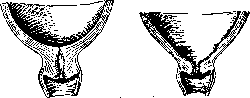
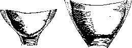
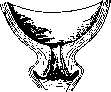
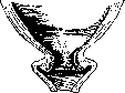

the possibility of the head of a full-term fetus passing through. Under normal conditions, the end of the dilation period usually coincides with the moment of rupture of the fetal membranes and the outflow of the forewaters, but this is observed far from always (in approximately 55% of cases): the fetal membrane may rupture before and after the end of the dilation period (premature, early rupture of the membrane and delayed rupture-birth of the child in the fetal membrane, "in a caul"). If the membrane ruptures too early, childbirth has a prolonged course ("dry labor"). Delayed rupture of the fetal membranes may lead to premature separation of the placenta and death of the fetus from asphyxia. The duration of the dilation period varies with different positions and presentations of the fetus. It also depends on the strength and frequency of uterine contractions. With normal occipital presentation, the average duration of the first stage of labor is calculated for primiparas as 16-18 hours, and for multiparas as 10-12 hours. Complete dilation of the uterine cervix, i.e., the beginning of the expulsion period, is clinically marked by the onset of bearing down, i.e., the contraction of the striated musculature of the abdominal press. Thanks to bearing down, the expulsive forces of labor increase almost twofold (Schatz). In this period, the head, resp. the presenting part, makes a series of progressive movements along the axis of the pelvis (translation according to Selheim) and a series of additional movements-rotational movements around the longitudinal axis of the body and movements around the frontal axis (see below). The action of bearing down consists in the fact that the parturient, holding her breath during a contraction (as during defecation), activates the abdominal press and thereby, by increasing the intra-abdominal pressure, assists the progressive movement of the fetus forward. During the expulsion period, under the action of contractions and bearing down, the head enters the pelvis first with the smallest segment. Its greatest circumference is still above the pelvic inlet, the head is mobile or slightly fixed, on internal examination linea terminalis, s. innominata is not entirely free, as a certain segment (pole) of the head has already entered the pelvic inlet, but the promontory is still accessible to the examining finger, the sagittal suture is closer to the transverse diameter of the pelvis. Then the head enters the pelvis with the largest segment. Above the pelvic inlet, a certain pole of the head can still be palpated, it covers only a small part of the symphysis and sacrum; on internal examination, without pushing the head away, the promontory can no longer be reached, the sagittal suture is slightly in an oblique diameter, the sacral hollow is free. Further, the head enters the cavity of the pelvis. Above it, it is not determined, on internal examination the sagittal suture is in one of the oblique diameters, the sacral hollow is completely filled, spina ischii is not determined. The head reaches the pelvic floor. This is indicated by: the gaping anal opening, the sensation in the parturient of an urge to the low part, the passage of feces, and sometimes also cramps in the calf muscles. The head is at the pelvic outlet. In the gaping vulvar ring, a small segment of the presenting part becomes visible, appearing during a contraction-bearing down and hiding again during a pause ("crowning" of the head). Following crowning comes "crowning": the crowning head, advancing more and more forward, during a pause no longer retreats back, the vulva remains gaping. Contractions and bearing down during "crowning" and "crowning" reach maximum tension ("shaking" bearing down). The crowning of the head and its birth is accompanied by circular stretching of the vulvar ring. Following the birth of the head, the birth of the body quickly follows. The born head turns with its face, depending on the position, to one or the other thigh of the mother (in the first position-to the right, in the second-to the left). The birth of the shoulders occurs by the same mechanism as the birth of the head. The crowning of the shoulder girdle produces no greater stretching of the vulvar ring than the head does. This circumstance is important to consider when protecting the perineum (see below). With the birth of the fetus, the expulsion period ends and the placental period begins. The expulsion period lasts on average for primiparas 2-3 hours, for multiparas-half an hour to an hour. The placental period is the shortest (15-20 min.), but at the same time the most dangerous and responsible period of the act of labor. In it, it is difficult to distinguish: the moment of separation of the placenta from the walls of the uterus and the moment of its expulsion from the birth canal. Normally, separation of the placenta occurs after the birth of the child. Separation of the placenta before the birth of the fetus is a very serious complication, which leads to the death of the fetus and can have the most fatal consequences for the mother (see Placenta, premature separation of the placenta). Separation of the placenta occurs due to contractions of the uterus ("placental" contractions). Separation occurs at the border of the compact and spongy layers of the shedding membrane, it does not occur immediately and not in the same way. A distinction is made between separation of the placenta according to Duncan (the placenta separates laterally, goes forward with one of its edges and only then with its maternal side, the membranes together with the umbilical cord retain their position on the amniotic side of the placenta), according to Schultze (first the central part of the placenta separates with the formation of the so-called retroplacental hematoma-a collection of blood between the placenta and the uterine wall-the placenta exits with the amniotic, resp. fetal side, forward goes the central part of the placenta together with the umbilical cord, the membranes turn inside out, covering the maternal surface of the placenta) and according to Franz
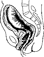
Figure 9. Beginning separation of the placenta (Duncan's method).
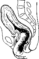
Figure 10. Exit of the placenta through the vagina with the lower edge forward (Duncan's method). (separation of the entire surface of the placenta simultaneously) (figs. 9-12). The obstetrician must know well and clearly the clinical picture of the course of the placental period, as complications often occur here requiring medical intervention. It is especially important to know the clinical signs of a separated placenta. The most important of them are: 1) Schröder's sign (occurs in 95-97%)-the fundus of the uterus rises above the navel, usually to the right, the uterus becomes narrower and flatter (the anteroposterior diameter decreases); 2) Alfeld's sign-lengthening of the external segment of the umbilical cord (a clamp placed on the umbilical cord moves away from the vulva by 10-12 cm); 3) Küstner's sign (95-98%)-when pressing with a hand above the symphysis, the external segment of the umbilical cord in an unseparated placenta goes back into the vagina and remains in place if the placenta has separated from the uterine wall; 4) the appearance of a bulging above the pubis: the anterior wall of the lower segment, into which the separated placenta descends, bulges forward under the pressure of the latter, lifting the abdominal wall forward; between this bulge and the hollow muscle (upper segment) a distinctly expressed groove can sometimes be traced (a kind of "retraction" or "contraction" ring); 5) the sign
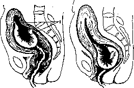
Fig. 11. Central separation of the placenta (Schultze's method) 1 - retroplacental hemorrhage.
Figure 12. The placenta has been expelled from the uterine cavity and is in the cervix in a folded state (Schultze's method). Strassmann's method - light tapping with the fingertips in the area of the uterine fundus with an unseparated placenta is felt as a slight wave (blood in the umbilical vein is in contact with the mother's blood), under opposite conditions this wave is absent; with more energetic pressure on the uterine fundus, along with a slight wave, tension of the umbilical vein with its twisting (overfilling with blood) into a spiral can be obtained (Hochenbichler's sign); b) Klein's sign - when the woman strains or pressure is applied to the uterus, the umbilical cord protruding from the vulva is pulled out and in case of placental separation it does not retract back; c) Mikulicz-Radecki and Calmann's sign - the sensation of a urge to the low that women complain of, due to pressure from the descending placenta on the rectum. The expulsion of the separated placenta occurs either spontaneously (contractions and bearing down) or with the use of obstetric aids (see Obstetric manual maneuvers). In the placental period, the woman loses a certain amount of blood. When the placenta separates according to Duncan's method (from the edge), bleeding may be observed throughout the placental period, while with Schultze's method, the retroplacental hematoma empties after complete separation and expulsion of the placenta. Blood loss varies within 200-600 cm3 (see Postpartum period, postpartum hemorrhages). Bleeding after the birth of the placenta - stops due to 1) contractions of the uterus (the lumen of the torn vessels decreases), 2) the formation of clots in the vessels (especially in the atonic state of the uterine musculature) and 3) the twisting of the thin-walled partitions in which the vessels pass. The average duration of the placental period does not exceed 30 minutes in normal conditions. According to Varnegros (radiograms), the placenta separates from the uterine walls 5 minutes after the birth of the child. Weibel on his X-ray films only in 1/3 of cases found the placenta separated at this time, in 2/3 of cases it still remained in situ. According to Stöckel, final separation of the placenta occurs in the first quarter hour. In any case, within the first half hour after the birth of the child, the placenta in normal conditions is always separated from the uterine walls. Expulsion of the placenta occurs in 70% of cases within 11/2 hours after the birth of the child, in 20% - within the first hour, in 10% separation and expulsion of the placenta continues for more than an hour. Variations (deviations) of the main normal mechanism. Occipital presentations (flexion type). Occipital presentation with the anterior position (occiput and back facing forward) is the only one when the mechanism of childbirth and its clinical course should be considered normal, physiological. But even with this main normal mechanism, deviations (variations) are observed that have great practical importance. 1. Childbirth with the occiput and back facing backward (occurring in approximately 1% of all cephalic presentations). There are two varieties of such childbirths: a) posterior position of occipital presentation and b) anterior-head (anterior-cranial) presentation. The essence of these two variants is that the back of the fetus, sometimes during pregnancy, but more often during childbirth, turns backward (incorrect rotation of the occiput). With the development of labor, the position can correct itself: often the head makes a strong turn, and the occiput together with the back turns forward. In the etiology of stationary (remaining throughout the entire process of labor) anterior-head presentation and posterior position of occipital presentation, the decisive role is played by: the condition of the soft birth passages, resp. the pelvic floor, pelvic anomalies (change in shape and size), as well as the shape and size of the head (small head). Among the predisposing (stimulating) factors, one can note premature rupture of membranes, polyhydramnios, pendulous abdomen, cord entanglement, etc. The clinical course of childbirth with posterior position of occipital presentation in the dilatation period is usual (as with the anterior position). The difference begins with the expulsion period. Here the mechanism of childbirth proceeds in a peculiar way. First, the head performs strong flexion due to the descent of the small fontanelle, which at first plays the role of the guiding point (see below - mechanism of labor). At the second moment of the mechanism (rotation), the occiput turns backward, toward the sacral cavity, and the area of the large fontanelle approaches the symphysis pubis. In this case, if speaking precisely, the point between the large and small fontanelles should be considered the guiding point. For purely practical considerations, with posterior position of occipital presentation, the large fontanelle can be conditionally considered the guiding point. During crowning, the head, being born in posterior position of occipital presentation, has two points of fixation (two hypomochlia) - one to enhance flexion and another to deflex (for extension). At the vulvar ring, the head, as with the anterior position, engages with the small fontanelle. Reaching the pubic arch, it is fixed here by its anterior edge in the area of the hairy part of the head (first hypomochlion), flexion occurs, thanks to which the occiput rolls over the perineum to the suboccipital region. Then the suboccipital fossa is fixed in the area of the coccyx (second hypomochlion), the head extends, freeing the forehead, face, and chin from under the pubic arch. Thus, in general, the mechanism of childbirth with posterior position of occipital presentation consists of: 1) flexion (when the head enters the pelvis), 2) incorrect rotation in the pelvis (the small fontanelle is directed backward) and 3) additional flexion and extension (during crowning). The presenting circumference of the head is planum suboccipito-frontale (33 cm), corresponding to the average oblique diameter. The configuration of the head with posterior position of occipital presentation is dolichocephalic, with a sharp depression in the area of the large fontanelle. The birth tumor is located on the presenting parietal bone, closer to the large fontanelle. The configuration of the head here occurs extremely slowly, with a huge expenditure of labor forces. Very often, after the head has passed the pelvic plane, its forward movement stops. Anterior-head presentation, externally resembling posterior position of occipital presentation, differs from it in principle in that the head here is not flexed, but on the contrary, is in a slight degree of extension (the chin somewhat retracts from the chest). Anterior-head presentation occurs as a transient (temporary) condition in flat pelvises (see Narrow pelvis). In it, the back and occiput, as well as with posterior position of occipital presentation, are directed backward. The head, performing its mechanism, enters the pelvis with the sagittal suture in the transverse diameter (partially in the oblique). Due to the extension of the head, the large fontanelle stands in the same horizontal plane with the small one or even slightly below it. During rotation, the anterior part of the parietal bone (area of the large fontanelle) turns forward, at the same time descending lower. The head passes through the pelvic cavity, entering one of its oblique diameters, and rotates with the occiput backward. The mechanism of crowning proceeds according to the type of posterior position of occipital presentation, only the guiding point, points of fixation, the presenting circumference through the vulvar ring and the configuration of the head will be different. During engagement in the vulva, the large fontanelle and forehead appear first. Gradually crowning, the head moves forward until the glabella approaches the pubic arch. Fixed in this way, the head performs flexion, during which the occiput to the suboccipital tubercle emerges through the perineum. Fixed last in the area of the coccyx, the head extends, freeing the face and chin from under the symphysis. Thus, with anterior-head presentation we have: 1) slight extension when the head enters the pelvis (descent of the large fontanelle), 2) incorrect rotation of the head (with the occiput backward), 3) flexion and 4) extension of the head. The guiding point - undoubtedly the large fontanelle. Two points of fixation (two hypomochlia): 1) glabella and 2) occipital tubercle. The presenting circumference of the head - planum fronto-occipitale (34 cm), corresponding to the direct diameter of the head. The configuration of the head - brachycephalic (tower shape). The head tumor is located in the area of the large fontanelle on the presenting parietal bone. Bumm considers anterior-head presentation as the initial stage of face presentation, i.e. he attributes it not to the flexion, but to the extension type of the mechanism of labor. Timely recognition of the described variant of the main occipital mechanism does not always succeed. More often such recognition is made only at the moment of crowning. In diagnosis, one must first of all keep in mind that one can speak of posterior position of occipital presentation and anterior-head presentation only when the head is already on the pelvic floor, while it is at the entrance to the pelvis or in the wide part of the pelvic cavity, correction of the presentation is always possible.
During internal examination, main attention should be fixed on the position of the fontanelles (fig. 13 and 14). In the posterior view of occipital presentation (under the pubic area the region of the large fontanelle), the small fontanelle (posteriorly) is low-lying,
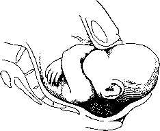
Figure 13. Crown presentation in posterior occipital presentation. The head is in maximum flexion, with the occiput in the genital fissure, the region of the large fontanelle under the pubic symphysis. As soon as the frontal eminences pass the edge of the pubic arch, extension of the head occurs, thanks to which the occiput quickly appears one after another over the perineum, while the face is under the pubic arch.
the large fontanelle is higher (anteriorly), sometimes difficult to reach, the anterior angle of it clearly lies higher than the posterior, there is marked flexion of the head. In anterior-occipital presentation under the pubic area the large fontanelle is also determined, and all its angles lie in one horizontal plane, sometimes the frontal suture can be traced quite far; on the other side (posteriorly) one can always reach (sometimes with difficulty) the small fontanelle. Prognosis. Childbirth with the spine and with the occiput turned backward usually ends spontaneously. There are still some moments which cloud the prediction. First of all, it should be noted that the prognosis for the mother and child in anterior-occipital presentation, all other conditions being equal, is worse than in the posterior view of occipital presentation. Mortality of children due to asphyxia in anterior-occipital presentation is three times greater than in the anterior view of occipital presentation. The expulsion period is significantly prolonged. If, in addition, there is a narrowed or narrow pelvis, which is not infrequently observed, or unyielding soft parts, the prognosis understandably worsens significantly for both the parturient and the fetus. Often there is entrapment of the anterior lip of the cervix and its edema (engagement of the head by its large circumference). Due to great resistance, contractions often are unproductive. As a result, secondary weakness of labor pains often occurs. In addition, in
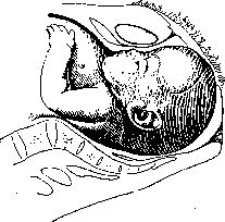
Figure 14. Engagement of the head in anterior-occipital presentation.
Childbirth with the occiput backward presents great difficulties in crown presentation (under the pubic arch the head must perform strong flexion). As a result, there is always great danger of damage to the perineum and even tearing of the m. levatoris. Management of childbirth should be strictly expectant. Protection of the perineum is carried out according to general rules (see below - management of childbirth). One should only remember that according to the mechanism, flexion of the head here should be done not toward the perineum, as in the anterior view of occipital presentation, but toward the symphysis. In the presence of appropriate indications (fetal asphyxia, rigidity of tissues, etc.) one has to resort to one or another operative intervention. The described variant of the flexion mechanism of childbirth more often requires and operation of applying forceps (according to Hogler in 14%). When applying forceps, one should remember that traction here should never be done downward (backward): unfavorable extension (see Obstetric forceps). 2. The second variant of the basic mechanism in occipital presentations - medium and low, resp. deep, transverse position of the sagittal suture. In this anomaly, for one reason or another, the head does not perform the second moment of the mechanism (rotation), positioning itself with its sagittal suture in the transverse dimension of the pelvic cavity (medium transverse position of the head) or even in the pelvic outlet (low, resp. deep, transverse position of the head). There is also a high transverse position of the head (prolonged standing of the sagittal suture in the transverse dimension of the pelvic inlet); it is observed in a flat pelvis as one of the moments of the mechanism of childbirth in the period of head configuration
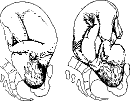
Figure 15. Positio occipitopubica.
Figure 16. Positio occipitosacralis. (see Narrow pelvis).-Transverse position of the head, low and medium, most often occurs in the so-called Deventer pelvis (simple flat pelvis - see Narrow pelvis). In all cases, during the expulsion period, with good contractions and a head low-lying in the pelvis, if childbirth does not progress forward, one should think, along with the possibility of narrowing of the pelvic outlet and the presence of the posterior view of occipital presentation, resp. anterior-occipital presentation, also about low or medium transverse position of the head. During internal examination, the head is usually on the pelvic floor, the sagittal suture is in the transverse dimension, the small fontanelle lies on one side, the large one on the other. The large fontanelle often stands at the same level as the small one (insufficient flexion of the head).-Childbirth in medium and low (deep) transverse position of the head rarely end naturally. They are prolonged, the fetus is threatened with asphyxia, which is why one often has to resort to artificial delivery. In such cases, forceps are usually applied. Before applying forceps, it is recommended for the parturient to lie on the side toward which the small fontanelle is looking, as in this variant it should be considered the guiding point. In general, in managing childbirth in the described anomaly of the mechanism, they should be managed according to the type of normal occipital presentation. 3. High direct position of the head (insertion of the head in the direct dimension of the pelvis) - a rarely occurring variant of the normal mechanism, in which the head at the pelvic inlet is established with its sagittal suture in the direct dimension corresponding to the true conjugate. The head can be inserted in such a way that its occiput is turned toward the pubis - positio occipitopubica (fig. 15) (it occurs more often and mainly in multiparas); the opposite insertion - positio occipitosacralis (fig. 16), when the occiput is turned

Figure 17. Beginning extension, frontal insertion at the onset of labor in face presentation.

Figure 18. Descent of the chin during labor in face presentation.
backward (toward the sacral promontory), is observed extremely rarely, and almost always in primiparas. The etiology of high direct insertion of the head remains unexplained to this day. Most obstetricians see the cause of this anomaly in changes in the shape and size of the pelvis (a greatly narrowed pelvis has a great influence). Direct insertion of the head before labor, sub graviditate (primary), is hardly encountered (the only, and that doubtful case belongs to Glocker), it more often occurs already during labor. In positio occipitopubica (anterior insertion) childbirth can end spontaneously, but in protracted cases one has to resort to forceps. Initially, in the absence of threatening phenomena, one can try Walcher's position (see Walcher's hanging position). With a movable head, one should perform rotation and extraction, but with an inserted head - apply forceps. In posterior direct insertion of the head, childbirth almost always has to be ended by operative means (rotation, forceps, perforation, cesarean section). Henkel for this rare anomaly of head insertion recommended manual correction (under anesthesia): with the help of 4 fingers of the hand introduced into the uterine cavity, placed Fig.19. Face presentation, posteriorly of the head, extension is completed, the chin is to the right and anteriorly should turn only 45°, to approach under the pubic arch.
and the large one - to the right and backward. 4. Excessive rotation of the shoulders - the shoulders pass through the pelvic cavity by the same oblique dimension as the sagittal suture (normally they pass in the opposite oblique). This anomaly has no practical significance. Extension (deflexion, facial) type of the mechanism of childbirth. The antithesis of the mechanism of childbirth in the anterior view of occipital presentation is face presentation, in which the head, being in a state of maximum extension, passes through the pelvis by the other pole of its ellipsoid - with the chin forward. Extension (extensio, defleXio) in this presentation is so marked that the occiput of the fetus lies on the shoulder girdle. Face presentations occur


Figure 20. Face presentation, extension completed, the chin is to the right and posteriorly should turn 135°, to approach under the pubic arch.
approximately in 1:200 births (0.52% according to Demuth). Extremely rarely, as an exception, the head is established in face presentation during pregnancy (primary face presentation), much more often, as a rule, such presentation arises during labor (secondary face presentation). In the origin of face presentations, numerous and heterogeneous etiological factors play a role. Winkel collected up to 33 hypotheses on the question of the etiology of the presentation under discussion. All of them, however, do not withstand strict criticism. Among the factors that may play a role in the etiology of face presentations, usually noted are: narrow pelvis, which, according to Demuth, occurs in 44.6% (Leopold's triad: narrow pelvis, primipara, face presentation), one or another abnormal shape of the head, oblique position of the uterus, unyielding lower segment of the uterus (Bumm), tumors of the neck (hygroma), congenital goiter in the fetus
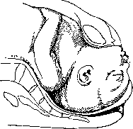
Figure 21. Engagement of the face.
etc. Kernauner sees the cause of the extension type mechanism of Childbirth in a violation of the function of the atlanto-occipital joint. Face presentations, upon external and internal examination, present a completely different picture compared to occipital presentations (fig. 17-24). The diagnostic points for face presentation are given in the accompanying table. Some (Fenomenov, Malinovsky) suggest determining the position in face presentations not by the spine, but by
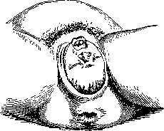
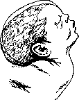
Figure 23. Configuration of the head in face presentation.
Fetus and its parts First position External examination Head and spine Buttocks and small parts Heartbeat Facial line Chin Forehead Presents Shoulders External rotation Birth tumor Left occiput, forms a sharp angle with the spine Lie together on the same side where the heartbeat is heard To the right of the midline under the navel (on the chest side) Internal examination In the transverse diameter, synclitically or in a slight Naegele's inclination To the left To the right Right half of the face In the left oblique diameter of the pelvis To the mother's right thigh More on the right half of the face In face presentation, the same positions and types are distinguished as in cephalic presentations Figure 22. Emergence of the head in face presentation. to the chin (chin forward-anterior view and vice versa). In internal examination, one can only speak of face presentation if the examining finger reaches the tip of the nose, and with further advancement-the chin, with one side of the face being accessible to palpation, and on the other only the forehead. It should be borne in mind that internal examination gives a clear picture only shortly after the rupture of the fetal membranes, before the birth tumor has had time to form. In cases of long-standing head engagement, with long-ago ruptured waters, recognition of face presentation becomes difficult, as on the face, due to the birth tumor, all its irregularities are smoothed out, the cheeks and lips become edematous (only the nose retains __________________. its characteristic shape), the depression of the mouth upon palpation resembles the anus. Face presentation in this case can be mistaken for a pure breech presentation. For recognition, it is recommended to carefully introduce a finger into the existing depression on the presenting part. If it is the mouth, one can feel the jaws, tongue, one can catch sucking movements of the fetus; in breech presentation, this data is not available, and in addition, the extracted finger is found to be stained with meconium, which does not happen in face presentation. Once again, it should be emphasized that internal examination in face presentations should be performed with extreme caution Second position Right occiput, forms a sharp angle with the spine Lie together on the same side where the heartbeat is heard To the left of the midline under the navel (on the chest side) In the transverse diameter, synclitically or in a slight Naegele's inclination To the right To the left Left half of the face In the right oblique To the left thigh More on the left half of the face (one can easily damage such delicate organs as the eyes), aseptically, necessarily with the pulp of the finger, avoiding contact with the nail. One should also refrain from introducing a finger into the mouth of the fetus (one can cause premature respiratory movements). Course of Childbirth in face presentation. The dilatation period proceeds the same as in other head presentations. With its completion, the head enters the pelvis
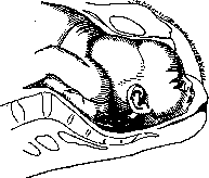
Figure 24. Face presentation. Abnormal backward rotation of the chin. Arrest of labor.
so-called facial line (analogous to the sagittal suture), running from the frontal suture along the bridge of the nose to the chin. This line corresponds to the transverse diameter of the pelvis, and in this position, the head, without making a screw-like movement, reaches the pelvic floor and only here begins rotation with the chin forward. The mechanism of the face passing through the pelvic brim will be as follows. First, the chin passes under the symphysis. As it passes further, the face reaches the sublingual area against the pubic arch. The sublingual area takes on the role of the hypomochlion. Fixing in the area of the hyoid bone, the head performs flexion, during which the forehead, the anterior and posterior parts of the head roll over the perineum. Thus, the entire mechanism of childbirth in face presentation consists of three moments: 1) extension of the head (deflexion), which occurs throughout from the entrance to the pelvic floor, 2) proper rotation-with the chin forward (in the pelvic floor) and 3) flexion (when passing through the pelvic brim). The guiding point is the chin. The fixation point is the area of the hyoid bone; the presenting circumference-planum trachelo-bregmaticum (34 cm)-corresponds to the vertical diameter; the birth tumor-on one side of the face (a terribly disfigured appearance); the configuration of the head-marked dolichocephaly. Unlike occipital presentation (in which the head also has a dolichocephalic shape), the configured head in face presentations has a disfigured side of the face (birth tumor), and in addition, due to habit, characteristic extension is maintained for 4-5 days. The swelling on the face also lasts for 4-5 days. Management of childbirth in face presentation should be strictly expectant with extremely careful observation of the mechanism of labor. About 95% (according to Demuth) of births in this presentation end spontaneously. In view of this, at present most obstetricians do not correct face presentation to occipital, as was formerly proposed by Baudelocque, Thorn, Schatz, and others. The transformation of face presentation to cephalic, according to Baudelocque, is performed by internal maneuvers: the entire hand is inserted into the vagina, positioned against the occiput, which is grasped and pulled downward directly. Schatz proposed for the same purpose an external manual maneuver, the essence of which is that by external methods an attempt is made to transform the S-shaped curvature of the spine (face presentation) into a C-shaped curvature (cephalic presentation). Correction of face presentation, according to Thorn (combined maneuver), is performed as follows: the internal hand, inserted into the uterus, attempts to flex the head, the external hand presses on the fetus's chest in the backward direction, and the assistant's hand, placed on the fetal breech, promotes flexion of the trunk. l In face presentation, premature rupture of the membranes is often observed (in 41% according to Demuth). Protection of the perineum requires particularly careful attention, although it is carried out in the same way as in occipital presentations. It is often necessary to resort to episiotomy (see). Lacerations of soft tissues in face presentations occur in 28.6% (Masticy). Careful attention should be paid to the fetal heart rate (frequent asphyxia). Operative assistance is indicated in the same cases as in occipital presentation. The application of forceps sharply increases the danger of lacerations (avulsions of the m. levatoris). (For peculiarities in the application of forceps-see Obstetrical forceps.) Prognosis. Maternal mortality in face presentations is twice as high as in occipital presentations (3%); mortality in children is three times higher (13-15% according to Winkel and 21.4% according to Demuth). Variants of the basic extension type mechanism of labor, 1. So-called posterior type of face presentation. Normal rotation in face presentation occurs on the pelvic floor, in such a way that the chin rotates forward (toward the pubis), and the forehead backward. Only with such rotation is spontaneous labor possible in the extension type mechanism. But sometimes rotation occurs in the opposite direction: the chin rotates backward, and the forehead forward. Under these conditions, labor is completely impossible for purely mechanical reasons. The posterior part of the head must pass through the pelvic ring together with the shoulders, and however wide the pelvis and however small the head, labor under these conditions is still impossible (the distance between the sternal notch and the anterior fontanelle is at least 13.4 cm). It is impossible to think of changing the position of the head when the face is impacted in the pelvis. Forceps cannot be applied either, and even an attempt to apply forceps (Runge) should be considered a mistake. Only perforation of the fetal head or cesarean section is possible, if there are no contraindications to them and if the mother wishes to have the child and the child is alive. 2. High direct insertion-and medium, resp. low (deep), station of the head in face presentations, in terms of course and management, are analogous to the same anomalies in the flexion type mechanism. 3. Breech presentation-a rather rare phenomenon (0.06-0.1% of all labors). The mechanism of labor in it is far from being clarified. One can speak of breech presentation only in that case if on internal examination the forehead and parts of the face are palpated: on one side one can reach the root of the nose, and on the other 14S reach the anterior angle of the large fontanelle. Among the various etiological factors contributing to the occurrence of breech presentations, one of the most important is pelvic narrowing. The head usually enters the pelvis with the frontal suture in the transverse diameter. Such insertion it maintains throughout its advancement to the pelvic floor. Here rotation occurs with the occiput backward and the orbital area forward. The mechanism of delivery in breech presentations occurs as follows: when the frontal suture stands in the anteroposterior diameter of the outlet, some point between the bridge of the nose and the chin (more often the upper jaw) is fixed under the pubic arch, and around it the entire head rolls over the perineum. Thus, as a whole, the mechanism of labor in breech presentations can be characterized by the following moments: 1) slight flexion (moderate degree of extension) of the head, 2) rotation with the orbital area forward, 3) slight flexion and 4) final very slight extension (when the chin is delivered). The guiding point is the root of the nose. The hypomochlion-the upper jaw. The presenting circumference of the head-planum maxillo-parietale (35 cm). The configuration of the head-triangular (with the apex at the forehead). The prognosis in breech presentations is uncertain. Maternal mortality is 5-10%, mortality in children-30-50%. Labor in breech presentations proceeds with great complications (lacerations of the perineum, avulsions of the mm. levatorum, vesicovaginal fistulas, ruptures of the uterus, fetal asphyxia, etc.); it is often necessary to resort to operative intervention, in particular to destructive operations. Extreme caution must be exercised with forceps. Under appropriate conditions, it is better to perform a version to a foot. On the contrary, in fixed breech presentation, it is better (especially for the beginning obstetrician) not to apply any corrective operations (Solviev's proposal to transform breech presentations to face presentations by extending the head with a finger inserted into the mouth, and Kholmogorov's-to correct to occipital presentations by flexing the head). Breech presentation with the orbital area turned backward is rarely observed. Such cases have been observed in labor with small (premature) fetuses. In a full-term fetus, labor in the posterior type of breech presentation (orbital area backward) is impossible (perforation or cesarean section) (fig. 25-27). Breech presentations. They occur on average in 3.5% of the total number of labors. They are divided into breech presentations (flexion type) and foot presentations (extension type). Breech presentations in turn are divided into pure (only the breech presents) and mixed (the breech presents together with the soles of the feet). Foot presentations are divided into complete (both legs present) and incomplete (one leg presents, usually the anterior one) (fig. 28-30). Foot presentations occur in 33% of all breech presentations. Knee presentations are very rare. The reason why the fetus, instead of the head, presents with the sw pelvis is Figure 30. Incomplete foot presentation. The anterior leg is bent upward.
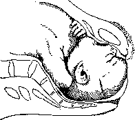
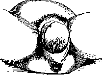
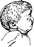
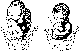
Related articles
Cite this page
“Childbirth.” Soviet Medical Encyclopedia. English translation of Bolshaya Meditsinskaya Entsiklopediya, 1st ed. (Moscow, 1928–1936), ed. N. A. Semashko. https://sovietmedicalencyclopedia.pages.dev/article/childbirth/