Microcephaly
Historical document, translated for reference. It reflects medical knowledge of the 1920s–30s and is not medical advice.
Summary
Microcephaly is a condition characterized by abnormally small head size with relatively normal growth of other body parts. The Soviet medical encyclopedia describes its causes, manifestations, and classification as either primary (true) or secondary (false) microcephaly.
Encyclopedia article (1928–1936)
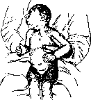
MICROCEPHALY (from Greek mikros-small and kephale-head), abnormally small size of the skull with normal or nearly normal growth of all other parts of the body. With this size of the skull is associated a correspondingly small brain (micrencephalia). Microencephalies are however observed with normal skull size. For adults, a head circumference below 48 cm and a brain weight below 950-1,000 g give grounds to speak of M. For the most part, the brain weight in microcephalics varies between 250 and 900 g, and the head circumference-between 30-40 cm. A brain with significantly smaller weight and volume has however been described. Thus, in the well-known from the literature Becker family, where among 9 children 5 were microcephalics, an 8-year-old girl's brain weighed 219 g, another, 15-year-old-152 g; Rohon described a microcephalic with a brain weight of 17.3 g. In the microcephalic described by Monakov, the head circumference was 24.5 cm.-M. rarely develops as a result of brain disease in extrauterine life. Most often the issue concerns intrauterine lesions of the brain. Giacomini divides all cases of M. into primary, or true, and secondary, or false, M. (microcephalia vera and pseudomicrocephalia). In the first case, it is a matter of arrested development of the brain, in the second-of disorders of growth due to severe diseases of it. The degree of brain damage can be very diverse; sometimes retardation of individual parts of it is noted, most often-the occipital lobes, as a result of which the normally developed cerebellum is left uncovered or the brain as a whole remains undeveloped. The number of convolutions in these cases is often very small, and sometimes they turn out to be very voluminous (macrogyria, see); in other cases, on the contrary, the convolutions are very small (microgyria, see); finally in some cases individual parts, especially the cerebrum, develop only rudimentarily. Histologically, the cortex appears immature, often with abnormal, difficult to explain cellular forms. The cortex is generally poor in cells, sometimes heterotopias (displacement of individual parts of the cortex into the white matter) are observed. The underlying parts of the brain usually have a normal structure. Careful path-anatomical research has shown that cases of true M. are observed much less frequently than is assumed. Often it is not a matter of a normal brain in a reduced, miniature form, but of various anomalies, such as retardation in the growth of the forebrain compared to the middle and hindbrain, abnormal convolutions, etc. In most cases, sclerotic and degenerative processes, cysts, hydrocephalus are found, so that even in such cases the observed changes must be considered remnants of intrauterine lesions.-The general appearance of microcephalics is very characteristic. Changes of the skull are often observed, consisting not only in a change of its size but also in its peculiar structure. Usually the skull is round, markedly brachycephalic in shape, the forehead is flat, the frontal bone slopes into the parietal bone, the occipital bones are poorly developed. Since the facial skeleton is usually developed to a sufficient degree, a very peculiar shape of the head is created (bird face, Aztec head) (see figure). At birth the skull may have normal size. Usually however the fontanelles close very early, and the sutures bulge in a ridge-like manner. Cases of M. are also observed in which late closure of the sutures and fontanelles, as well as late ossification of the skull bones are noted. The physical development of microcephalics may not suffer at all. They can live quite long (up to 50 years or more). In a psychological respect the majority of microcephalics are more or less severely feeble-minded. In many of them already in infancy an inadequately developed sucking reflex, constant restlessness, inability to fixate, absence of pain reaction are noted. Often they learn to hold their head up very late. Significant rigidity of muscles is most often noted. The upper extremities are pressed to the chest, bent at the elbow joints, the hands are clenched, the fingers are indented, the lower extremities are extended, adducted, and have a tendency to cross. The abdomen is strongly retracted, boat-shaped. The facial expression is very poor. Sometimes this picture is complicated by athetoid movements, disorders of swallowing, pseudobulbar symptoms and epileptic seizures. A whole series of autonomic symptoms is also observed: increased salivation, marked cyanosis, persistent constipation, etc. In these cases the issue usually concerns embryonic diseases of the brain with atrophic-inflammatory changes, especially in the area of the central convolutions. Other microcephalics are distinguished by significant motor excitement. Their movements are correct, coordinated, facial expression is lively. Sometimes they show some ability to develop. They learn to speak a little, recognize people around them, are clean, capable of self-care and even learn to perform some household chores. The latter forms usually belong to the so-called true M.
According to the opinion of Vogt, based on the study of microcephalic skulls, M. is an atavistic formation. Virchow put forward the theory of premature closure of the sutures, due to which the development of the brain is delayed. Both theories at the present time can be considered refuted. The cause of M. is now seen in the cessation of growth of the skull and brain, with the brain being primarily affected, which secondarily causes retardation of skull development. The decrease in pressure inside the skull due to various reasons leads to the premature cessation of growth of its bones, and therefore with underdevelopment of the brain the skull remains small. Cases of true M., according to Monakov, are explained by primary general or partial dysgenesis or dysplasia of the medullary tube with the resulting consequences in terms of the tectonic differentiation of individual parts of the brain. According to Monakov's opinion, the genetic process in case of insufficient differentiating force of the medullary tube for the full development of the central nervous system nevertheless continues in accordance with the existing predispositions, but it leads to incorrect development and often to premature (local) termination of the process with fixation of embryonic stages and to a general decrease in the volume of the organ. Individual parts however can continue to develop to almost normal size; in this case the embryonic material is used for phylogenetically older and more important for the maintenance of life formations. The parts of the medullary tube that are weak in terms of development are usually the parts that are intended to serve higher nervous processes in the future. The special localization of genetically weak parts of the medullary tube can vary greatly, but almost always it can be noticed that in these parts there are attempts at further development. In addition to this local subsequent development, morphological compensations originating from normally acting parts are also noted. They can lead to the fact that formations phylogenetically old and directly important for life accelerate their development and reach a strength that sometimes gives them the possibility to contribute to the beginning of regeneration in hypoplastic areas. Under such conditions, tectonically simpler types of brain can be created, encountered in lower mammals or vertebrates. But in addition to the indicated regenerative and postgenerative processes, secondary processes of decay and dissolution of nerve elements also occur. Macroscopically more coarse, mechanical adaptations (abnormal stretchings and shifts in the sense of saving space) and similar processes on the part of the secretory organs and organs of circulation (dilation of the ventricles, perivascular lymphatic pathways, hydrocephalus ex vacuo, etc.) are also noted. Such more coarse moments in development can lead to changes in the form of the brain, having a remote similarity with some lower types of animals; however they never correspond to these types in their internal structure. As an etiological factor, early occurring intoxication, thermal or mechanical damage to the embryo is put forward. In some cases, severe alcoholism of the father is noted.
The prognosis for M. is unfavorable.--There is no therapy. Operations of partial removal of skull bones, performed on the basis of the above-mentioned theory of Virchov about too early ossification of the skull bones, gave no results and have been abandoned. Educational measures can give only very insignificant results. Such children need to be placed in appropriate institutions. Sometimes the disease manifests itself familially. The type of hereditary transmission is apparently recessive. The above-mentioned Becker family is especially well known. Bernstein also described a family where among 10 children there were 5 microcephalics. Families with two microcephalics are observed comparatively often.
Related articles
Mentioned in
Cite this page
“Microcephaly.” Soviet Medical Encyclopedia. English translation of Bolshaya Meditsinskaya Entsiklopediya, 1st ed. (Moscow, 1928–1936), ed. N. A. Semashko. https://sovietmedicalencyclopedia.pages.dev/article/microcephaly/