Elbow Joint
Historical document, translated for reference. It reflects medical knowledge of the 1920s–30s and is not medical advice.
Summary
This article from the 1928–1936 Soviet Great Medical Encyclopedia provides a detailed anatomical description of the elbow joint (articulatio cubiti). It covers the structure of the humerus, ulna, and radius, the joint capsule, and the various ligaments that stabilize the joint.
Encyclopedia article (1928–1936)
ELBOW JOINT (articulatio cubiti), connects the bones of the arm and forearm, forming a so-called true (diarthrosis) joint, which includes the distal end of the humerus (bearing the head), the proximal ends of the ulna and radius (bearing the cavities), and is thus a complex joint. By the form of movements it allows, it represents a combination of a hinge joint (ginglymus) with a ball-and-socket/pivot joint (articulatio trochoides). The articular surface of the humerus is located on its lower, widened, and flattened end between the two epicondyles and breaks down into two parts passing into one another—medial and lateral. The medial part articulates with the ulna, has the shape of a pulley (trochlea), and forms an almost complete (320°) circumference with a radius of 9.8 mm (Fick). Its articular surface is covered with cartilage, extending beyond the lateral edges of the pulley, the notch of which excludes the possibility of the ulnar head sliding in a transverse direction. Above the pulley is the coronoid fossa (fossa coronoidea) and the deeper olecranon fossa (fossa olecrani); they are intended for the corresponding processes of the ulna (Figs. 1 and 2). The septum separating them is thicker in the fetal period and in children; with age, it becomes thinner and in adults (more often in men) often has an opening, which exists as a norm in some animals. The lateral part of the articular surface of the humerus represents a capitular elevation (eminentia capitata, s. capitulum humeri), which forms a spherical surface with a radius of 11 mm for articulation with the head of the radius; its center lies on
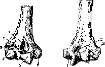
Figure 1.
Figure 2.
Figure 1. Trochlea (5) from the front: 1-fossa coronoidea; 2-fossa radial.; 3-epicond. lat.; 4-capit. hum.; 6-epicond. med. Figure 2. Trochlea from the back: 1-epicond. med.; 2-fossa olecr.; 3-epicond. lat. on the same axis as the pulley. The capitular elevation is covered with cartilage, passing from the pulley; above it, on the anterior surface, is a shallow fossa (fossa radialis). The epicondyles of the humerus located on the sides of the articular surface serve as the attachment site for the muscles of the forearm: the medial, more pronounced one, for the flexor group, and the lateral one for the extensor group. The bony process (processus supracondyloideus) sometimes encountered (2.7%) in humans above the medial epicondyle, directed downward in the form of a hook, serves for the attachment of an additional head of the pronator teres and is an atavistic formation, existing as a norm in carnivores, monkeys, and reptiles, in connection with well-developed pronation movements; in humans, it is closed by a ligament into a complete ring, through which the neurovascular bundle passes. The proximal end of the ulna, which is part of the joint, bears two processes: the anterior—coronoid (proc. coronoideus) and the posterior—olecranon (olecranon), which together form a deep notch (incisura semilunaris) (Fig. 3), encompassing the pulley in the form of a semicircle with a radius
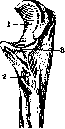
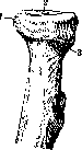
Figure 3.
Figure 4.
Figure 3. Ulna: 1-incis. semilun.; 2-tuber. ulnae; 3-proc. coronoid. Figure 4. Head of radii (1): 2-fovea capit.; 3-collum radii. of 1 cm; the articular surface corresponding to the notch of the pulley has a ridge in the middle. The cartilaginous covering of the olecranon is separated from the cartilaginous covering of the coronoid process by a transverse groove filled with connective tissue. It appears by the time the epiphyseal cartilage disappears, being in connection with the processes of ossification. On the lateral side of the coronoid process is the so-called radial notch (incisura radialis) (for articulation with the circumferentia articularis radii), having a circumference of up to 90° with a radius of 15 mm. The processes of the ulna, forming the joint, simultaneously serve as brakes limiting the range of motion, and on their outer surfaces bear the attachments of the main muscles of the joint: the olecranon—for the m. triceps, the coronoid process—for the m. brachialis. The head of the radius (Fig. 4) has the shape of a disk and bears a saucer-like depression on the upper surface, representing a segment of a spherical surface for articulation with the eminentia capitata humeri, having the same radius as the latter, but half the extent. On the medial side of the lateral surface of the radial head is an articular facet, representing a segment of a cylindrical surface for articulation with the radial notch of the ulna (circumferentia articularis), with a circumference equal to 120–180°, and a radius of 12 mm. The capsule of the elbow joint (Figs. 5 and 6) attaches
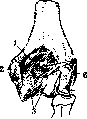
attachment of the capsule (1); 2 - epic. med.; 3- trochlea; 4-rad.; 5- emin. capit.; 6- epic. lat.; 7- fossa olecr.
on the humerus, encompassing the pulley, the capitular elevation, and the fossae, leaving both epicondyles outside the joint cavity. On the side of the ulna, it encompasses the articular fossa, attaching along the cartilaginous edge. On the radius, it has a loose attachment on the neck below the annular ligament, including the entire head in the joint cavity; on juvenile bones, the capsule extends beyond the epiphyseal line. The capsule has the greatest volume posteriorly in connection with the large range of motion of the olecranon. At the ends of the transverse groove and below the annular ligament of the radius, it forms eversions (on the radius—recessus sacciformis radii for the free rotation of the radial head). The thinnest part of the capsule is located posteriorly along the edges of the tendon of the m. triceps. The m. brachialis, triceps, and anconaeus covering the capsule anteriorly and posteriorly provide fibers to it, protecting it from being pinched between the articular surfaces. On the inner surface of the capsule, a series of synovial folds is formed: above the posterior and anterior fossae, between the radius and the capitulum humeri; the latter plays the role of a meniscus. Strengthening ligaments (lig. ant. and post. cubiti) (Figs. 7 and 8). On the anterior surface of the capsule, the ligamentous fibers run in longitudinal and oblique directions. The fibers running from the medial epicondyle to the outer edge of the radial head weave into the lig. annulare radii and are more pronounced than the others. Posteriorly, the capsule is reinforced by fibers running from the upper edge of the fossae olecrani downward into its thickness, and by transverse fibers connecting the edges of the fossa between
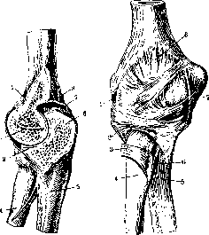
Figure 6.
Figure 7.
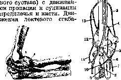
Figure 6. Elbow joint in cross-section: 1-fossa coronoidea; 2-trochlea humeri; 3-proc. coron. ulnae; 4-radius; 5- ulna; 6-olecranon; 7- capsula articularis; 8-fossa olecrani. Figure 7. Ligaments of the elbow joint from the front: 1-lig. collat. radiale; 2-lig. annulare rad.; 3-tendom. bicipitis; 4-radius; 5-ulna; 6- chorda obliqua; 7-lig. collat. ulnare; 8-capsula articularis. themselves and the olecranon. The lig. collaterale ulnare is a powerful ligament starting on the medial epicondyle and attaching along the inner edge of the incis. semilunaris, divided into three portions (Fig. 9). 1) A thinner, fan-shaped part (pars posterior; Braus), expanding downward, attaches along the inner edge of the incis. semilunaris; it forms the lateral wall of the osteofibrous receptacle of the ulnar nerve. 2) Pars anterior, or the main portion, of parallel fibers; ends on a special tubercle of the edge of the incis. semilun.; it is more powerful and is tense in any position of the joint. 3) Fascia transversa, or lig. Cooperi, connects the lower attachment points of both previous ones, ne
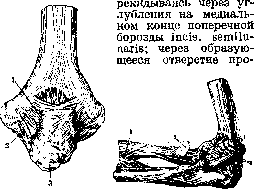
Figure 8.
Figure 9.
Figure 8. Elbow joint from behind: 1-capsula articularis; 2-lig. collat. ulnare; 3-olecranon. Figure 9. Elbow joint from the side of the ulna: 1-membrana interossea; 2-tendo m. bicipitis; 3-lig. collateral ulnare. The nutrient artery enters here. - Lig. collaterale radiale (lateral ligament of the elbow joint) begins on the lower and anterior surface of the lateral epicondyle, extends downward, widening, and divides into two branches, which pass in front of and behind the head of the radius and attach to the corresponding edges of the incisura radialis ulnae, connecting here with the fibers of the lig. annulare (Figure 10). The posterior branch of the lateral ligament of the elbow joint gives fibers to the crista supinatoria ulnae, which weave into the origin of the supinator muscle. - As a special ligament, the lower part of the capsule is described, the recessus sacciformis, in the area between the neck of the radius and the lower edge of the incisura radialis (Denuce's quadrate ligament). The lateral ligament is closely connected with the origin of the extensor muscles of the forearm. Due to the fan-like shape of the lateral ligaments of the elbow joint, one or another of their portions is tensed in any position of flexion, which prevents lateral deviation and limits movement in extreme positions. - Lig. annulare radii (annular ligament) encircles the neck of the radius with a tight and strong ring, forming a funnel-shaped depression open upwards, and attaches to the anterior and posterior edge of the incisura radialis ulnae.
Mucous bursae. Bursa olecrani is located in the subcutaneous tissue above the olecranon and sometimes consists of several chambers; the bursa intertendinea in the thickness of the tendon of the m. triceps above the olecranon and the bursa subtendinea between the tendon and the posterior surface of the capsule are inconstant. Between the m. anconaeus and the joint capsule, there is a mucous bursa, which in adults often communicates with the joint cavity. Two bursae on the sides of the tendon of the m. bicipitis-bursa bicipito-radialis and cubitalis interossea-protect the tendo m. bicipitis from friction against the radius and ulna. On the surface of the m. supinator, separating it from the lateral extensor group, a mucous bursa is also formed.
The nutrition of the elbow joint occurs at the expense of the arterial network (rete cubiti) (Figure 11). The network is especially developed on the dorsal side, which is connected with the need for collateral circulation during compression of the main trunk during flexion at the elbow. The collaterals themselves consist of branches running in longitudinal and transverse directions and bypassing the central, most mobile part of the joint capsule (Lesgaft). The aa. collateralis ulnaris superior and inferior, recurrens ulnaris ant. and post., recurrens radialis, recurrens interossea volaris and dorsalis, collateralis radialis, and collateralis media participate in the formation of the network. The network is located in two layers: above the tendon of the m. triceps and below it on the joint capsule. The epiphysis of the humerus is nourished by vessels penetrating it in the place where it lies outside the capsule (epiphyseal vessels), as well as from vessels entering at the level of the attachment of the capsule and from there penetrating into the epiphysis, piercing the epiphyseal cartilage in young bones (Braus).
Innervation. The anterior wall of the capsule is innervated by the nn. radialis, medianus, musculo-cutaneus; the posterior wall-by the nn. radialis and ulnaris, which gives a branch at the place of its adherence to the capsule.
Statics and dynamics of the elbow joint. Functionally, the elbow joint represents two independent sections: the hinge humeroulnar joint with movements of flexion and extension, and the cylindrical humeroradial joint, which is functionally inseparable from the proximal radioulnar joint.
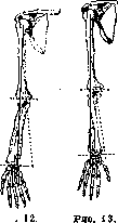
Movements occur around a transverse axis passing through the lower end of the humerus from a point on the medial epicondyle, located somewhat above its lower edge, to the lateral epicondyle, immediately below its most prominent point. This axis, in relation to the long axis of the humerus, is inclined from above, from the outside, inward and downward (Figure 12). The range of flexion movement is equal to an arc of 130° in men and 140° in women. Maximum extension gives an angle of 180° in women and 175° in men. The larger arc in women and children and the hyperextension more frequently encountered in them are connected with less developed musculature and less strongly expressed olecranon and coronoid processes, which are the 'absolute' (Fick) brakes of movement.
Relative brakes are: 1) muscle activity and tension of the capsule on the side opposite to the movement, 2) the action of the corresponding sections of the lateral ligaments, 3) pinching of soft tissues between the contacting surfaces of the bones (during flexion). Relative braking takes place mainly during flexion, whereas extension is braked mainly by the collision of the olecranon with the fossa. The inclined position of the transverse axis causes the formation of the so-called cubital angle, open outward and equal to 162-170° (Fick). In extreme forms of its expression, the limb acquires a type similar to genu valgum. The reason for its formation (expressed more weakly in children) lies in the greater pull of the forearm in the radial direction (abduction) by the lateral extensor group of muscles. The presence of the cubital angle is useful in that lifting a weight with the forearm, which is deviated outward, causes the pressing of the articular surfaces and an increase in support (Lesgaft).
A number of factors cause a deviation from the purely hinge movement of the elbow joint. Thus, the screw-like movement of the ulnar notch around the trochlea, depending on the spiral twisting of the latter, causes a deviation of the distal end of the ulna from the plane of movement (up to 2 mm), just as the pull of the muscles does. Pronation and supination occur in the humeroradial and in both radioulnar joints, proximal and distal. This movement occurs around a diagonal axis, one end of which passes through the head of the radius, the other through the head of the ulna, and the direction of which coincides with the direction of the fibers of the lig. interosseum; the part of the ligament between the proximal radial and distal ulnar attachment points remains tense during the entire rotation, not allowing the bones to diverge in the longitudinal direction; the head of the radius moves in this movement like a wheel around its own axis (art. trochoides-wheel joint) in the incisura radialis ulnae.
Braking of movement occurs due to the tension of antagonistically acting muscles (during supination, the pronators, and vice versa) and ligaments: during supination, the anterior wall of the elbow joint capsule with its oblique ligaments is tensed, and in the distal radioulnar joint, the anterior wall of its capsule is tensed; during extreme turns, the Denuce's quadrate ligament has a braking effect; the interosseous ligament, being located along the axis of movement, does not represent a brake, just as the chorda obliqua does not; during pronation, the pinching of muscle masses between the bones and the tension of the skin on the dorsal side have a braking effect. The diagonal axis of the forearm represents a continuation of the axis of rotation of the humerus and passes through the caput et eminentia capitata humeri (Figure 13). A full turn of the entire forearm with the hand and the humerus is equal to almost 360°. The action of the main group of muscles on the elbow joint is expressed in the following scheme (W. P. Bowen): Elbow joint Radioulnar joint
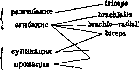
supinator, pronator teres, pronator quadratus. Flexion. The brachialis muscle simultaneously with flexion pulls the lower end of the humerus posteriorly. The biceps flexes better in the position of supination; the abducting action, caused by its attachment to the radius, is partially compensated by the pull of the lacertus fibrosus; the biceps possesses greater strength than the brachialis muscle. The brachioradialis is a powerful flexor. The pronator teres, in contrast to the biceps, flexes better in the position of pronation; it usually acts together with its antagonist, the supinator, which strengthens the radius. The flexor digitorum sublimis, flexor carpi radialis, extensor carpi radialis longus and brevis possess a weak flexing action. Extension. The triceps is the only extensor. Its tendon sends processes to the lateral and medial sides to the fascia of the forearm, enveloping the elbow joint like an accessory extensor apparatus of the knee joint (Schultz). Usually, its extensor functions are not fully manifested due to the action of an absolute brake. Its mass and strength are less than those of the flexor group. The anconeus muscle has the significance of a muscle that, along with the biceps, brings together the articular surfaces of the proximal end of the radius and ulna. Rotational movements. Supination. The biceps, the tendon of which is wrapped around the diaphysis of the radius in the position of pronation, is the main supinator; it supinates only in the flexed position of the elbow joint; for isolated supination, the joint action of the triceps, which strengthens the elbow joint, is required. The supinator muscle acts in any position and possesses a strength equal to half the strength of the biceps. Pronation. The pronator teres muscle, the true antagonist of the supinator, produces isolated pronation when acting together with the triceps, which strengthens the elbow joint. The brachioradialis muscle does not take part in isolated rotational movements, performing pronation (as well as supination) secondarily during flexion to bring the radius into the middle position (Bowen). The pronator quadratus, besides isolated pronation, brings the articular surfaces of the distal radioulnar joint into full contact. Rotational movements are usually combined with rotation of the shoulder. Supination is performed with greater force due to the preponderance of the corresponding muscle group. This explains why working movements associated with rotation usually occur outward when using the supinator group (e.g., working with a chisel when screwing). In any work, movements in the elbow joint occur in combination with movements in the shoulder joint and the shoulder girdle as a whole. The middle position in the elbow joint with a hanging arm is the position of initial flexion and the middle between pronation and supination in the radioulnar joint; the former is explained by the preponderance of the normal tone of the flexor group over the force of gravity; the latter by the equilibrium between the groups of supinators and pronators. In the middle position, there is maximum rest and the greatest readiness for any movement. Topography. The region of the elbow joint is divided into the posterior (extensor) and anterior (flexor) surfaces. The external contours of the flexor surface are determined by the bulges of three muscle groups: on the radial side—the extensors, on the ulnar side—the flexors, and the group of flexors of the arm wedged between them in the middle (Figure 14). In the subcutaneous layer, along the outer groove limiting the flexors of the arm, the cephalic vein with the lateral antebrachial cutaneous nerve is located; on the inner side—the basilic vein with the medial antebrachial cutaneous nerve; and in the middle—the significantly varying median vein, which has a constant anastomosis with the deep veins (Fig. 15). The fascia covering the entire region has reinforcement from the tendon of the biceps in the form of the lacertus fibrosus, which is stretched over the location of the cubital artery and gives a septum deep into the tissue. The latter, together with the tendon of the biceps, separates the inner part of the deep subfascial space from the outer one. In the former, bounded on the inside by the entire group of flexors of the forearm and on the outside by the tendon of the biceps, the cubital artery is located on the surface of the broad belly of the brachialis muscle; it gives off the ulnar artery lower down and continues into the radial artery; medial to it lies the median nerve (Fig. 16). The belly of the brachialis muscle covers the entire anterior surface
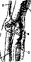
Figure 14. Relationship between bones and soft tissues: 1- lateral bicipital groove; 2- radial groove; 3- elevation of flexor muscles; 4- cubital fossa; 5- medial bicipital groove. (From Corning.)
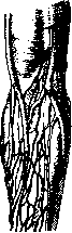
Figure 15.
of the joint capsule, corresponding to the position of the coronoid fossa, the trochlea, and the coronoid process, attaching with a short tendon to the surface of the coronoid process. The flexor muscle group originating from the medial epicondyle does not have direct contact with the joint capsule. The outer subfascial space is located laterally from the tendon of the biceps; it is bounded on the side by the mass of the brachioradialis muscle, under the edge of which the radial nerve passes on the surface of the brachialis muscle, dividing lower down, on the surface of the supinator muscle, into two branches, of which the deep one goes into the canal in this muscle and wraps around the neck of the radius
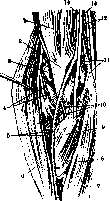
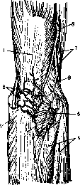
Figure 16.
Topography of the cubital fossa: 1- biceps muscle; 2- tendon of the biceps; 3- brachialis muscle; 4- deep branch of the radial nerve and radial recurrent artery; 5- radial artery and superficial branch of the radial nerve; 6- brachioradialis muscle; 7- flexor carpi ulnaris muscle; 8- flexor carpi radialis muscle; 9- pronator teres muscle; 10- ulnar artery and recurrent branch of the ulnar artery; 11- inferior ulnar collateral artery and medial intermuscular septum; 12- ulnar nerve and superior ulnar collateral artery; 13- median nerve; 14- brachial artery. Figure 17. Region of the elbow joint; posterior view: 1- triceps muscle; 2- ulnar nerve and superior ulnar collateral artery; 3- flexor carpi ulnaris muscle; 4- extensor muscles; 5- anconeus muscle; 6- dorsal antebrachial cutaneous nerve; 7- brachioradialis and extensor carpi radialis longus muscles; 8- posterior radial collateral artery. (From Corning.) anteriorly, upward, downward, and outward; the brachialis muscle also covers the anterior surface of the capsule here, corresponding to the capitulum of the humerus and partly the head of the radius. On the extensor side, the joint is covered by a less powerful muscle layer, which, together with the bony parts, is clearly contoured through the skin. The olecranon, protruding in the middle, serves as the attachment for the broad band of the triceps, which covers the posterior surface of the capsule corresponding to the olecranon fossa (Figure 17). On the radial side, the clearly palpable lateral epicondyle serves as a center for: 1) the group of dorsal extensors (extensores: digitorum, ulnaris), starting at the epicondyle and partly covering the capsule of the radial head from behind with its upper end; 2) the group of lateral extensors, covering the capsule from the side and partly fusing with it; 3) the anconeus muscle. The latter, expanding in a fan shape, attaches to the crest of the ulna and covers the entire surface of the capsule between the base of the epicondyle and the elbow. The lateral epicondyle and the head of the radius directly below it, which are clearly palpable right under the skin and well visible as tubercles when the joint is semi-flexed and as a retracted dimple when extended, serve as good landmarks for approaching the joint cavity between the head of the radius and the humerus. The groove between the medial epicondyle and the olecranon serves as a bed for the ulnar nerve, located under the skin and fascia directly on the joint capsule and entering lower down into the canal between the two heads of the flexor carpi ulnaris muscle. The projection of the articular cleft on the flexor side corresponds not to the transverse skin fold located 1.5 cm higher, but to a line inclined inward, passing 2 cm below the lateral and 3 cm below the medial epicondyle (Fig. 18). When
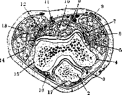
Figure 18. Cross-section through the elbow joint: 1- subcutaneous olecranon bursa; 2- olecranon; 3- ulnar nerve; 4- medial epicondyle; 5- flexor carpi ulnaris muscle; 6- pronator teres muscle; 7- median nerve; 8- antebrachial fascia; 9- brachial arteries and veins; 10- brachialis muscle; 11- tendon of the biceps; 12- radial nerve; 13- brachioradialis muscle; 14- extensor carpi radialis longus muscle; 15- lateral epicondyle; 16- anconeus muscle; 17- tendon of the triceps muscle. (From Corning.)
In the position of full extension, both condyles are on the same horizontal line as the upper edge of the olecranon; in the flexed position, it forms with the condyles the apex of a triangle pointing downwards. The joint capsule lies most superficially on both sides of the olecranon and the tendon of the triceps. Architectonics of the bones of the elbow joint. In the lower end of the humerus, the trabeculae of the spongy substance are arranged parallel to the articular surface, intersecting with the trabeculae starting from the sides. Ulna. The trabeculae in the olecranon are arranged in a direction transverse to the diaphysis of the bone, leaving narrow slits between them, while those starting from the front and back intersect towards the apex of the olecranon and the processus coronoideus. Under the coronoid process, a plate of compact bone is noted; from it, trabeculae run to the articular surface of the semilunar notch, crossing on the way the trabeculae arranged concentrically, parallel to the articular surface. Radius. The bottom of the articular fossa consists of thick compact substance, the side walls of a thin layer; parallel to the articular surface, slightly concave trabeculae are arranged concentrically, intersected by others extending from the side walls. Development. The basic forms of the elbow joint are outlined as early as the second month of fetal life. The separation of the arm from the forearm in the upper limb bud occurs in the eighth week. The cleft, forming from the general mesenchymal mass located between the three still cartilaginous primordia in a 3-10 mm fetus, extends from the periphery to the center. The capsule is formed from mesenchyme; the lateral ligaments are formed earlier than the capsule and outside it; the ligamentum annulare radii is created from the lateral ligament and the upper attachment of the musculus supinator (Mutel); the anterior and posterior ligaments are formed from the fasciae of the musculi brachialis, triceps. Initially, the depression of the supratrochlear fossae is absent, and the depression of the trochlea itself is weakly expressed. In postnatal development, there is a thinning of the septum under the action of the olecranon. The formation of the trochlea, the depression of which is initially weakly expressed, is linked to the great pressure experienced by this half of the articular end of the humerus (Thompson). The spiral direction of the trochlea is explained by the direction of the pull of the flexors towards the radial side (Fick). The olecranon of the newborn is relatively well developed, due to which the angle of extension in it is smaller (150-160°). The rapidly growing head of the radius causes a flattening of the elbow and the formation of the radial notch on it. Ossification. The ossification center of the eminentiae capitatae of the humerus appears in the second half of the 1st year of life, ossification of the trochlea occurs between the 2nd and 5th years. By 14 years, both centers fuse with each other, and by the 17th year, the epiphysis fuses with the diaphysis. The center of the medial condyle appears at the age of about 4-6 years, the lateral one only by 12-13 years. The lateral condyle soon fuses with the eminentia capitata and together with it with the diaphysis. The medial condyle remains isolated until 18 years. The center of the head of the radius appears around 4-5 years in the form of a central one, with which many small ones fuse, forming the shape of a disk; fusion of the head with the diaphysis occurs by 16-17 years. A large center at the base of the olecranon appears at 12 years, of the apex of the olecranon at 14 years; fusion with the diaphysis by means of a third intermediate center, having the shape of a plate (apophysis conjunctiva - Schwegl), occurs at 16-17 years.
Comparative anatomy and phylogenesis. The function of the limbs depending on the way of life is reflected in the form and degree of rotational and lateral mobility. In crawling animals (crocodile, lizard), whose trunk lies on the ground, the limbs are directed to the side, the transverse axis of the elbow joint is parallel to the axis of the trunk. Where the forelimb plays the role of a support and serves only for forward movement on a flat surface, there is pure hinge movement; the ulna moves backward and plays a secondary role (dog) or disappears completely, representing only a small process (horse). In predators (cat, tiger), the forelimb serves for grasping and holding prey; for this, rotational movements of the hand (paw) are needed: the ulna is a support and a place of attachment for the rotating muscles. The radius moves to the side, next to the trochlea a spherical surface for rotation appears for it. The highest degree of development of rotational movements in connection with the variety and volume of functions is found in climbing animals (monkey) and in humans. The origin of the human form is more likely not from quadrupeds, but from tree-dwelling forms of animals, in which only the hind limbs served to support the trunk, and the forelimbs (in connection with the need to climb, catch, tear off food) acquired special development. In quadrupeds, in which the limbs move under the trunk with the lifting of the trunk, the transverse axes of the elbow joint become perpendicular from being parallel to the axis of the trunk; at the same time, the flexor surfaces turn to the side opposite to the hind limbs, and an angle open anteriorly is formed. In humans, in connection with the movement of the scapula to the posterior surface of the thoracic cage, a rotation of the transverse axis of the head of the humerus from the sagittal direction to the frontal one occurs, and the lower end of the humerus rotates together with the transverse axis of the elbow joint inward; as a result, the angle between the transverse axes of the head of the humerus and the elbow joint decreases from 90° to 14° (in an adult human); this decrease in the angle of torsion (Torsionswinkel) is also observed in the order of individual development. Yu.
B. Pathology. Congenital defects (deformities) of the elbow joint are rare. (Absence of the arm or forearm - see Phocomelia, Hemimelus.) Among congenital abnormal positions of individual bones of the elbow joint, the most frequent is an isolated dislocation of the radius forward. This deformity is considered a developmental defect, which is clear from the fact that it is usually accompanied by synostosis of the radius and ulna. The head of the radius is in a state of pronation (as in quadrupeds), supination is absent, and complete separation of the ulna and radius (ontogenetic) does not occur. In these cases, the connection of both bones is preserved at the peripheral end as well. This deformity functionally entails the absence of supination and a restriction of flexion movements in the elbow joint. The underdeveloped head of the radius, by adhering to the ulna, restricts flexion movements. Nevertheless, one happens to see children who perfectly mask the absence of these movements with corresponding rotational movements of the shoulder. Surgical intervention is possible (but not mandatory) - resection of the part of the radial head that has adhered to the ulna. Congenital developmental defects also include congenital ankylosis of the elbow joint, usually associated with an abnormal position of the wrist joint (Klumphand) (see Clubhand). Deformity of the elbow, the so-called cubitus valgus and cubitus varus (by analogy with genu valgum and genu varum), relates not so much to developmental defects as to growth defects (diseases) and is most often the result of rachitic curvatures of the humerus. A slight deviation of the forearm outward in the sense of cubitus valgus is physiological and ranges for men from 1 to 9° (from an angle of 180°); in women, this curvature is normally even greater - from 15 to 25°. Cubitus varus is always a pathological symptom, the result of curvature of the humerus on the basis of rickets or the result of trauma - a fracture of the internal epicondyle; dislocation of the radius gives cubitus varus, fracture of the external condyle - cubitus valgus. In small degrees, the indicated curvatures of the elbow joint have no significance even from a cosmetic point of view. High degrees, especially cubitus varus, are subject to correction with the help of appropriate splints and plaster casts. However, one should not get carried away with the prolonged use of plaster casts in a straightened position (remember the possibility of ankylosis). If the curvature is severe and does not yield to bloodless correction, an osteotomy of the humerus should be applied. Since cubitus valgus and cubitus varus are often the consequence of a fracture or avulsion of the corresponding epicondyles, it is clear that by timely suturing or screwing of the avulsed lateral epicondyles, the above-mentioned curvature can be fully prevented. The elbow joint, being poorly covered by soft tissues, is easily subjected to various kinds of injuries. Closed injuries include contusions, sprains, dislocations, and fractures; open ones include stab, incised, lacerated wounds, gunshot wounds, machine injuries of the skin and soft tissues, and finally penetrating wounds of the joint itself, open fractures, and dislocations. In wounds of the anterior surface of the elbow joint region, serious damage to vessels and nerves is possible. Stab and gunshot wounds of the vessels (ulnar artery and vein) can lead to the formation of aneurysms, which are recognized by the presence of a pulsating elastic tumor in the region of the elbow joint; the tumor, when auscultated with a stethoscope, gives a blowing (murmuring) sound, sometimes determined by touch. In long-standing aneurysms, a change in the color of the limb, slight cyanosis, filling and dilation of the cutaneous veins, sharp pains from compression of nerves, and muscle atrophy are observed. Aneurysms of the vessels of the elbow crease were encountered quite often in former times during unskilled bloodletting (aneurysms - see below). In wounds of the elbow, damage to nerves is possible, which is confirmed by the loss of cutaneous sensitivity in the corresponding areas of the forearm, hand, and fingers and the loss of motor ability of the corresponding muscle groups. Wounds of the anterior surface of the elbow joint region easily penetrate into the joint cavity, infecting it and causing subsequent functional disturbances. In wounds of the dorsal surface, the joint cavity is as if protected by the olecranon, and therefore the possibilities of penetration into the joint are fewer; however, one should not forget about the ulnar nerve located superficially in the groove on the internal condyle, damage to which is accompanied by a disturbance of sensitivity of the ulnar side of the forearm and a disturbance of the movement of the IV and V fingers. A fresh wound in the elbow region should be enlarged after excision of the edges and examined to the very bottom to ensure the integrity of the vessels and nerves. Then, layered sutures should be applied to the freshened joint capsule, muscles, nerves, aponeurosis, tendons, and skin. Gunshot and machine injuries of the elbow joint produce extensive destruction both in the joint itself and in the tissues surrounding it. When the hand gets into fast-moving parts of a machine, along with injuries to the fingers and hand, characteristic injuries of the soft parts of the forearm and the elbow joint region occur; these injuries are accompanied by tearing and stripping of large areas of skin (the so-called decollement is obtained). Large circular and spiral wounds of the forearm and elbow with huge skin defects, not treated in time, healing per granulationem, give extensive, easily ulcerating dense scars that compress the underlying soft tissues, disrupt blood circulation, and hinder the return flow of lymph. The appearance of such an edematous limb, as if constricted by a scar ring, clearly speaks of its complete unsuitability for work. The same picture is given by extensive burns of this area. In this case, there is also a complete disruption of blood and lymph circulation due to the coarse scar constriction. To make such a limb workable again, it is necessary to perform excision of the scars and secondary plastic surgery, for which one has to take a large skin flap on a pedicle. However, timely primary treatment with the application of a primary suture, and with appropriate indications - with the use of primary plastic surgery, gives incomparably better results in a shorter time. A dangerous and always possible complication in injuries of the elbow joint is suppuration, with the worst prognosis in this sense being given by lacerated, crushed wounds with bruised edges (gunshot, shrapnel, machine injuries). Nevertheless, with correct, timely (within the first 6-12 hours) radical primary treatment of such wounds, one can hope for their smooth course. In cases where such treatment was not performed in time, one has to deal with an infection, sometimes very severe. In such cases, one has to apply all the basic methods of subsequent treatment of infected wounds and joints. In closed injuries of the elbow, contusions occur most often, which are the consequence of a more or less strong direct blow during a fall on the elbow, a push, etc. A contusion is accompanied by subcutaneous hemorrhage with subsequent edema and swelling of the entire joint region. Most often, the region of the mucous bursa of the elbow is subjected to a contusion with subsequent hemorrhage, which entails traumatic bursitis. Symptoms: sharp pain, swelling, redness, and fluctuation in the region of the olecranon. The disease is dangerous because, due to the superficial position, the bursa is very easily infected and gives purulent bursitis. Stronger contusions entail hemorrhages into the joint, the so-called hemarthrosis, which is accompanied by sharp pains, swelling of the joint, and prolonged restriction of mobility. Sprains, distortions of the elbow joint occur extremely rarely during excessive extension, when the anterior ligaments of the joint are damaged - a moment preceding an anterior dislocation. Treatment: short rest, ice at first, then early movements, warming compress, baths, massage. One can finally settle on a diagnosis of contusion or distortion in the region of the elbow joint only when, after a thorough clinical (and sometimes radiological) examination, the possibilities of an intra-articular fracture or dislocation of the elbow joint are excluded. Fractures of the bones of the elbow joint arise under the influence of trauma acting either directly (a blow or fall on the elbow) or along the length - a fall on the hand 1) with a straightened arm or 2) with a bent elbow. In addition, open fractures are possible, crushing of the elbow during its twisting and crushing, e.g., when the hand gets into a transmission or moving parts of a machine (into a centrifuge, thresher, etc.). Intra- and supra-articular fractures of the elbow joint region can relate to fractures of 1) the humerus (supra- and intra-articular fractures), 2) to fractures of the epiphyses of the forearm (ulna and radius). These fractures can be periarticular or intra-articular, isolated - chipping or avulsion of individual epicondyles, and combined - simultaneous fracture or crushing of several epicondyles. Fractures of the humerus in the region of the elbow joint (according to Kocher's experimental studies) are caused either by excessive extension (hyperextensio) or excessive flexion (hyperflexio). These moments contribute in strong, muscular people (in adults) to a dislocation (posterior or anterior).
As for children with an unhardened epiphyseal line and the elderly with thinned, brittle bones, in these cases, such a mechanism most often results in a supracondylar or intra-articular fracture of the humerus. Fractures of the lower epiphysis of the humerus are especially common in children under 15 years of age and in the elderly. In children, along with this, epiphyseolysis often occurs, i.e., the detachment of the peripheral part of the bone along the epiphyseal line. Fractures of the elbow joint include: I. Fractures of the lower epiphysis of the humerus: 1) supracondylar-diaphyseal and para-epiphyseal (fractura humeri supracondylica); 2) intra-articular—fractures of the external condyle (fractura condyli externi) (falling on an outstretched arm), fractures of the internal epicondyle (fractura epicondyli interni), the internal condyle (fractura condyli interni) (Fig. 19), a combined fracture of both condyles—T- and Y-shaped (falling on the elbow from a height), transcondylar fracture (fractura diacondylica), partial fracture of the trochlea (fractura trochleae partialis). II. Fractures of the upper epiphyses of the forearm (Fig. 20): 1) fractures of the ulna: fracture of the coronoid process (fractura proc. coronoidei ulnae), of the olecranon (fractura olecrani); 2) fractures of the radius: fracture of the head of the radius (fractura capituli radii) (falling on the hand with the forearm in a pronated position), fracture of the neck of the radius (fractura colli radii). All these fractures can occur in various combinations depending on the force and severity of the injury, and fractures of the condyles, the head of the radius, and the coronoid process of the ulna often accompany dislocation of the forearm. Isolated and avulsion fractures of individual condyles, epicondyles, and the olecranon are not uncommon. Diagnosis of an intra-articular fracture of the bones of the humerus or forearm does not present difficulty in severe cases where, along with a change in the configuration
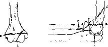
Figure 20.
Figure 19. a-fractura diacondylica; b-fr. condyli int. Fig. 20. Typical lines of intra-articular fractures: a-paraepiphyseal; b-diaphyseal; c-colli radii; d-processus coronoid. ulnae; e-olecrani. In the configuration of the joint, abnormal mobility of the fragments is determined with easily palpable (very painful) crepitus. Huge bruises, appearing after a few days on the inner surface of the arm and forearm, confirm the diagnosis. To distinguish an intra-articular fracture from a dislocation, one should remember that in the latter, limitation of movement and a forced position of the limb come to the fore. Attention should be paid to the following sign. As can be seen in Figs. 4 and 5 [see separate table (pp. 371-372)], in a person with an extended elbow, the middle of the olecranon and the middle of both epicondyles are on one straight line; with a bent elbow, these three points lie in one sagittal plane. The lines connecting these points form a triangle. In a dislocation, the indicated relationship of these points is disturbed. In intra-articular fractures, the relationship of these points is usually preserved, since both the condyles and the olecranon usually shift in the same direction. In isolated fractures of the epicondyles, condyles, or olecranon, the normal relationship of these points changes accordingly; at the same time, it is often possible to palpate a mobile free fragment of the humerus or elbow, which, however, is sometimes hindered by severe pain and the presence of swelling. Fragments lying inside the joint cannot be palpated; they can only be judged by sharp pain and limitation of movement. On the other hand, one should not forget that a simple intra-articular hemorrhage also causes sharp pain and limitation of movement. It is even more difficult to make a diagnosis in cases where partial fractures of the humerus and forearm are combined with a dislocation of the forearm, which is by no means rare. Hence it is clear that, although one always strives to make a diagnosis of an intra-articular fracture of the elbow and distinguish it from a dislocation of the elbow, hemarthrosis, contusion, etc., confidence in the correctness of the diagnosis is provided only by an X-ray taken in two projections. - Course and treatment. Intra-articular fractures of the elbow joint, accompanied by extensive intra-articular hemorrhages, displacement, and interposition of fragments, result in fusion in an incorrect position with impaired joint function. Striving at all costs to restore normal anatomical relations after intra-articular fractures of the elbow, surgeons of the past, after setting these fractures under anesthesia, tried to keep the fragments in such fractures in the correct position using circular plaster casts, all kinds of immobilizing splints, and hardening bandages. The results obtained in this way, which might be not bad in an anatomical sense, left much to be desired in a functional sense. In most cases, after the removal of the cast, there was a great limitation of mobility, which sometimes did not yield at all to subsequent development of the joint on apparatuses. Complete ankyloses were not uncommon, and the best that they tried to achieve in those days was ankylosis at a right angle in the position of supination (the most advantageous position in a functional sense). At present, when the greatest importance is attached to the restoration of joint function, most surgeons, therefore, in the treatment of intra-articular fractures of the elbow joint, have abandoned circular plaster casts, allowing the use of immobilizing splints only for a short period. If in the treatment of fractures of tubular bones, plaster is increasingly giving way to functional treatment using traction and early movements, then in the treatment of intra-articular fractures of the elbow joint, this method should have dominant importance. At present, timely reposition of fragments is given no less importance than before, but it is done not under general anesthesia, but under local anesthesia (Gorinevskaya et al.): 1% Novocain (20-30 cm3) is injected into the fracture sites, after which the reposition of the fragments is achieved completely painlessly, and the arm is left at rest in a physiological position in a light cardboard splint or simply on a sling (depending on the severity of the injury). Then, if no detached bone fragments that have penetrated into the joint cavity, interfere with movement, and are subject to removal are found on the X-ray, light active, strictly dosed movements are started from the 2nd-3rd day, the strength and amplitude of which gradually increase. After three weeks, they switch to passive-active exercises on pendulum apparatuses of the Krukenberg-Kornev Leningrad State Trauma Institute (see Mechanotherapy). With such a method of treatment, the patient can freely perform flexion and extension of the elbow, pronation and supination, and all movements in the wrist and shoulder after a month. A similar method has been carried out over the last two years (15 cases) in the traumatic department of the Orthopedic-Prosthetic Institute in Moscow with good success. - The meaning of treatment with early movement: 1) movement improves blood and lymph circulation in the area of the elbow joint. 2) Movement contributes to the most rapid resorption and absorption of blood poured into the joint and around the joint. 3) It promotes the correct circulation of synovial fluid inside the joint, which prevents the joint from becoming empty and the capsule from shrinking, which is observed with prolonged rest of the joint. 4) Movement maintains active muscle tone, preventing their atrophy. There is no need to fear secondary displacement of fragments due to early movements, since uniform movements of muscle groups (antagonists) hold the fragments in a relatively correct position. As for minor lateral and anterior-posterior displacements, they are partially corrected by subsequent exercises, and even if complete correction does not occur, a slight deformation of the elbow joint does not matter much: it is fully compensated by the excellent function and mobility of the elbow joint obtained by treatment with early movement. Volkovich, Wagner, Matti, and others insist on early movements in intra-articular fractures of the elbow joint. In cases where there is a sharp displacement of detached epicondyles (T- or F-shaped fractures of the humeral epiphysis), avulsion of the coronoid or olecranon process of the ulna, avulsion of the head of the radius, and bone fragments freely moving inside the joint in an intra-articular fracture, one should immediately resort to surgical intervention. In these cases, it is possible to reposition the fragments and subsequently screw them with a screw, if it concerns the condyles of the humerus. Bone fragments lying freely in the joint cavity should simply be removed, otherwise, they, getting stuck between the articular surfaces, will constantly interfere with movements and eventually can give rise to intra-articular bone fusions [see separate table (pp. 371-372), Fig. 3]. Fracture of the olecranon (fractura olecrani) occurs most often under the influence of a direct blow, a fall on the elbow; fractures can also be avulsion fractures due to rapid and sharp contraction of the triceps muscle of the arm (such cases are described as a sports injury in fencers). The detached part of the olecranon, due to the contraction of the triceps muscle attached to it, in a complete fracture and rupture of the lateral parts of the aponeurosis, moves far upward to a distance of several cm (this fracture is analogous to a fracture of the patella, where, due to the contraction of the extensors [B. M. E. Vol. XVI.], the fragments also diverge over a large distance). In order to relax the triceps muscle and put the displaced olecranon fragment back into its place, the arm is given an extended position and the displaced fragment is fixed with adhesive plaster. However, this method does not always achieve the goal. It also has the disadvantage that, due to the long stay of the arm in an extended position, it is subsequently difficult to achieve sufficiently full flexion in the elbow joint, which badly affects the working capacity of the victim. At present, in fractures of the olecranon with large displacement (as in fracture of the patella), most surgeons consider immediate bloody intervention indicated. A semilunar skin incision in the elbow area, the skin flap is separated, the fracture site is exposed, blood clots and shreds of soft tissues that interfere with the reduction of fragments are removed; approximation of fragments and suturing of the periosteum and lateral ruptures of the aponeurosis. With such treatment, the result is significantly better than with conservative treatment. Articular bodies (joint mice) of the elbow joint are relatively rare. Although, according to Weil (Breslau), they are found in the elbow joint 2-3 times more often than in the knee joint and are described by him as an occupational disease of apprentice carpenters and locksmiths, this opinion is not confirmed by other authors. The small number of joint mice of the elbow joint may be due to the difficulty of diagnosis and insufficient use of X-rays for this purpose (For the causes of formation, see Joint mouse). Articular bodies of the elbow joint mostly consist of cartilage; some of them also contain bone tissue. Symptoms: limitation of movement, sharp sudden pains that disappear as unexpectedly as they appear.
X-rays confirm the diagnosis, although the absence of a corresponding shadow on the X-ray does not yet indicate the absence of a joint body. Joint bodies of the elbow joint, causing severe pain and restricting movement, interfere with work and therefore are subject to surgical removal, which is a difficult task. The radial posterior incision recommended by Weil for the removal of joint bodies is not always convenient, as it provides poor access to the joint (it is suitable only for joint mice located in the posterior part of the capsule). The anterior incision, proposed by Simon and König, is more anatomical and provides free access to the joint. The incision is longitudinal along the anterior surface of the elbow bend: the skin, subcutaneous tissue, and superficial fascia over the sulcus bicipitalis medialis are incised, and the lacertus fibrosus is incised (and later sutured). The neurovascular bundle is retracted medially with a hook, the m. biceps (belly and tendon) is retracted laterally; the underlying m. brachialis, covering the joint capsule from the front, is incised, and its fibers are separated and stretched with hooks; the underlying capsule is incised, and with the help of Pean forceps, the joint mouse is extracted from the joint. This incision is complex due to the proximity of vessels and nerves, but it provides good access to the joint and is convenient for extracting free fragments from the joint cavity. To chronic diseases of the elbow joint of traumatic origin should be attributed the so-called epicondylitis of athletes, developing on the basis of overexertion in fencers, boxers, and from the abuse of tennis. Symptoms: severe pain in the region of the epicondyli upon touch and movement; objectively, neither swelling nor thickening of the bone is determined; in older cases, swelling of the periosteum is noted on X-rays. Massage is harmful; complete rest and prolonged abstinence from the corresponding sports are necessary. Inflammation of the elbow joint can be, depending on the course, acute or chronic, according to the pathological-anatomical picture—serous, purulent, or hyperplastic, and finally specific: gonorrheal, tuberculous, syphilitic. Inflammatory processes in the elbow joint can be of purely local origin, e.g., as a result of trauma, or one of the manifestations of a general process (tuberculosis, syphilis, general infection, etc.). Symptoms: the initial stages of the inflammatory process in the elbow joint are expressed by pain, restriction of movement, and swelling, whereby the intra-articular exudate, stretching the joint capsule, protrudes mainly its posterior parts along the sides of the olecranon, where fluctuation can be detected. This picture of synovitis acuta with periarticular swelling of the joint is observed to a greater or lesser extent in all inflammatory diseases of the elbow joint. To establish a more accurate diagnosis, knowledge of the etiology, an accurate anamnesis, and observation of the course of the local process in connection with a clinical study of the patient's general condition are necessary. Serous-inflammatory effusions in the elbow joint appear during trauma (contusions, distortions, fractures, dislocations, etc.) or as a sympathetic process during a purulent-inflammatory process in the surrounding tissues, during osteomyelitis of the epiphyses of the bones of the arm or forearm. In addition, synovitis of the elbow joint forms during all specific chronic and acute inflammations of the elbow joint. However, it should be noted that the elbow joint, in contrast to the knee, has a greater tendency toward adhesive inflammation than toward exudative; therefore, as a result of trauma, as well as a result of prolonged inflammatory processes, immobility of the joint is more often observed in the elbow joint than dropsy or looseness of the joint. Purulent inflammation of the elbow joint arises 1) as a result of infected penetrating wounds of the elbow joint; 2) upon the spread of a purulent process from purulent foci of surrounding tissues—ostitis and osteomyelitis of surrounding bones; 3) as metastases during general septicopyemic processes. Treatment of acute inflammatory diseases is carried out according to the general principles of treating intra-articular inflammatory processes. Where the inflammatory or purulent process in the elbow joint is the result of a general disease, treatment of the general disease is applied first of all.

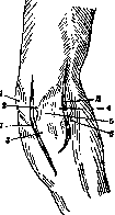
Figure 21. 1-epicondylus lat.; 2-olecranon; 3-capitulum radii. [Treatment] is joined by the treatment of the local process. In all inflammatory diseases of the elbow joint in the initial stages, the elbow joint is given, first of all, complete rest in a physiological position: a sling, an immobilizing cardboard splint bandage, and in more severe cases, suspension of the forearm in a vertical position (to improve blood circulation and to avoid edema). Further, Bier's passive hyperemia, heat, and dry-air baths are applied, depending on the indications. With symptoms indicating a transition to suppuration, treatment is applied according to the general principles of treating purulent arthritis (see). Puncture of the elbow joint is performed at the site of fluctuation in the posterior sections of the joint capsule on the sides of the olecranon (Fig. 21). Immediately there, on both sides of the olecranon, wide incisions are made for drainage or irrigation of the joint. In a progressive purulent process, one resorts to resection of the joint. Pirogov was the first to widely apply resection of the elbow joint for purulent processes, primarily in gunshot wounds. During the Crimean War, he used this method with great success before surgeons of the English and French armies, who did not recognize any other method of treating severe purulent arthritis of the elbow except amputation of the arm, began to resort to it. At the present time, partial resection of the epiphysis of the humerus is more often performed, by which a wide opening of the joint is achieved. Due to its anatomical structure, the elbow joint is very difficult to drain, therefore its resection in a number of cases is the only way to widely release the accumulated pus. At the present time, amputation of the arm for purulent arthritis of the elbow joint is resorted to extremely rarely, only in cases of a rapidly progressing local and general septic process threatening the life of the patient. Among gonorrheal diseases of the joints, the elbow joint occupies the sixth place. This disease is encountered most often in the form of monoarthritis, but sometimes it accompanies primary gonorrheal gonitis. An acute onset, high temperature, sharp pains, the appearance of serous effusion (smoothing of the periarticular folds), difficulty in movement due to sharp pains, with a complete absence (especially at the beginning) of any data from X-rays—this is the typical picture of gonorrheal arthritis, which in the elbow joint proceeds just as it does in other joints (see Arthritis, gonorrheal arthritis). A carefully collected anamnesis and examination of the genital tract (bacterial examination of secretions) confirm the clinical diagnosis. Treatment is according to the general principles of treating gonorrheal arthritis: local treatment—initially rest, Bier's passive hyperemia; general treatment—vaccine therapy, internally—urotropin, salol, etc. Tuberculosis of the elbow joint [arthritis tuberculosa cubiti, olenitis tuberculosa (Kocher's clinic)] constitutes 12% of all tuberculous arthritis (Tikhov) and occupies the fourth place among tuberculosis of large joints. Of the three joints of the upper extremities, tuberculous arthritis of the elbow joint constitutes, according to Garré, 51%, and according to Valtancoli, 47.7%. According to data from other authors, tuberculosis of the elbow joint is encountered relatively even more often (Kornev, Vindavsky sanatorium, etc.). While osteoarticular tuberculosis generally affects men more often than women, the opposite relationship is observed in tuberculosis of the elbow joint: in women—51.4%, in men—48.6% (according to Tikhov and König). According to Tikhov, trauma in the etiology of tuberculosis of the elbow joint is noted in 22% of cases, and tuberculous heredity in 39%. The distribution by age, according to the data of the same author, who collected 645 cases, is as follows: from 1 to 10 years—17.7%, from 11 to 20 years—31.5%, from 21 to 30 years—18.4% of cases; in older ages, this disease is encountered less frequently. In this sense, tuberculosis of the elbow joint differs sharply from syphilis of the same joint: the latter, not being encountered at all before the age of 10, is more common in adults and at a more advanced age. This circumstance should not be forgotten in the differential diagnosis of these two diseases. Tuberculous involvement of the elbow joint is revealed anatomically primarily either in the form of a synovial or in the form of a bone (osseous) form; mixed forms are also encountered, where it is impossible to accurately indicate whether the tuberculous involvement began with the synovial bursa and secondarily passed to the bone or vice versa. In any case, the primary bone form is encountered significantly more often than the synovial one, with the latter being more common in adults than in children. According to Tikhov, the synovial form is encountered in 21%, the bone form in 63%, and the mixed form in 16%. According to König, the bone form accounts for 71%. Hydrops tuberculosa, dropsy of the elbow joint, is encountered extremely rarely; fungous forms are more common. Bone forms initially have a focal character, subsequently breaking through either into the joint or periarticularly and forming external fistulas. The lower epiphysis of the humerus is most often affected in isolation. Isolated lesions: humeri—32%, ulnae—56% (most often), radii—6%. In 10% of cases, simultaneous tuberculous lesions of all three bones of the elbow joint are encountered. The diagnosis of tuberculosis of the elbow joint in typical cases does not present difficulties. In childhood, it is necessary to differentiate it from acute osteomyelitis (difficult in small children); the stormy onset, characteristic of acute osteomyelitis, excludes tuberculosis, which in this joint, as in others, begins gradually. However, it must be remembered that in children the onset often proceeds unnoticed, and the disease is discovered only upon the onset of severe pain. In youth and middle age, it is necessary to differentiate it from syphilis of the elbow joint; the question is decided by the anamnesis, general clinical examination of the patient, the Wassermann reaction, and an X-ray. It is especially difficult to sort out cases of mixed infections. The prognosis for isolated tuberculosis of the elbow joint is not dangerous to life; it worsens with the generalization of the process, in the presence of a tuberculous process in other organs and joints, and also in those cases where a secondary purulent infection is added to the tuberculosis of the elbow joint. In terms of function, the prognosis is worse, because even with a favorable course, tuberculosis of the elbow joint often ends in ankylosis. However, ankylosis in the correct position (at a right angle and in a state of supination) does not deprive the patient of the ability to work in many professions. As for the methods of treating tuberculosis of the elbow joint, in this matter, as in the treatment of tuberculosis of other joints, two trends are struggling in surgery: 1) conservative, proposing long-term sanatorium treatment, medicinal (iodoform emulsion, creosote, etc.), immobilizing bandages, helio- and thalassotherapy, Bier's passive hyperemia, quartz lamp, X-ray, etc.; 2) operative—resection or amputation. All authors incline toward conservative, sanatorium treatment in childhood (Krasnobaev, Rollier, Velyaminov, and others). Views on the treatment of tuberculosis of the elbow joint in adulthood diverge: while recognizing the desirability of conservative treatment in mild cases of tuberculosis of the elbow joint, many authors consider resection of the joint indicated in more severe cases, believing that resection of the elbow joint for tuberculosis shortens the duration of treatment and gives more stable long-term results, ridding the organism at once of a long and persistent struggle with the infection, a struggle from which it does not always emerge victorious. Some surgeons (Krasnobaev, Velyaminov, and others) set exclusively social indications for resection of the elbow joint for tuberculosis, taking into account 1) the lack of funds and objective possibilities for long-term and correct implementation of conservative sanatorium-resort treatment; 2) long-term disability, so burdensome for an adult working person, associated with the implementation of conservative treatment of the elbow joint. From this point of view, even such supporters of conservative treatment as Krasnobaev, Velyaminov, and others allow resection of the elbow joint for tuberculosis based on social indications. Spizharny, Gedroyts, and others set these indications even more broadly. As for the indications for amputation of the arm for tuberculosis of the elbow joint, at the present time they are sharply narrowed and are set only in the case of a severe local process associated with a general infection and a severe general condition; here the indication is the possibility of generalization of the process, complete exhaustion, increasing decline in strength, in a word—phenomena threatening the life of the patient.
Syphilis of the elbow joint occurs predominantly in the working age (from 26 to 50 years); moreover, it is rarer in women than in men (in women 12%, in men 20%). Late congenital syphilis of the elbow is encountered less frequently in men than in women, manifesting late in mature age (in children up to 15 years it is encountered only as an exception). In gummatous acquired syphilis, the disease of the elbow belongs to the early forms of gummatous syphilis. After 50 years, syphilitic arthritis is encountered very rarely. Bilateral involvement of the elbow is rare. Of the epiphyses, the epiphysis of the humerus is most often affected. Bone involvement is sometimes accompanied by serous synovitis, but hydrops fibrinosus et villosus develops more often; osteoarthritides with hyperplastic synovitis are most frequent; due to the thinness of the bone, the gummas of the bone epiphyses easily disintegrate and form fistulas. The differential diagnosis between this disease and the fungous form of tuberculosis of the elbow presents great difficulties. The question is resolved by anamnesis and specific reactions. Tabetic arthropathies in the elbow joint are rarer than in other large joints; they are characterized by abnormal movements (hyperextensio and lateral movements), a sharp change in the shape of the articular ends, an increase in protrusions and the formation of osteophytes, thickening of the articular ends due to their flattening, enlargement of the olecranon, etc.; spontaneous painless intra-articular fractures, detectable by simple palpation, complete the characteristic picture; however, the disease is definitively confirmed by the presence of general symptoms of tabes. Serous luetic synovitis and hydrops fibrinosus et villosus can be confused with syringomyelia. In syringomyelia, the elbow joint is affected quite often; characteristic looseness, large swelling, rapid accumulation of exudate, painlessness during extensive destruction of bones, and the presence of general symptoms (muscle atrophy, paresis, trophic disorders, skin anesthesia) facilitate the diagnosis.
Besides inflammatory purulent processes of local origin and inflammatory processes of a specific nature (gonorrhea, tuberculosis, lues, etc.), inflammatory diseases of the elbow joint (both serous and purulent) arise as complications of a whole series of infectious diseases: scarlet fever, measles, diphtheria, dysentery, typhoid, smallpox, pneumonia, acute articular rheumatism, etc. (see corresponding articles). Diseases of metabolism (gout) and blood (hemophilia, etc.) also cause destructive processes in the elbow joint, although in these diseases the elbow joint is affected significantly less often than others. It should not be forgotten that arthritides associated with anomalies of internal secretion and all kinds of autointoxications also affect the elbow joint; but, proceeding as polyarthritides, they affect the elbow joint only incidentally; therefore, the general symptomatology of the given disease is decisive. Of other forms of chronic joint diseases, the elbow joint is quite often affected by osteoarthritis deformans. The disease develops secretly, without any general symptoms, progresses chronically over several years, never causes suppuration, ankylosis, or loose joints; however, due to the deformation of the articular ends of the bones, flexion contractures form in the elbows (for more details, see Arthritides, arthritis deformans). As already stated, the elbow joint, prone to adhesive inflammation both after trauma and after inflammatory processes, easily develops contractures and ankyloses, which are caused not only by intra-articular but also by periarticular adhesions. Contractures of the elbow joint are sometimes caused by cicatricial changes in the muscles, which have a great tendency even to ossification. Myositis ossificans of the m. brachialis internus, m. biceps, developed after trauma (old dislocation) or after surgical intervention, is often the cause of ankylosis. Early movements are the only way to combat these complications. Massage is harmful because, by irritating the young torn periosteum, it contributes to its overgrowth. Contractures of the elbow joint as a result of cicatricial skin contractions due to burns and wounds are subject to removal and replacement by plastic surgery. Tumors and cystic formations of the bones of the elbow joint do not present anything peculiar. Operative approaches to the elbow joint. Operations on the elbow joint are performed for acute purulent diseases (arthrotomy or resection of the joint), for tuberculosis of the joint (resection), for suppuration in connection with gunshot and other penetrating wounds, for old dislocations (open reduction), and finally for ankylosis of the elbow (arthrolysis, arthroplasty). Proceeding from the anatomy of the joint, one should remember during every surgical intervention the necessity to spare first of all the nourishing vessels (a. fossae cubiti), as well as the nerves (n. medianus, n. ulnaris, and nervus radialis), muscles, and tendons representing the motor apparatus of the elbow, forearm, and hand. In view of the fact that the main nourishing artery, the median nerve, and the entire group of flexors are located on the anterior surface of the elbow, covering the joint area with a powerful layer of muscles, one should avoid the anterior surface of the elbow during surgical intervention (with the exception of certain indications). All typical proposed approaches to the elbow joint relate to the posterior and lateral surfaces of the elbow joint. However, even here one must remember the need to spare the ulnar and radial nerves, of which the former lies directly on the bone in the sulcus ulnaris, and the latter is hidden in the thickness of the muscles. During surgical intervention in the area of the elbow joint, approaching from behind or from the outside, one can damage the extensor apparatus. Thus, the m. triceps is damaged in all methods; the m. supinator longus suffers due to shortening of the skeleton, the m. anconaeus due to damage to its nerve apparatus, but not equally in all methods. The most common and convenient method for access to the elbow joint is the Kocher method. Next come the methods of Langenbeck (Fig. 22) and Ollier (Langenbeck, Ollier). The Kocher method (represents a modification of the old incision by Chassaignac).
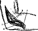
Figure 22. I and II—incisions according to Langenbeck and Kocher: 1—epicondyl. med.; 2—olecranon; 3—base of olecranon; 4—fold of the capsule; 5—capitulum radii; 6—epicond. lat.
Figure 23. Resection according to Kocher: 1—epicondylus lat.; 2—m. anconaeus; 3—mm. brachioradialis & extensor carpi radialis; 4—m. extensor carpi ulnaris.
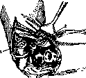
The Kocher method, which provides the widest access for resection of the elbow joint (it is also used for open reduction of an old dislocation, arthrolysis, arthroplasty), consists of the following: with the elbow flexed at 150°, an incision is made along the outer ridge of the humerus, starting 3-5 cm above the epicondylus lateralis, descends vertically to the head of the radius, and turns to the dorsal surface of the forearm along the anterior edge of the m. anconaeus to the edge of the ulna, ending 4-6 cm below the tip of the olecranon. The incision passes between the m. triceps on one side and the m. supinator and m. extensor carpi radialis on the other; further into the fossa pulchritudinis between the m. anconaeus quartus and the m. extensor carpi ulnaris (Figs. 23 and 24). The incision according to Langenbeck is made 10 cm long along the medial edge of the olecranon, in a straight line along the axis of the limb, and, dissecting the musculus triceps longitudinally, reaches the bone immediately. The bayonet-shaped incision of Ollier begins between the m. triceps and the m. supinator longus 6 cm above the joint line, is carried vertically downward almost to the epicondylus lateralis, and turns from here at an obtuse angle medially in a transverse direction to the olecranon; from the olecranon, the incision goes vertically downward for 5 cm. In the latter two methods, the branch of the nervus radialis going to the m. anconaeus is damaged. In purulent processes, where it is necessary to perform an arthrotomy to widely open and drain the elbow joint, two lateral incisions are used: an incision according to Kocher (upper or middle part) from the lateral side and a medial incision passing through the septum intermusculare along the anterior surface of the epicondylus medialis (cave n. ulnaris!). For extensive resections performed for severe purulent arthritides with the aim of widely draining and opening the joint, both incisions are good: both according to Kocher and according to Langenbeck. To the former, in some cases, it is recommended (Bier) to add a medial incision. A complete resection of the elbow joint with removal of the articular ends of the humerus, ulna, and the head of the radius is performed for extensive purulent processes, extensive tuberculous processes, when the resection is performed together with extirpation of the entire joint capsule, and also for malignant tumors. In other cases—for comminuted fractures, old dislocations. Figure 24. Resection of the elbow joint according to Kocher; dislocation of the bones after detachment of the epicondylus lateralis (2) from the humerus (1).
In cases of ankylosis, arthroplasty, etc., it is often sufficient to remove only the articular surface of the humerus, preserving the olecranon and its articular surface; in some cases, only the head of the radius is resected, specifically in isolated dislocations of the radius with a simultaneous fracture of the ulna (in these cases, the protruding head of the radius interferes with flexion movements and hinders pronation and supination). For old dislocations of the elbow, open reduction is usually undertaken, which, however, is successful only within one or two months after the dislocation; in cases of dislocations of longer standing, secondary changes in the joint—shriveling of the joint capsule, shortening and atrophy of muscle groups, etc.—make even open repositioning extremely difficult. Moreover, even after returning the dislocated fragments to their place, the necessary joint cavity is not obtained; the joint space is absent, and upon healing, a complete limitation of movement occurs, and sometimes ankylosis. Therefore, in late open reduction of the elbow, in most cases, it is necessary to perform a partial resection, most often of the articular surface of the humerus and the head of the radius. For ankylosis of the elbow joint resulting from trauma or inflammatory processes, operations of arthrolysis and arthroplasty are resorted to. An absolute indication is ankylosis in a straight position; contraindications are childhood and a fresh inflammatory process. The operation consists of partial or complete resection (most often according to the Kocher method) with the creation of a large gap (about 4 cm) between the humerus and the ulna; after resection, both ends of the bone are covered with fascia (free transplantation of the fascia lata or fat). Some authors (Wier and others) are content with interposing a small muscle flap taken from the m. triceps or anconaeus and secured between the bones so that it prevents their fusion. Other authors (Mouchet) do not consider it necessary to perform muscle or fascial interposition, attaching primary importance (as in any arthroplasty) to early movements. The latter is, in any case, the most important condition for success. Therefore, in arthroplasty of the elbow joint, it must not be kept in a rigid dressing for long (no more than 7-8 days), but it is necessary to immediately adapt traction in a flexed position in such a way that active movements can be performed immediately. The success of arthroplasty depends entirely on subsequent treatment. Arthrodesis. Operations of arthrodesis, i.e., complete immobilization of the elbow joint, have to be resorted to extremely rarely: in cases of flail elbow joint in connection with complete paralysis of all flexors. This operation does not present any particular difficulties; it consists of freshening the articular ends and securing the joint with a bone lock or (even simpler) fastening it with a nail or wire. However, much simpler and with better success in these cases is prosthetics—a brace with a lock. After unsuccessful resections of the elbow joint that have caused excessive flailness, there are also no indications for arthrodesis, as simple prosthetics can provide quite satisfactory results. V. Gorinevskaya.
Related articles
Mentioned in
Cite this page
“Elbow Joint.” Soviet Medical Encyclopedia. English translation of Bolshaya Meditsinskaya Entsiklopediya, 1st ed. (Moscow, 1928–1936), ed. N. A. Semashko. https://sovietmedicalencyclopedia.pages.dev/article/elbow-joint/