Temporal Region (regio temporalis, has boundaries that precisely)
Historical document, translated for reference. It reflects medical knowledge of the 1920s–30s and is not medical advice.
Summary
The temporal region corresponds to the boundaries of the temporal muscle and consists of multiple layers including skin, subcutaneous tissue, fascia, and the temporal muscle itself. This anatomical area contains important neurovascular structures and is clinically significant for various surgical approaches.
Encyclopedia article (1928–1936)
TEMPORAL REGION, regio temporalis, has boundaries that precisely correspond to the outline of the temporal muscle (m. temporalis), which is easily identifiable in a living person if they are asked to clench their teeth. Anteriorly, the temporal region barely extends beyond the hairline, superiorly it extends for 8 cm, posteriorly it reaches the auricle, and inferiorly it extends to the zygomatic arch. On the skeleton, the boundary of the temporal region is the linea temporalis superior of the frontal, parietal, and temporal bones and the zygomatic arch. This lateral surface of the cranial vault, as is known, forms with the base of the skull an obtuse angle, open inward. Crista infratemporalis separates them. The lateral wall of the skull deviates medially in relation to the zygomatic arch, so that between it and the zygomatic arch there remains a spacious space (fossa temporalis), filled with the organs of this area. Layers: 1. Skin, covered with hair that turns gray early. 2. Loose subcutaneous fatty layer, in which pass muscles--mm. attrahens and attollens auriculae, vessels--artery and vein temporalis superficialis and their branches, and nerves; the nerves of this layer are as follows: a) n. auriculo-temporalis (see figure 1-3) originates from the third branch of n. trigemini, emerges from the foramen ovale as the posterior bundle, surrounds the a. meningea media with two prongs, and, curving under the zygomatic arch, passes behind the mandibular joint, in front of the external auditory meatus; sometimes gives an anastomosis to n. facialis; above the zygomatic arch it divides into several branches (3-4), of which one goes (not always) to the angle of the mouth, the second in a transverse direction, and
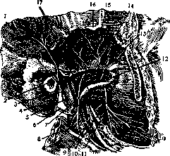
Figure 1. N. auriculo-temporalis (superficial part; zygomatic arch and coronoid process of the mandible removed): 1-n. auriculo-temporalis; 2-meatus acusticus externus; 3-n. meatus acustici externi; 4-processus mastoideus; 5-rami anastomotici cum n. facial.; 6-n. facialis; 7-n. massetericus; 8-m. masseter; 9-n. alveolaris inferior; 10-n. buccinatorius; 11-n. lingualis; 12-a. maxillaris interna; 13-n. temporalis profundus anterior; 14-n. temporalis profundus medius; 15-n. temporalis profundus posterior: 16-m. temporalis: 17-m. pterygoideus externus (according to Spalteholz).
two-to the skin of the temple, corresponding to the two branches of a. temporalis superficialis; lies behind the artery; b) branches of n. facialis go to the angle of the eye and frontal muscles; c) in the upper part of the area are branches of n. zygomatico-temporalis, piercing the aponeurosis and emerging through the corresponding canal in the zygomatic bone from n. infraorbitalis (second branch of n. trigemini). 3. The next layer-fascia temporalis-constitutes a continuation of the galea aponeurotica, gradually disappearing in the inferior direction. Under it is
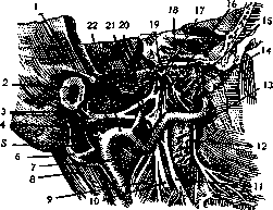
Figure 2. N. auriculo-temporalis and its branches (deep part); its relation to a. maxillaris int., to other branches of n. mandibularis and to n. facialis: 1-n. auriculo-temporalis; 2-meatus acusticus externus; 3-n. meatus acustici externi; 4-rami anastomotici cum n. facial.; 5-ramus digastricus; 6-n. facialis; 7-a. carotis externa; 8-m. digastricus (posterior belly); 9-m. pterygoideus internus; 10-n. alveolaris inferior; 11-n. lingualis; 12-n. buccinatorius; 13 and 14-m. pterygoideus externus; 15-n. pterygoideus externus; 16-n. buccinatorius; 17 and 19-n. mandibularis; 18-ganglion semilunare (Gasser); 20-a. meningea media; 21-chorda tympani; 22-a. maxillaris interna (according to Spalteholz).
a very small amount of very loose connective tissue, under which lies 4. Fascia temporalis propria s. profunda. The latter begins as a single sheet from the periosteum of the lineae temp. sup. and near the zygomatic arch divides into 2 sheets (see figure 5); the superficial one attaches to the upper edge of the arch and the outer surface, while the deep one attaches to the inner surface; as a result, a triangular space in cross-section is formed between the sheets of the fascia, filled with connective tissue and containing a small segment of a. temporalis mediae. The fascia consists of strong bundles and forms a strong, unyielding plate. Between it and the bones of the skull, which curve medially, there remains a large triangular space, narrowing and closed above and freely open below. The space is called fossa temporalis. 5. In fossa temp, is placed the subaponeurotic layer of connective tissue, developed mainly around the zygomatic arch, and m. temporalis with vessels and nerves. The muscle originates from the linea temporalis inferior and attaches to the edge and inner surface of the proc. coronoidei mandibulae; the direction of its fibers is fan-shaped, with the posterior ones running at an angle of about 30° to the horizontal. The site of attachment of the tendon to the bone is the strongest part of the muscle, and its separation is so difficult that some surgeons prefer to chop off the proc. coronoideus with a chisel. The vessels-aa. temporales profundae (2)-enter the muscle from its deep surface, the nerves-nn. temporales prof. post. and ant.-originate from the third branch of n. tri
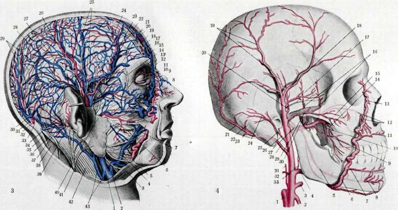
Figure 3. 1-v. temporalis post.; 2 and 4-v. facialis ant.; 3-platysma; 5-a. maxillaris superficialis; 6-ductus parotideus; 7-ram. buccales n. facialis; 8-areas tarseus inf.; 9 and 10-a. v. angularis; 11-n. infratrochlearis; 12-n. supratrochlearis; 13-arcus tarseus sup.; 14-n. zygomatico-facialis; 15-n. frontalis; 16-a. n. frontalis; 17-n. frontalis; 18 and 19-a. n. supraorbitalis; 20-rami n. supraorbitalis; 21-a. zygomatico-orbitalis; 22-n. zygomatico-temporalis; 23 and 24-galea frontalis v. et a. temporales superf.; 25-n. auriculo-temporalis (rami temporales superf.); 26-a. et v. temporalis superf.; 27-n. auricularis superf.; 28-rami n. occipitalis majoris; 29-v. occipitalis; 30-m. occipitalis major; 31-a. occipitalis; 32-n. occipitalis; 33-m. auricularis post.; 34-m. trapezius; 35-n. occipitalis minor; 36-n. auric. post. (n. facialis); 37-m. splenius capitis; 38-a. auricularis post.; 39-v. jugularis; 40-n. facialis (rami zygomatici et temporales); 41-n. auricularis magnus; 42-m. sterno-cleido-mastoideus; 43-anastomosis to vena jugularis externa. Fig. 4. 1-a. carotis interna; 2-a. thyreoidea sup.; 3-a. lingualis; 4 and 5-a. maxillaris externa; 6-a. alveolaris inf.; 7-a. temporalis inf.; 8-a. meningeal; 9-a. buccinatoria; 10-a. palatina descenders; 11-a. temporalis sup.; 12-a. infraorbitalis; 13-a. auricularis; 14-a. alveolaris superior post.; 15-a. sphenopalatina; 16-aa. temporales profundae; 17-a. zygomatico-orbitalis; 18-ram. frontalis a. temporalis superf.; 19-ram. parietalis a. temper. superf.; 20-a. temporalis media; 21-a. occipitalis; 22-ram. internasens; 23-ram. descendens; 24-ram. occipitalis; 25-a. stylo-mastoidea; 26-a. auricularis post.; 27-n. maxillaris int.; 28-a. occipitalis; 29-a. transversa faciei; 30-a. meningea media; 31-a. pharyngea ascendens; 32-a. sterno-cleido-mastoidea; 33-a. palatina ascendens (according to Sonotta). K CT, Височной области. gemini and form its anterior bundles, separating from the trunk near the foramen ovale. Further follow: 6. periosteum and 7. bone. The latter in most people is very thin, translucent, and due to its fragility, often undergoes fractures. On the inner surface of the bone are grooves for the trunk and branches of a. meningeae mediae, sometimes (in about 40%) turning into a canal. The lymphatic vessels of the temporal region collect in lymphoglandulae auriculares anteriores. The temporal region is supplied with blood from the temporal arteries and veins (see figures 3 and 4). Three temporal arteries are distinguished: a. temporalis superf., media and profunda. The first represents one of the terminal branches of a. carot. externae; arises at the level of the neck of the mandibular joint and between
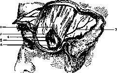
Figure 5. Superficial layers of the temporal region: 1-aponeurosis temporalis; 2-a. tempor. superficialis; 3-aponeurosis temporalis (lamina superficialis); 4-m. temporalis; 5-aponeurosis temporalis (lam. profunda); f-fatty layer. The parotid gland emerges onto the surface in front of the condylus mandibulae and behind the external auditory canal, curving around the arcus zygomaticus. Above the latter, it lies in the subcutaneous tissue immediately in front of the ear and soon divides into terminal branches-ramus frontalis and ramus parietalis. Veins of the same name, one or two, lie irregularly-sometimes behind, sometimes in front; n. auriculo-temporalis-usually behind. At a distance of 1 cm forward, branches of n. facialis pass. Branches of the artery: a) within the parotid gland, 2-3 thin rami parotidei depart; b) slightly below the zygomatic arch, parallel to it and Steno's duct (above the latter), passes a. transversa faciei, supplying the mimetic muscles and anastomosing with terminal branches of a. infraorbitalis; c) backward, 2-3 branches to the external auditory canal and auricle depart (rami auriculares anteriores); d) immediately above the zygomatic arch, in a medial direction, goes a. temporalis media; it pierces the outer lamella of fascia temporalis, lies for some distance in the tissue above the zygomatic arch, pierces the deep lamella of the fascia and enters the temporal muscle, within it it ascends upward; e) ramus zygomatico-orbitalis departs above the zygomatic arch and directs itself to the margin of the orbit, where it anastomoses with branches of a. frontalis and a. lacrimalis and sometimes with the following; f) ramus frontalis s. anterior-the anterior terminal branch, thicker, rises from the place of division of the trunk, in an oblique direction forward and upward, gradually divides into branches anastomosing with a. frontalis and supraorbitalis, with branches of rami parietalis s. posterioris a. temporalis, as well as with arteries of the opposite side; g) ramus parietalis s. posterior-the second, usually thinner, terminal branch ascends almost vertically or slightly inclines backward into the parietal region and with its branches anastomoses with ramus anterior a. occipitalis and the artery of the opposite side. A. temporalis superficialis and its branches, due to their superficial position, are easily accessible to palpation, which is sometimes used to determine the state of cardiac activity. It is more often affected by arteriosclerosis than others, and in such case its tortuosities are well visible under the skin. Another disease of the artery is angioma s. aneurysma cirsoideum, having the appearance of a cone-shaped or cord-like pulsating tumor; it is often accompanied by neuralgias due to the accompanying lesion of n. auriculo-temporalis. If there is communication between the artery and vein, the tumor is called angioma arterio-venosum. The characteristic symptom should be considered the presence of a buzzing noise and the presence of varicose veins. Treatment of these diseases can be conservative (pressure bandages, cauterization, injection of blood-clotting substances) and active (removal of the tumor and ligation of the feeding vessels). A. temporalis profunda ant. and post. arise from a. maxillaris int.-the second terminal branch of a. carotis ext., alongside and in front of a. meningea media; ascending upward, they pass laterally from m. pterygoideus ext. and, bending over crista infratemporalis, enter the muscle from the medial side, at its anterior and posterior border. The temporal veins accompany the arteries of the same name. V. temporalis superficialis arises from the venous plexus of the cranial vault; it consists of two veins corresponding to the anterior and posterior branches of a. temporalis superficialis; above the zygomatic arch it receives v. temporalis media, which carries blood from the temporal muscle and pierces the aponeurosis separately from the artery; still lower into it flow v. auricularis ant., transversa faciei, etc. The v. facialis posterior, formed by their confluence, receives branches of the plexus pterygoideus, located between mm. pterygoideus externus and internus. Some authors call v. temporalis superficialis and media the superficial branches of v. facialis posterioris. Venae temporales profundae flow into the plexus pterygoideus.
Related articles
Mentioned in
Cite this page
“Temporal Region (regio temporalis, has boundaries that precisely).” Soviet Medical Encyclopedia. English translation of Bolshaya Meditsinskaya Entsiklopediya, 1st ed. (Moscow, 1928–1936), ed. N. A. Semashko. https://sovietmedicalencyclopedia.pages.dev/article/temporal-region-2/