Lymphogranulomatosis
Historical document, translated for reference. It reflects medical knowledge of the 1920s–30s and is not medical advice.
Summary
Lymphogranulomatosis is a distinctive disease of the lymph nodes that can affect numerous glands and is currently classified as an inflammatory process. The disease was first described by Hodgkin in 1832 and was later classified as a pseudoleukemic process.
Encyclopedia article (1928–1936)
LYMPHOGRANULOMATOSIS (syn.: lymphomatosis granulomatosa, Paltauf-Sternberg's disease), a distinctive disease of the lymph nodes, which sometimes affects numerous lymph glands, is currently classified as an inflammatory process. The history of this disease begins in 1832, when Hodgkin (England) first described a series of cases of widespread swelling of the lymph glands, which proceeded with fever and ended in death. Hodgkin's disease was subsequently classified as the pseudoleukemic processes described by Cohnheim in 1860, and was often denoted
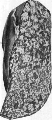
lymphoma malignum. However, with the passage of time, it became clear that the group of pseudoleukemias and lymphomas included several different disease forms. One of them, identified by Paltauf in 1898, was thoroughly studied by C. Sternberg and characterized in detail by him in 1905 under the name lymphogranulomatosis. The old Hodgkin's disease turned out to be L., and not the pseudoleukemia of Conheim. Pathological anatomy. L. is the most common systemic disease of the lymphatic apparatus. The disease usually begins with the swelling of one gland or a group of lymph nodes in the neck; they are painless in themselves, but can cause pain due to pressure on blood vessels and nerves. Initially, the glands do not fuse together, but over time they can merge into large masses. The swollen glands are initially soft, later they harden and decrease in volume. The swelling gradually involves other groups of glands, for example, axillary, inguinal, mesenteric, but unlike leukemic forms, in L. it is often observed that some groups of lymph nodes remain completely unaffected. On cross-section, the glands are grayish-pink in color and often contain irregularly shaped (potato-like) yellowish-white foci of necrosis (see separate table, figure 5) - this is an extremely important feature for the pathological-anatomical diagnosis of L. At the beginning of the disease, the glands on cross-section are juicy and translucent, later they become fibrously dense. In some cases, the glandular involvement is limited to the formation of one mass; in others, it can gradually involve all glands. With widespread lymph node involvement, a characteristic change in the spleen is also observed. It enlarges, and multiple yellowish-white nodules, ranging in size from a millet seed to a walnut, form in its tissue. Bulging under the capsule, the nodules give the spleen a coarse-grained appearance, unlike in leukemia, and on cross-section they bear some resemblance to anemic infarcts, as they often contain the same foci of necrosis that form in the lymph nodes. The combination of the color of the nodules with the red background of the splenic pulp led to a comparison of its cross-section appearance with porphyry (Porphyrmilz) (see separate table, figure 3), with 'sausage with bacon' or with almond hardtack (Hardbake-Spleen of English authors). Besides such the most common cases with lymph node and spleen involvement, cases have been described where L. manifested as an isolated tumor, for example, in the mediastinum, lung, stomach, intestine, and other organs. The similarity with a neoplasm on the one hand, and with systemic lesions of the lymphatic apparatus (leukemias, pseudoleukemias) on the other, is also expressed in that numerous miliary or larger foci of the same tissue as found in L. in the lymph nodes can form in the liver, kidneys, lungs, bone marrow, heart, and other organs. These foci have the structure of lymphogranulomas. In appearance, they are extremely similar to metastatic nodes of tumors. Sometimes L. is accompanied by general amyloidosis. The microscopic picture of L. also shows many characteristic features and diversity. Usually, the disease begins with hyperplasia of the entire lymphadenoid tissue of the gland, and then the proliferation of reticular cells begins to predominate (first stage); in this way, elements with light, bubble-like nuclei are formed, which are significantly larger than lymphocytes. Often, along with them, a small emigration of multinucleated leukocytes is observed, eosinophilic leukocytes and plasma cells appear. Their presence is considered especially important in the diagnosis of early stages of L. The appearance of leukocytes gives reason to consider L. an inflammatory disease from the very beginning. With the passage of time, granulation tissue is formed from the newly formed reticular elements, which gradually replaces the lymphadenoid tissue (second stage). A characteristic feature of this granulation tissue is the diversity of its composition: alongside epithelioid cells of various sizes, elongated fibroblasts, lymphocytes, plasma cells, and eosinophils are visible. In addition, some reticular cells transform into special large elements with a large cell body and very diverse nuclei - sometimes in the form of a tangled sausage, sometimes lobulated or even disintegrated into parts; the nuclei are sometimes pale, on the contrary, rich in chromatin, pyknotic, but mostly bubble-like and transparent. These cells, first described by Sternberg, were named giant cells of Sternberg (see separate table, figure 4), as they sometimes reach large sizes and bear some resemblance to the giant cells of tuberculous granulomas. On the other hand, the giant cells of L. have an undeniable resemblance to megakaryocytes of the bone marrow, precisely in the whimsicality of the nuclear contours. Sternberg cells are a very important diagnostic feature of L. and facilitate recognition of the process, as they sometimes appear very early, when other elements of the granuloma have not yet fully developed. Subsequently, the elements of the granulation tissue transform into ordinary fibroblasts, bundles of connective tissue appear, and secondary fibrosis of the gland occurs (third stage), due to which it changes its consistency from soft to dense and decreases in volume. Finally, it should be noted that necroses are a very frequent phenomenon in L.; they develop either according to the type of caseous necroses of tuberculous granulomas or as the death of areas of already fully developed granuloma containing already fibrous connective tissue. The presence of necroses allows for easy differentiation of gland swelling in L. from lymphadenoses, in which necroses do not form. The histological picture described above represents a typical lesion; in practice, however, various deviations from it are encountered, which gives reason to speak of atypical forms of L. First, the changes in the lymph nodes in L. can remain for a relatively long time in the initial stage of the process, presenting a picture of simple hyperplasia of lymphadenoid tissue; in other cases, hyperplasia of reticular cells of the reticulosis type occurs without the formation of granulation tissue. Sometimes in L., the proliferating reticular cells give rise to growths of the endothelioma or polymorphic sarcoma type. Modifications with the formation of a large number of plasma cells (plasmocytomas) are known. All these cases, of course, can be very difficult to recognize. It is also necessary to mention that in the literature, several cases of systemic hyperplasia of the reticulo-endothelial apparatus have been described, which histologically differ from L., but are classified by Sternberg as atypical L. This shows that L. can manifest extremely diversely. Rare variants of L. are acute cases with a predominantly exudative character of changes, as well as those in which only widespread formation of miliary nodules of L. (miliary L.) is observed. In addition, L. sometimes combines with tuberculosis of the lymph nodes. In these cases, it is difficult to distinguish what belongs to L. and what to tuberculosis, as the formation of tubercles with caseous necrosis and true multinucleated giant cells of the Langhans type is observed in the lymph nodes. Sometimes Koch's bacilli can be detected in these caseous foci. The etiology of L. remains unclear. Sternberg initially considered L. a special manifestation of tuberculosis. This view was based on a number of observations in which lymph nodes affected by L. contained tubercle bacilli; they could be detected both microscopically and by inoculation into guinea pigs. However, in view of the frequently observed involvement of lymph nodes in the tuberculous process and even the finding of tubercle bacilli in lymph nodes of completely healthy-looking people, it was natural to think of a combination of these diseases. On the other hand, many cases of pure L. have been noted in the literature, in which neither microscopically nor by inoculation of L. nodes into guinea pigs could the participation of Koch's bacilli in the development of the process be proven. This forced the search for other causative agents. In 1911, Frankel and Much published their research, which seemed to prove that the cause of L. is a rod that is insoluble in antiformin, not acid-fast, but staining by Gram's method. It appears in the form of chains of Gram-positive grains or individual grains. In cultures, it was rarely possible to isolate it; inoculation into animals gave contradictory results. The virus of Frankel and Much was found by several researchers; however, most authors after Frankel and Much failed to either see in the tissue or isolate in cultures their virus. On the other hand, various other microorganisms (Negri, Kuczynski and others) were found in tumors in L.; the very diversity of these findings proves that attributing them an etiological role is hardly possible (for example, rods recently isolated by Buzni and resembling tubercle bacilli). H. Hirschfeld, based on both his own research and literary data, comes to the conclusion that one cannot completely deny any connection between tuberculosis and L. He believes that tuberculosis of the lymph nodes favors the development of L. in them, without being its cause.
Thus, up to the present time, the causative agent of L. remains unidentified, although the disease itself, by its nature, is undoubtedly an inflammatory lesion of the lymphatic apparatus, and not a neoplasm. Tsypkin argues against the inflammatory nature of L. According to him, there is no fundamental difference between lymphoma malignum Orth's (under this term are combined leukemia, aleukemia and lymphosarcomatosis) and granuloma malignum Benda, i.e., L., but whereas in lymphoma malignum we are dealing with the loss of differentiation (Entdifferenzierung of German authors) of the parenchyma of hematopoietic organs, in L. the first stage of the anatomical process has the same character, and in the second stage the embryonalization of the stroma of these organs predominates. The third stage, then, represents the result of reverse differentiation of the stroma that has lost differentiation, or in other words - should be considered as an anatomical remission.
f. Chistovich. Forms. Depending on the extent of the lymphatic gland lesions, two forms of L. are distinguished: localized and generalized. In the first form, the process is localized in any one group of external or internal glands. Due to the exceptional localization within a particular group, a particular clinical variant of the disease is established. The pathological process may be concentrated exclusively in the lymphatic apparatus of the eyelids, salivary, and lacrimal glands (Mikulicz's disease), in the retroperitoneal glands (typhoid form), in the skin, as well as within the gastrointestinal tract. The course of some localized forms is often prolonged. Thus, Westphal's patient lived for 17 years. The temperature in these cases is usually normal, and the changes in the blood are limited to a slight leukocytosis and moderate eosinophilia. The course takes on a malignant character in cases where the tumor begins to grow rapidly and in its rapid growth compresses neighboring vital organs, large vessels and nerves (Mediastinaltumor formed on the basis of enlargement of mediastinal glands or thymus). Around such rapidly growing tumors, small nodules usually appear. The disease also takes on a malignant course when the localized form passes into the generalized form, which occurs in most cases - and especially in childhood. Symptomatology. Among the most important symptoms of L. should be mentioned fever. Cases are often encountered where the temperature curve takes the form of recurrierendes Fieber, or in other words, where there is a regular alternation of periods with elevated temperature with periods of normal, and even subnormal temperature (see curves). The temperature curve of the recurrierendes Fieber type undoubtedly has pathognomonic significance for L., and whenever one encounters such a fever, one should first think of L., even when there is no enlargement of the external lymphatic glands. Unfortunately, not all cases of L. are accompanied by the type of fever described. In many cases, a remittent type is observed, while in other cases the temperature is so irregular that it loses all diagnostic significance. Blood. In L., one cannot speak of any specific hematological picture. Here we encounter all kinds of deviations from normal leukopoiesis. First of all, mention should be made of absolute and relative neutrophilic leukocytosis, accompanied by eosinophilia. The total number of leukocytes can reach very high figures [240,000 in Schur's case, but leukopenia (Fabian) may also occur]. Then mention should also be made of cases of leukopenia with a clearly lymphemic character. Finally, occasionally L. may be accompanied by a leukemic blood picture (M. A. Skvortsov). With a prolonged course of the disease, anemia of the hypochromic type and cachexia always develop, from which patients die. This occurs mainly in the generalized forms of the disease. The erythrocyte sedimentation rate is sharply increased, and this occurs even in the initial period of the disease, which is of great importance for differential diagnosis. Urine. In severe cases, the diazo reaction is often positive. The traces of protein frequently noted are associated with the febrile condition of the patient. Large amounts of protein with a small amount of hyaline cy
sh
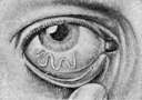
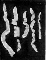
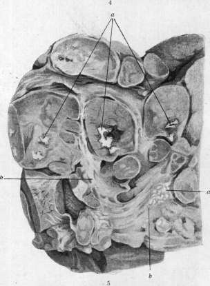
Figure 1 and 2. Loa in the subcutaneous tissue (1) and in the conjunctiva of the eye (2). Figure 3. Porphyric spleen in lymphogranulomatosis. Figure 4. Sternberg cells in lymphogranulomatosis. Figure 5. Enlarged lymphatic glands with necroses (a) in lymphogranulomatosis; b-sclerosed fields. Figure 6. Fragments of the parasite Ligula, found in canned fish [natural size].
To the article Ligula, Lymphogranulomatosis, Loa. The presence of 18 Lindro's bodies and a relatively low specific gravity should always suggest amyloid of the kidneys, which, as well as amyloid of other organs, sometimes complicates lymphogranulomatosis. The skin is very frequently affected, and this affection occurs in the most diverse forms. First of all, mention should be made of pruritus, which is concentrated mainly on the extensor surfaces of the extremities, but sometimes is universal and often represents the initial symptom of the disease. Among other disorders, marked sweating should be noted, and sometimes, conversely, extreme dryness of the skin. To the disorders of nutrition of the skin should be included the frequently observed loss of hair, atrophy of the skin, and hyperkeratosis. On the other hand, the skin sometimes is the site of deposition of granulomatous formations in the form of nodules of various sizes, which form either in the cutis itself or in the subcutis. The granulomatous nodules of the skin may undergo disintegration, forming ulcers with elevated edges. To the cutaneous form of L. apparently belongs the skin disease known under the name mycosis fungoides. In conclusion, mention should be made of limited edemas of the skin, which form mainly as a result of stagnation of the lymph. The localization of these edemas depends entirely on the localization of the granulomatous nodules, which impede the flow of lymph. The gastrointestinal tract is very frequently affected in lymphogranulomatosis, and it is not uncommon for it to be the exclusive site of deposition of granulomatous formations. Usually, in one or another segment of the gastrointestinal tract, tumors of various sizes appear, which may narrow the lumen of the given segment and even cause symptoms of ileus and intussusception. In addition to limited tumors, L. of the gastrointestinal tract also manifests in the form of small nodules, diffusely scattered throughout the tract. The granulomatous nodules sometimes undergo disintegration, leaving behind ulcers with collared edges. These ulcers bleed easily, and the hemorrhages may take on a profuse character and consequently become the direct cause of death. In other cases, the ulcers spread deeply up to perforation with subsequent peritonitis, leading to rapid death. The clinical picture of L. may be complicated by symptoms caused by the pressure exerted by conglomerates of enlarged lymph glands on neighboring organs. These include dyspnea, transudates into the pleural and pericardial cavities, ascites, jaundice, neuralgias, paralyses, etc. Diagnosis. There are relatively few symptoms that allow for the certain diagnosis of the disease. These include the finding of groups of enlarged lymphatic glands in various places, enlargement of the spleen, the intermittent type of fever, and then polymorphonuclear neutrophilic leukocytosis. In doubtful cases, it is advisable to resort to biopsy of some superficially situated enlarged lymph gland. Instead of an excision biopsy, puncture of the affected gland is also recommended. The finding of a large number of eosinophils in the punctate necessitates the diagnosis of lymphogranulomatosis. Diagnosis is extremely difficult when there is no enlargement of the external glands. Such cases (latent form) can easily be confused with acute miliary tuberculosis or typhoid fever. An excision biopsy is a reliable diagnostic measure only where the disease has reached a certain degree of development, but even it often fails to achieve its purpose in the initial stage. In this stage, the anatomical changes consist of lymphatic hyperplasia, which also occurs in aleukemic lymphadenosis and lymphosarcomatosis. Here only additional excision biopsies performed at later periods of the disease can help. L. must also be differentiated from generalized tuberculosis of the lymph glands with a weak tendency to regressive metamorphosis and absence of the histological changes characteristic of tuberculosis. In these cases, the tuberculin test often gives a negative result. The differentiation of luetic affection of the lymph glands may also prove very difficult. Here only the Wassermann reaction, as well as the result of antispecific treatment, for which luetic granulomas represent an extremely grateful object, can help. Course of the disease. L. occurs mainly in the age between 40 and 60 years. According to the statistics of Fabian and Ziegler, 1/4 of all observations fall on the youthful age. The disease spares not even infants. In men, it is 4 times more frequent than in women. The course of the disease is chronic. The average duration is 12-18 months. Cases have been described where the disease lasted from 8 to 10 years. In children, it runs more rapidly than in adults. On the other hand, cases have been observed where death occurred after 3 months. The possibility is not excluded, of course, that in these cases there was an exacerbation of a hitherto latent process. Remissions accompanied by decrease in the glands are very rarely observed during the course of the disease. In most cases, the disease continuously progresses. When the spleen and liver are found to be involved in the pathological process, the possibility of remission is excluded and the disease rarely lasts longer than 6 months. The existence of fever constitutes a bad sign, especially when it has a constant character. Cases with exclusive affection of the internal glands, especially retroperitoneal, give a very poor prognosis. In these cases, cachexia leading to death develops relatively quickly. The very fact of the existence of a lymphatic-hyperplastic period casts doubt on the inflammatory nature of the pathological formations in L., resp. granuloma malignum. Treatment of L. should be undertaken as early as possible, since it can be successful only during the period of lymphatic hyperplasia. The therapy of L. has developed in various directions. Partly by medicinal and partly by surgical means, attempts have been made to combat this disease. During the last 23 years, roentgen therapy has also been applied. Unfortunately, the results from all these measures are in most cases unsatisfactory. In a number of cases, improvement in the general condition and regression of the hyperplastic glands have been described after roentgen therapy. In children, great caution in the dosage of roentgen rays is necessary. Of medicinal agents, arsenic in the form of subcutaneous injections is most frequently used (up to 100 injections within 6 months). A striking effect was observed by Naegeli in two cases from the internal use of Ar'sazetin (3-4 times a day, 0.05). Salvarsan was also tried. Wolff recommends intravenous infusions of Jodrocid (Jod-Cerium). Surgical intervention in generalized forms of L. is, of course, contraindicated. Another matter in strictly localized forms of the disease. Here operation is quite appropriate, especially when a conglomerate of enlarged lymphatic glands presses on blood vessels, nerves, and generally on organs vital for life. The fear of a recurrence at the site of the operation has no foundation.
S. Tsyopkin. Roentgen Diagnosis and Roentgen Therapy. Roentgenological investigation, along with clinical data and biopsy, plays a significant role in the early recognition of L. of the mediastinum and lungs. Roentgenological investigation is of no less importance when L. spreads to the gastro-intestinal tract and the skeletal system. Sometimes in the complete absence of any clinical data and complaints from the patient, fluoroscopy or a roentgenogram of the chest gives indications of the presence of L. Kruchen divides L. of the mediastinum and lungs based on the roentgenological picture, according to the character of localization, form, and size of shadows into 4 groups. 1) Enlarged tracheal glands on both sides appear as homogeneous shadows with even edges (separate plate, fig. 1). Sometimes above the aortic arch in this form there is a protrusion, little noticeable in the first period of the disease. 2) In the second group, the most frequently encountered, the nodes appear as densities merging with the shadow of the vessels (fig. 3). 3) Large massive shadows occur with tumors located in the anterior mediastinum (fig. 4). This form for a long period may not be accompanied by any clinical data. 4) In the fourth group (fig. 6) the density appears as an isolated shadow against the background of the lung field. Tumors usually originate from the bronchial glands or hilus. When the capsules of the glands are intact, the edges of the shadows are sharply defined. With violation of the integrity of the capsules and penetration of the process into the lung tissue, the contours of the shadows are smoothed. A rarer miliary form of L. is characterized by the presence on the roentgenogram of sharply limited small shadows, distributed throughout the entire extent of both lungs or part of them. In addition, a form of L. of the lungs is described in the form of single or partially merging large nodes. L. of the bronchial mucosa manifests itself roentgenologically as bronchostenosis with atelectasis of the lungs and bronchiectasis. None of the roentgenological signs gives sufficient data for the differential diagnosis of L. from other tumors, for example, cancer, sarcoma, and lymphosarcoma. Sometimes L. can be confused with an aneurysm of the aorta. In some cases, to finally resolve the question, it is useful to resort to bronchography. The disease of the bones and L. is characterized by the formation in them of roundish clearings resembling myeloma. Sometimes the process is accompanied by periostitis and osteitis. Roentgenological signs of L. of the stomach resemble the picture of an ulcer or tumor. Roentgenotherapy of L. sometimes gives results. Improvement occurs for a more or less prolonged period, after which a recurrence appears. A favorable result is expressed in improvement of the patient's well-being, reduction of tumors, and subsidence of temperature and all clinical phenomena associated with pressure of the tumors on the surrounding tissues. The result of treatment is in direct dependence on the stage of the disease and on whether roentgenotherapy is conducted for the first time or repeatedly. In addition, a certain individual sensitivity of each patient to roentgen rays is noticeable. It is not possible to achieve complete cure of patients. The maximum achievable at the present time is to restore working capacity for 3-4 years. According to some authors, the duration of life of a patient after the first exposure to roentgen rays should be counted as 3 years (Chaoul, Lange). However, in recent times, thanks to better treatment techniques, there are observations in the literature with favorable results lasting 5-7-9-11 years. Attempts to question whether the duration of life of patients with L. subjected to roentgenotherapy increases compared with untreated ones are poorly justified; at the present moment among all methods of treatment of lymphogranulomatosis, roentgenotherapy is the most reliable means. Since the sensitivity of L. to roentgen rays is quite great, treatment with them should be conducted with a certain caution. In chronically occurring forms, 50-75% H. E. D. is given to each affected gland. Soft glands respond more quickly and strongly to treatment. To avoid harmful general reaction of exposure on the body, it is necessary to give a partial dose for the first time, approximately 25% N. E. D. This is also necessary because in some cases improvement occurs already in the first days after exposure with a small dose (fig. 2 and 5). If temperature rises after treatment, it is stopped. It is resumed (with a smaller dose) when the temperature returns to normal or approaches normal. Some authors propose, in addition to the obviously affected glands, to irradiate neighboring ones, as well as the retroperitoneal, which, as is known, are mostly affected by the process. Such treatment is justified only for those cases which initially do not respond to treatment, or for the addition of the remote action of rays, or finally in repeated treatment. In chronic forms, the choice of treatment technique is relatively easy. Treatment in acutely occurring cases is much more difficult. It is necessary to carefully observe the general condition of the patient, his temperature, and blood composition. Sensitivity to roentgen rays in such patients can vary at different periods of the disease. The higher the temperature, the more widespread the process, the more caution is required; cases were observed where after sharp local improvement, patients died from general exhaustion. The most correct thing is to make a trial small exposure and, based on its results, to plan further treatment. This is all the more necessary because after roentgenological treatment, alarming phenomena of suffocation were also observed due to pressure from the affected enlarged glands in the upper part of the chest. In cases of cachexia and complications, it is necessary to be even more cautious in the application of roentgenotherapy. Except for sharp cachexia, there are in essence no contraindications to the application of roentgenotherapy in all cases of L., if the above-mentioned instructions are observed. As for the change in the blood composition under the influence of treatment--decrease of Hb, erythrocytes and decrease in the number of leukocytes--in most cases regeneration occurs rather quickly. Microscopic changes in the lymph glands under the influence of roentgen rays consist in the disappearance of affected cells and their replacement by connective tissue. The lipoids released during the breakdown of cells are absorbed by phagocytes. All processes that arise spontaneously are also observed in roentgenotherapy (Prym), but their cycle of occurrence is significantly accelerated. Roentgenotherapy can play a certain role in diagnosis in doubtful cases where biopsy is not accessible. In terms of sensitivity to roentgen rays, L. is situated between the very sensitive lymphosarcoma and the less sensitive fibrosarcoma. Trial exposure can indicate the nature of the process. Some authors prefer treatment with radium to roentgenotherapy (Lazarus). Burman in 173 cases treated with radium had an average duration of life of the patient of 6th/4 years--a period significantly longer than that of most authors who used roentgenotherapy. Therefore these observations deserve great attention.
S. Frenkel. Skin lesions in lymphogranulomatosis ("lymphadenitis," according to Arnat) are of two types: 1) specific, tumor-like lesions of typical lymphogranulomatous structure, of various sizes and round shape, usually brownish-dark red, sometimes superficial and flattened, sometimes larger and hemispherical, which may superficially ulcerate, sometimes deep, more noticeable to the touch, of the diffuse type without sharp borders of infiltration; 2) "toxic," types of urticaria, prurigo, diffuse neurodermatitis, purpura, erythema (either focal spotted or universal), pemphigus (Hoffmann), pigment spots, sometimes accompanied by patchy alopecia (N. Fox). Men get sick more often than women (according to Strartel and Hazen-80% of patients are men). Kuhlman (Knhlmarm) describes a case of complete cure from combined treatment with X-ray and ultraviolet rays. Under the term "benign L." Schau-mann describes granulomas of the sarcoid type (see) and lupus pernio (see). Lymphogranulomatosis inguinalis subacuta (Nicolas-Favre). The disease designated by this name is now often called Nicolas-Favre disease, as well as the fourth venereal disease. The latter is not entirely correct, since the name of the fourth venereal disease is claimed by erosive balanoposthitis and venereal granuloma (gra-nuloma venereum). The merit of Nicolas and Favre is that their work drew the attention of clinicians-venereologists to this disease, which was already fairly characteristic, although not fully described, in the middle of the 18th century by Chassaignac, Velpean (Chassai-gnac, 1859; Velpean, 1865), and later by Nelaton and Hardy (Nelaton, 1890; Hardy, 1895). The latter two noted the fairly characteristic property of lymphogranulomatosis inguinalis-the formation in the affected glands of numerous small miliary abscesses. Some use the name Hmphogranuloma in-guinale. Sobolev uses the name mi-croporoadenitis inguinalis suppurativa, which clearly and definitely distinguishes this disease from malignant L. Stern-berg-Paltau and from other inguinal adenitis. Most authors consider lymphogranulomatosis inguinalis identical to climate bubo. With the so-called strumous bubon, which develops in some people during soft chancre, lymphogranulomatosis inguinalis has nothing in common. The disease mainly affects men, less often women, does not affect children and old people not living a sexual life. Transmitted through sexual intercourse. Recently, individual cases have been reported where intercourse was absent' (Weiss, Gans), which is why the assumption is expressed about the possibility of development of inguinal poroadenitis as a result of generalization of infection from the throat. These cases are still controversial, as they differ from the usual clinical picture of lymphogranulomatosis inguinalis. Lymphogranulomatosis inguinalis is observed mainly in southern tropical and subtropical countries. It is believed that it was brought to Europe by colored troops during the last world war. At present, lymphogranulomatosis inguinalis is observed everywhere. In the USSR, cases have been noted in Crimea, Kharkov, Moscow, Odessa. The disease is preceded by the appearance of a small erosion, papule, vesicle or superficial ulcer on the genital organs (on the skin surface or mucous membrane of the navicular fossa), developing several days after intercourse. This ulcer has a tendency to rapid healing without any treatment; patients often overlook it, which is why they go to the doctor only when a tumor appears in the groin. It is called ulcus adenogenum venereum. Sometimes the development of lymphogranulomatosis inguinalis is preceded by the so-called lymphogranu-le-matous urethritis. But often neither the history nor a thorough examination of the patient reveals the port of entry of infection (1. i. d'amblee). In general, the incubation period from the beginning of infection to the tumor
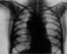
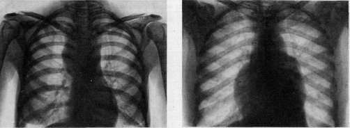
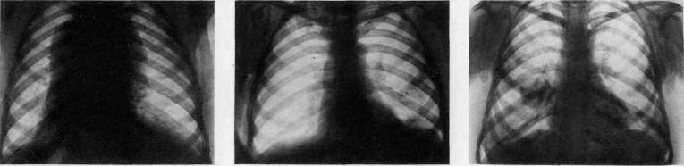
Fig 1 2 first group (I before treatment, 2-after X-ray treatment); fig. 8 second group; fig. 4 third group; fig. 5 the same case after treatment. fig. 6 fourth group. Classification according to Kruchen'y. To the article Lymphogranulomatosis. nia of the glands fluctuates within 2 to 4 weeks. The glands increase gradually, have a tendency to fuse with each other. In typical cases of lymphogranulomatosis inguinalis, there is a dense, slightly painful bundle of glands above Poupart's ligament, which subsequently gives rise to multiple foci of softening with the subsequent formation of fistulas, from which a small amount of viscous yellowish pus is exuded, which then becomes more liquid [see separate table (art. 247-248), figure 7]. A probe inserted into the fistula tracts does not find a large cavity. The accompanying swelling of the deep inguinal glands is considered characteristic, about which it was thought that they never suppurate. Apparently in severe cases they can also suppurate and give similar changes (cases of Nicolas, Favre and Lebeuf). Their swelling is by no means constant. With the appearance of swellings of the inguinal glands in many patients within 1-2 weeks, and sometimes longer, temperature fluctuations (37.5 - 38.5°) are observed; the general condition either changes little or progressive anemia, weakness, sweats, pains in the legs, etc. appear. The spleen sometimes enlarges. Polyadenitis may appear. In the blood, leukocytosis, some lymphopenia and sometimes monocytosis. Cerebrospinal fluid is unchanged. Sometimes temporary positive RW. The reverse development of lymphogranulomatosis inguinalis and healing of fistulas go extremely slowly- from 2 months to 1 year, on average about 6 months; after healing, retracted scars remain, and the resulting fibrous strands can cause disorders of lymphatic circulation in the corresponding areas. Path.-anat. changes are quite characteristic. The glands are succulent, fused with each other, cherry-red color, sometimes with visible grayish grains; on the cut, the entire tissue of the gland is penetrated by a mass of the smallest abscesses or necrotic nests, sometimes star-shaped, and presents the picture of a "wasp's nest" (Nicolas-Durand-Favre). Histologically: a granuloma, the individual nodules of which consist in the center of necrotic cells and disintegration, and at the edge of epithelioid cells, among which giant cells are also found; the afferent and efferent vessels are surrounded by an infiltrate, their inner membrane is thickened sometimes to complete closure of the lumen; the infiltrate is polymorphic, which prompted Nicolas and Favre to name it lymphogranulomatosis. Gamna bodies (Gamna), found in such granulomas, were initially considered as possible causative agents; apparently they are nothing other than nuclear-cellular disintegration (Bougu). The etiology of the disease is still unclear. The possibility of infection by non-sexual route has been proven. 2 cases of disease in surgeons who injured their hands during extirpation of diseased glands in the axillary region have been described. The pus of the glands is sterile, autoinoculation is unsuccessful. In closed buboes, various rods, coccobacilli TnnaCorynebacterium, pseudodiphtheritic rods, Bac. fluorescens, amoebas, fungi, etc. were found. Experiments on animals are unsuccessful. A supposedly complement-binding reaction with an antigen prepared from the corresponding found microbes was observed. During the process, allergic phenomena develop, proven by the Frey reaction (Frey). Intradermal injection of 0.1 vaccine from adenitis pus (the so-called "pus-vaccina") gives a typical papule about 1 cm in diameter, which does not occur in healthy people and other patients, e.g. with soft chancre bubo. Differential diagnosis is based on the clinical picture, such as: the formation of multiple foci of softening, packet-like swelling of the inguinal glands, and sometimes the inguinal and other, on the macroscopic appearance of the glandular tissue on the cut (picture of "wasp's nest"), on the Frey reaction with pus-vaccine. This reaction is especially important in cases of mixed infection with lymphogranulomatosis inguinalis and soft chancre, lymphogranulomatosis inguinalis and syphilis. The prognosis is favorable. One fatal case from complication with gangrene has been described. After radical surgical intervention, elephantiasis of the genital organs with subsequent disorders may develop, although the fear of this is apparently exaggerated. In individual cases, elephantiasis of the genital system with various disorders of lymphatic circulation can also be observed without surgical intervention. For treatment, the following have been proposed: extirpation of the affected glands, not always shortening the course of the disease, radiotherapy, large doses of iodine preparations orally or intravenously, intravenous infusions of 1% Tartar, stibiati from 0.05 to 0.1 per dose up to a total dose of 1.0; antimosan, stib-enol. Neosalvarsan, mercury, bismuth were tried with doubtful success. Xyloid-iodoform emulsion was injected into the gland. Apparently iodine preparations, repeated incisions of the glands with evacuation of pus, rest, warmth have their advantages. Sobolev saw success from intradermal application of pus-vaccine.
l. Sobolev.
Related articles
Mentioned in
Cite this page
“Lymphogranulomatosis.” Soviet Medical Encyclopedia. English translation of Bolshaya Meditsinskaya Entsiklopediya, 1st ed. (Moscow, 1928–1936), ed. N. A. Semashko. https://sovietmedicalencyclopedia.pages.dev/article/lymphogranulomatosis/