Pemphigus
Historical document, translated for reference. It reflects medical knowledge of the 1920s–30s and is not medical advice.
Summary
A comprehensive 1930s medical article on pemphigus, covering its classification, various forms, associated conditions, and clinical manifestations. The text details different types of pemphigus including pemphigoid eruptions, neonatal pemphigus, acute febrile pemphigus, and chronic pemphigus with its subtypes.
Encyclopedia article (1928–1936)
PEMPHIGUS (from Greek pemphix-bubble), bubble disease, in the narrow sense a severe dermatosis with the formation of blisters on the skin and mucous membranes. The term R. was used for a long time to designate various symptomatic blister eruptions. Because of this, by the beginning of the 19th century, up to a hundred different forms of R. were counted. And at the present time, the term R. is still used for completely different diseases. For orientation, it is advisable to divide all the material into several groups: A) Pemphigoid symptomatic eruptions in various diseases; B) R. neonatorum; C) P. acutus febrilis; D) Proper R., or P. chronicus (P. traumaticus, see Epidermolysis bullosa). A. Pemphigoid eruptions. 1) Blister eruptions have been described in organic (P. neuroticus) and "functional" diseases of the nervous system (P. hystericus). Among organic cerebral lesions, progressive paralysis takes first place, which, according to O. Fischer, in 1/3 of cases is accompanied by the eruption of groups of blisters on the distal parts of the extremities after "ictal attacks" or shortly before death. Individual cases of blister eruptions have been described in epidemic encephalitis, intracranial tumors, and hemiplegias. In the latter cases, blisters are located on both the healthy and paralyzed halves of the body. Among lesions of the spinal cord, blisters are most often observed in syringomyelia, often accompanied by other trophoneurotic and vasomotor phenomena (edema, necroses, dystrophy of nails, etc.). In diseases of the peripheral nerves, the formation of blisters is also observed. All pemphigoid eruptions observed in diseases of the nervous system have the following common features: regional localization, preferably on the upper extremities, lack of tendency to generalize; combination with sensory disorders and vasomotor skin changes. The question of "psychogenic" R. is controversial. Some authors explain the development of blisters in hysterical patients exclusively as artifacts. Others object to this. Indeed, in some observations, the possibility of artifacts is completely excluded. The development of "psychogenic" blisters is explained by reflex from vasomotor and sympathetic centers, irritated by impulses coming from the cerebral cortex. The participation of these same centers is assumed to some extent also in P. neuroticus. 2) Pemphigoid eruptions have been described in lymphatic leukemia and lymphogranulomatosis (see Leukemia, skin manifestations). 3) Some medications (antipyrine, iodine, salvarsan, etc.) can give allergic skin reactions of the bullous type and sometimes vegetating toxidermias. Iodine deserves special mention. Acting as an aggravating factor in Duhring's disease (see Duhring's disease) and sometimes in true R., iodine can occasionally provoke the onset of these diseases. Recognition can be difficult in such cases. 4) P. leprosus occurs either in the prodromal stage of leprosy or in its nervous form (see Leprosy). 5) P. syphiliticus occurs rarely and exclusively in congenital syphilis. The blistering eruption is either already present at birth or develops in the first days after birth. The characteristic, but not exclusive, localization is the palms and soles. The blisters are usually flaccid with cloudy, sometimes hemorrhagic content, located on relatively little inflamed skin, sometimes surrounded by a brown-red halo. 6) Pemphigoid eruptions in septic diseases are varied. In sepsis caused by the blue pus bacillus, hemorrhagic blisters have been described, after the opening of which deep defects remain. Staphylococcal sepsis can be accompanied by the formation of metastatic dermal pustules, which in exceptional cases turn into pustules of a bullous nature. The blistering eruptions in streptococcal sepsis are very similar to true R. (see below). 7) In various exudative skin diseases, pemphigoid blisters can develop. These include exogenous dermatitis, eczema, dyshidrosis, erythema multiforme, herpes zoster, ichthyosiform erythroderma of Brocq (Brocq), pyoderma (mainly staphylococcal), blastomycosis, premycotic rashes in mycosis fungoides, lichen planus pemphigoides, urticaria bullosa, hydroa vacciniformis. B. P. neonatorum, blister disease of newborns (synonyms: pemphigoid of newborns Jadassohn, p. infantum neonatorum epidemicus, etc.), is observed in newborns most often between the 3rd and 8th day of life and belongs to the group of bullous epidermal pyoderma (see Impetigo). According to most researchers, the causative agent is the golden staphylococcus, according to Sabouraud and others - diplococcus. Histologically - single-chamber intraepithelial blisters; vessels of the dermis are dilated and surrounded mainly by leukocytic infiltrate. Infectiousness has been proven by epidemics in obstetric departments and infant homes, as well as by the inoculability of blister fluid. In adults, inoculation proceeds as impetigo. In turn, adults suffering from impetigo can be a source of an epidemic of p. neonatorum. Transmission usually occurs through intermediate objects (linen) or through the serving personnel. The disease begins either suddenly or after a slight increase in temperature. The eruption initially consists of erythematous spots, on the background of which blisters rapidly appear, ranging in size from a pinhead to a walnut. The size and number of blisters are very varied. The content of the blisters is initially serous, then becomes cloudy. They can appear on any area of the skin, but their favorite localization is skin folds. On the palms and soles they are almost never present. The blisters burst quickly, leaving superficial erosions that are covered with a thin crust and heal without scar formation. Usually the disease ends with recovery within a few weeks and hardly affects the general condition, although sometimes in weakened children, septic phenomena are observed on the basis of R. Some cases, due to the fusion of numerous, mostly flaccid blisters or as a result of sheet-like exfoliation of the epidermis, proceed under the guise of exfoliative dermatitis of Ritter (Ritter v. Rittershain). Epidemiological facts indicate the identity of p. neonatorum and Ritter's disease, although one cannot deny that the latter has a peculiar clinical picture (beginning with cracks around the mouth, spread from top to bottom, general erythema, lesions of mucous membranes and internal organs), which may be explained in part by general sepsis. In diagnosis, it is necessary to take into account p. syphiliticus (localization on the palms and soles, presence of other signs of congenital syphilis) and epidermolysis bullosa. The prognosis is favorable. In Ritter's disease, mortality reaches 50%. Prevention: isolation of the sick, disinfection of linen, dishes and premises, as well as temporary removal from service of infants of persons suffering from pyoderma. When R. appears in a maternity home, a careful detailed review of all aspects of child care is required. Treatment: some authors recommend baths with an admixture of potassium permanganate or rivanol with subsequent disinfecting dusting, others, on the contrary, prohibit baths and limit themselves to dusting with sterile talc and the use of sterile linen. Good results from exposure to mountain sunlight are also reported. C. P. acutus febrilis, a disease belonging to the group of septicemias of various etiologies. Clinically, on the one hand, it is connected by transitional cases with pemphigoid septic rashes, and on the other, with subacute course (p. subaeutus malignus) it closely approaches true R. The etiology is different (streptococci, blue pus bacillus, anaerobes, etc.), but streptococcus probably predominates. The eruption consists of stretched, rapidly bursting blisters, which sometimes tend to bleed, deep suppurations and necroses. The general condition is septic; the prognosis is doubtful. Treatment is symptomatic. D. Proper R. (p. chronicus), blister disease, characterized mainly by the repeated formation of multiple blisters, usually over the entire surface of the skin and on mucous membranes. R. belongs to the most severe and agonizing skin diseases. Clinical symptoms and course are extremely varied. The division of R. into 3 main subspecies (p. vulgaris, p. vegetans and p. foliaceus) is generally accepted, but at the present time most authors agree that transitional cases speak for the close connection of these subspecies. The question of the relationship between R. and Duhring's disease (p. benignus) is still controversial, because here too transitional cases cast doubt on the possibility of strict demarcation. The distribution of R. in different localities is uneven, but it is everywhere a rare disease. In the clinic of Kaposi (Kaposi), who collected 320 cases in 35 years, it accounted for about 0.3% of skin patients. According to American statistics for 15 years, its frequency is determined at 0.16%. In Germany, Austria and Italy, p. vegetans is relatively not very rare, in France only 5 cases were observed in 25 years. In the USSR, R. is more common probably in the western and southwestern regions. According to some authors, men get sick more often than women.
Neisser's opinion that p. vegetans occurs exclusively in Jews was not confirmed. Initial and skin symptoms. 1. P. vulgaris in most cases develops gradually, often remaining localized for several weeks, months, and even years. Sometimes the onset of the disease can be associated with emotional shock or skin trauma, on the surface of which initial manifestations develop. The most frequent initial localization is the oral mucosa, where erosions and membranous deposits with various localization are observed, accompanied by pain and salivation. They are distinguished from stomatitis by their spontaneous appearance, persistence, and sometimes unusual localization (soft palate, arches, tonsils). Less frequently, the process begins on the mucous membranes of the larynx, eyes, nose, and sometimes on the external genitalia in women. The primary element is a tightly tense blister ranging in size from a pinhead to a chicken egg, filled with serous fluid. Blisters appear either on unchanged skin or against the background of an erythematous spot. They erupt either haphazardly over the entire surface of the skin (p. disseminatus) or are located in separate groups (p. circinati); sometimes new blisters erupt around old ones, forming garland-like figures (p. serpiginosus) [see separate table (pp. 223-224), Fig. 6]. Along with blisters, erythematous and urticarial elements may sometimes be observed. Cases have been described with exclusive eruption of the smallest blisters: (p. miliaris). Sometimes the contents of the blisters quickly become purulent. Healing of the blisters occurs without scarring, but sometimes a sharp pigmentation remains in their place. Subjective complaints during the eruption stage are usually insignificant, but ruptured blisters with their exposed, weeping base, sticking to clothing, are a source of the most agonizing sensations. Blisters appear either continuously one after another or in separate outbreaks, separated by free intervals of varying duration. 2. P. vegetans (syn. Neumann's disease) resembles p. vulgaris in its onset, but differs a) in more frequent localization around natural openings, especially in the area of the external genitalia, b) in the characteristic change of the base of blisters and erosions, which, being covered by proliferations, rise in the form of sharply limited plaques with a dirty-gray coating and moist crusts. New blisters appear around the plaques. Herpes vegetans is a special form of p. vegetans, characterized by the small size of the blisters, arranged in herpetiform groups. P. vegetans bears a great resemblance to vegetating syphilitic papules due to the appearance of individual plaques and their localization. P. vegetans has a purely external resemblance to the bullous vegetating erythema described by Unna, which apparently belongs to the group of polymorphous exudative erythemas. 3. P. foliaceus (syn. Casenave's disease) is characterized mainly by a special property of the entire skin, consisting of the weakening of the connection between the horny and granular layers (Nikolsky's phenomenon). This phenomenon is detected either by rubbing healthy skin with a finger, whereby the horny layer peels off, or by pulling on a torn blister cover, whereby the horny layer can be removed far beyond the limits of the blister. Due to this condition of the epidermis, the blisters in p. foliaceus are not very tense, have a creeping character, and are not prone to healing (see separate table, Fig. 7). Often new blisters arise in the places of former ones, as a result of which the skin is covered with constantly increasing and layered scales and crusts. Hair becomes sparse and falls out, nails become dull and brittle. Other symptoms and course. Mucous membranes are affected not only at the beginning but also in the further course of P. Here blisters burst almost immediately upon formation, giving sharply limited, bright red, round erosions, sometimes covered with a grayish coating. They are usually very painful, and when extensively distributed, they make swallowing impossible, lead to aphonia and difficulty breathing. When localized on the conjunctiva, the consequences may be adhesions and blindness. P. of mucous membranes is usually accompanied by P. of the skin, but isolated cases are also observed. The general condition suffers to varying degrees. Temperature is elevated in most cases, especially during the eruption of blisters. Often the fever has a long remitting or irregular character, with chills being not infrequent. There are no symptoms characteristic of P. from the internal organs. Sometimes there are diarrhea, rarely an enlarged spleen. Protein and formed elements are not always found in the urine, although functional tests almost constantly indicate damage to the renal parenchyma. In most cases, a sharp decrease in the excretion of NaCl with urine and a tendency to its retention in the body are noted, detectable by a salt load test. The excretion of urea is also decreased, while the content of uric acid is increased. The blood picture is relatively little studied. Most authors note almost constant eosinophilia; according to the available sparse observations, a nuclear shift of neutrophils is almost always noted. The course is extraordinarily diverse. Along with benign cases (p. benignus), sometimes lasting with long remissions of 5-10 or more years, malignant forms (p. malignus) are known, ending in death in several months. Most cases of p. vegetans run a malignant course, although with this form, cases of sudden and persistent recovery are known. P. foliaceus can give long remissions up to apparent recovery and usually lasts for many years [see separate table (pp. 155-156), Figs. 1 and 2]. P. vulgaris in adults almost always ends in death, while in children, on the contrary, it runs a benign course. As complications of P., erysipelas, gangrene of the skin, lymphadenitis, and pneumonia are observed. The causes of death are marasmus or various complications, especially pneumonia, sometimes sepsis. Pathological anatomy. In the internal organs, autopsies usually reveal non-specific phenomena in the form of diffuse degenerations and atrophy. Various, but not constant, degenerative and partly inflammatory changes have been described in various parts of the nervous system, central and peripheral, particularly in the intervertebral ganglia. However, cases without such changes have also been observed. Changes of one kind or another are often found in the endocrine glands, especially sclerosis of the sex glands. The histology of skin elements varies. In p. vulgaris, the blister is located either in the deep layers of the epidermis or subepidermally. In the blister contents, epithelial cells, single lymphocytes, and neutrophils are found initially. Later, the number of leukocytes increases due to neutrophils or eosinophils. From the side of the derma, edema, dilation of blood vessels and lymphatic vessels, and perivascular infiltration are observed. Tissue eosinophilia is often found. In p. vegetans, significant elongation (due to edema) of the papillae and interpapillary epithelial processes is found. In the epidermis—edema, leukocytes in the intercellular spaces, especially eosinophils, singly and in clusters. In the derma, edema and sharply expressed infiltration, partly of a granulation tissue nature, partly with the formation of small abscesses. The vessels are markedly dilated and surrounded by cuff-like infiltrates with a significant admixture of eosinophils. In old elements, the development of scar tissue is found. In p. foliaceus, due to the easy detachability of the horny layer, blisters always form directly beneath it. The Malpighian layer is thickened, with many mitoses in its cells. The granular layer is also thickened. Pathogenesis and etiology. 1. The theory of Leredde has purely historical interest, who, based on eosinophilia, associated P. with diseases of the blood-forming organs and classified P. as 'hematodermias.' 2. The hypothesis of Tommasoli, considering P. as auto-intoxication, also did not receive recognition. 3. The 'nervous' theory deserves more serious attention, based on findings of multiple anatomical changes in the nervous system (Nikolsky and others). Buschke emphasizes the importance of anatomical changes in the diencephalon and thinks of primary damage to the sympathetic center located there, which damage can occur from various causes. Sormani saw in P. an infectious disease with primary damage to the central nervous system. Petersen explained the increase in t° in P. by damage to the temperature centers. However, observations have been published showing the absence of anatomical changes in the central nervous system, contrary to these data and considerations. Moreover, the positive findings differ—
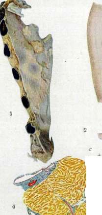

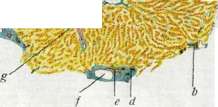
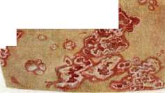
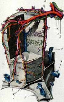
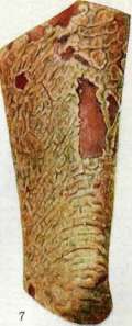
Figure 1. Reaction of Nikolsky. Figure 2. Reaction of Nikolsky. Figure 3. Histological picture: a - epidermis (staining with hematoxylin-eosin). Figure 4. Individual liver lobule with adjacent connections: a - central vein; b - hepatic artery; c - portal vein; d - bile duct; e - interlobular bile duct; f - hepatic artery; g - portal vein; h - hepatic artery; i - portal vein; j - interlobular connective tissue; k - central vein. Liver lobule with blood vessels and bile ducts: a - intralobular blood vessel; b - hepatic artery; c - portal vein; d - bile duct; e - interlobular connective tissue; f - central vein. Figure 5. Pemphigus vegetans. Figure 6. Pemphigus foliaceus. (After Pribot).
See also under St. Ohraz, Pemphigus. Liver, with the absence of regularity and constancy. Crinis described similar changes in diabetes, eclampsia, epilepsy, and burns and associates them with increased blood acidosis. It is also necessary to emphasize that clinically p. chronicus and p. neuroticus sharply differ from each other. The sometimes observed development of P. after severe emotional shocks can be easily explained from the point of view of both endocrine and infectious theories. 4. The delay in chlorides established by several authors, especially Kartamyshev, led to the hypothesis that the essence of the disease is reduced to a violation of salt metabolism. However, most authors see in the delay of chlorides not the cause of P., but a consequence, connected either with the neutralization of toxic products of protein breakdown (Ur-bakh) or with impaired kidney function (Rosenthal). In some cases, the delay may also be absent, especially in the stage of acute blistering. 5. The endocrine theory is based on observations that make the role of the endocrine factor in the pathogenesis of P. very probable. P. is observed predominantly in the age of sexual decline. In earlier age, it is often associated in women with pregnancy, lactation, and amenorrhea. Fibrosis of the sex glands is often found on autopsies. There are also reports of changes in the thyroid gland and adrenal glands. In some cases, especially with p. foliaceus, the success of organotherapy is undeniable, and Schreus reported a good effect from X-ray irradiation of the ovaries. 6. The greatest interest is presented by the infectious theory. Arguments for it are: a) cases with feverish onset and course, producing a clinical impression of general infection, b) nuclear shift in the white blood picture, c) transitional cases, which through P. acutus febrilis establish a connection between P. and sepsis, d) observations in which P. developed either on the soil of sepsis, or directly adjacent to streptogenic diseases, or finally began after trauma in the mouth (tooth extraction, wearing dentures). Opponents of the infectious theory point out that the content of fresh blisters is usually sterile, but this argument is not decisive, since even in erysipelas the content of blisters is sometimes sterile. In the origin of blisters, apparently, the microbe itself plays less of a role than its toxin or endotoxin. Apparently, a corresponding reactive readiness of the skin, depending on endocrine (or nervous) factors, is also a necessary condition for the development of a blister. This makes it possible to combine all the observations collected so far into a single whole, with P. being considered as an infectious (chronic-septic) disease, the development and course of which depends on many factors. The most substantiated of these is the endocrine moment. From this point of view, the unusual diversity of the course of P. becomes understandable. Bacteriological findings in P. (blood, blister content, internal organs) are varied. Many authors attribute the main role to streptococcal infection, but multiple etiology of P. is also possible. There are no facts indicating the contagiousness of P., except for the insufficiently traced observation of Maksimtseva, who injected herself with fluid from a blister. In several cases of P. in pregnant women, children were born with pemphigus blisters, which passed within a few days. Recognition may present difficulties in the initial period in the presence of localized phenomena. P. vegetans is often confused with syphilitic condylomas. In differential diagnosis of P., it is necessary to take into account all pemphigoid rashes (see above). Delay in chlorides may have auxiliary diagnostic value, but it is also observed in bullous toxidermias. The prognosis in adults is more serious than in children. Cases with high temperature and extensive lesions of mucous membranes and skin lead to death usually within a few months. However, it is necessary to keep in mind all the diversity of the course of P. Even hopeless cases of p. vegetans may end in recovery, and p. foliaceus sometimes lasts for more than 10 years. - T r e a t m e n t. Local treatment consists mainly of antiseptic procedures - baths with the addition of potassium permanganate, tannin or tar, compresses from Burov's solution and hydrogen peroxide, lubrication of the exposed bottom of blisters with a 5% solution of silver nitrate, disinfecting dustings, ointment dressings. The permanent bath proposed by Gebra is not always well tolerated by patients and is rarely used at present. From internal remedies, quinine in large doses (especially intravenously), plasmochin, arsenic are recommended. The latter can be used in the form of stovarsol and neosalvarsan. Organotherapy (ovarin, polyglandulin, thyroidin), blood transfusions, autogenous hemotherapy, irradiation with mountain sun, treatment with smallpox vaccination are recommended. X-ray therapy in p. vegetans gives a good symptomatic effect. Schreus and Esser recommended irradiation of the sex glands with X-rays (there are reports of good results). The development of P. during pregnancy is an indication for its termination. There are reports of improvements both from a salt-free diet and from the daily administration of several grams of salt. In the search for the focus of infection, carious teeth were sometimes removed, and there are known cases both of recovery and of deterioration. The same applies to smallpox vaccination. In general, although there is no single treatment for P., it should still be emphasized that each of the listed measures may turn out to be effective in one case or another, and therefore therapy is not hopeless.
From Rosenthal.
Related articles
Mentioned in
Cite this page
“Pemphigus.” Soviet Medical Encyclopedia. English translation of Bolshaya Meditsinskaya Entsiklopediya, 1st ed. (Moscow, 1928–1936), ed. N. A. Semashko. https://sovietmedicalencyclopedia.pages.dev/article/pemphigus/