Syringomyelia
Historical document, translated for reference. It reflects medical knowledge of the 1920s–30s and is not medical advice.
Summary
Syringomyelia is a relatively common disease of the spinal cord and often the medulla oblongata, characterized anatomically by proliferation of glial tissue in the central parts of the spinal cord with subsequent cavity formation, clinically manifested by atrophic paralysis, dissociated anesthesia, and trophic disorders of the skin, subcutaneous tissue, and joints.
Encyclopedia article (1928–1936)
SYRINGOMYELIA (from Greek syrinx-tube and myelos-marrow), or spinal gliomatosis (gliomatosis, s. gliosis spinalis, syringomyelia), represents a relatively common disease of the spinal cord, and often the medulla oblongata (syringo-bulbia), which consists anatomically of proliferation of glial tissue in the central parts of the spinal cord with subsequent formation of cavities, to which clinically corresponds a complex picture characterized mainly by the development of atrophic paralyses, dissociated anesthesias, and disorders of trophicity of the skin, subcutaneous tissue, and joints. The history of the isolation of this form is quite long. Individual cases where cavities were found in the spinal cord upon autopsy were published as early as the 16th, 17th, and 18th centuries. The name "syringomyelia" was proposed by Olivier d'Angers in 1825. For a long time, the study and description of these anomalies interested only pathologists. This continued until 1882, when Charcot and Schultze (independently of each other) managed to show that these patho-anatomical changes correspond to a very specific clinical picture, and the most characteristic clinical features were identified. Morvan in 1883, under the name "analgesic paresis with development of panaritium" (paresie analgesique a panaris), described what he thought was an independent disease, but it was later found that this disease is entirely encompassed by S. In 1902, a monograph on S. by Schlesinger appeared, which played a major role in the study of this disease. For a long time, there were disputes about the essence of the disease and its pathogenesis, during which they learned to separate from the true, "genuine", S. the secondary cavities of the spinal cord, developing as a result of meningo-myelitis, hemorrhages, syphilis, etc. When, in this way, S. was purified from the externally similar conditions that had confused it, the pathological mechanisms that lead to this disease and which were reduced to anomalies of embryonic development (the work of Bel'shovsky and others) became somewhat clearer. S. is now considered1 as one of the manifestations of a more general process, the anomaly of formation of raphe, which has other manifestations. Widespread, universal. The disease is well known in Europe, America, Australia, Japan. There are indications, however, that S. does not occur equally frequently in all countries. Thus, Austregesilo reports that S. is a great rarity in Brazil. In our country, it is one of the most common diseases of the spinal cord. The disease affects men 2 or 3 times more often than women. It has long been pointed out that S. is predominantly a disease of persons engaged in heavy physical labor. Authors working in bourgeois countries considered S. a disease of "the poorest strata of the population". The relative frequency of the disease in men and persons engaged in physical labor is probably connected with the circumstance that in the etiology of S. a certain role must be attributed to the traumatic factor. Pathological anatomy and pathogenesis. Even macroscopically, the spinal cord on sections appears altered. The dura mater is mostly normal, but sometimes shows a picture of pachymeningitis hypertrophica, and adhesions of the arachnoid and pia mater are often present. The spinal cord has various appearances: it is sometimes wide, flat, like a ribbon or a collapsed tube, and sometimes, conversely, especially dense and hard; in fresh cases, the spinal cord appears rather
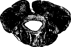
Figure 1. Syringomyelic cavity in the thoracic region of the spinal cord.
dense; in fresh cases, the spinal cord appears rather
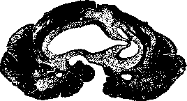
Figure 2. Syringomyelic cavity in the cervical region of the spinal cord.
If the cavity is very large, the spinal cord clearly fluctuates like a fluid-filled bag with thin walls. On cross-sections (Fig. 1 and 2), a cavity is found, sometimes in the form of a slit, located in the area of the posterior horns or posterior commissure. This cavity usually extends over a large distance, occupying many segments of the spinal cord, but is most developed in the cervical enlargement. The surrounding parts of the spinal cord that have been preserved are often compressed and deformed, so that their original structure is sometimes difficult to distinguish. The cavity is lined with glial tissue. The relationship between this glial proliferation and the cavity varies. Sometimes the cavity is very extensive, and its walls are lined with a thin layer of glial tissue; in other cases, the layer of proliferated glia surrounding the cavity is significantly thicker; sometimes the slit is weakly expressed, and the main change amounts to a long core of gliomatosis occupying the central parts of the spinal cord (the so-called glial plug). These different pictures correspond to different stages of the process: initially a glial plug forms, which then undergoes disintegration with the formation of slits and cavities. Sometimes more organized intramedullary gliomas develop in addition to the central gliomatosis; sometimes central gliomatosis is combined with an extramedullary glioma; in the cortex of the cerebral hemispheres, the development of multiple small gliomas was often found. Sylvius' aqueduct is usually dilated. Very often S. is accompanied by a mild degree of internal hydrocephalus, but sometimes the hydrocephalus of the brain ventricles can be expressed in a very severe degree. In approximately 2/3 of all cases, the process of central gliomatosis with cavity formation can also be traced in the brain stem; here the process is mostly unilateral, preferably involving the area of the loop, as well as involving the area of the nuclei of the vagus nerve, accessory nerve, hypoglossal nerve, glossopharyngeal nerve, and the spinal root of the trigeminal nerve. For the most part, the syringomyelic cavity cannot be traced further than the pons Varolii. In individual cases, however, the process was traced even further in the oral direction, for example up to the head of the caudate nucleus. Changes in the optic nerve (neuritis, atrophy) were often observed, but these changes should be considered as secondary, depending on the brain hydrocephalus. Histologically, the process is characterized as follows. True hydromyelitic changes are rarely observed. The starting point of the process is not the central canal, but the area at the ventral pole of the posterior columns, between them and the posterior commissure. Here more rudimentary pictures are also often encountered: simultaneously with various anomalies of the central canal (formation of diverticula, doubling, etc.), a more or less significant proliferation of ependymal cells (ependymatitis proliferans cum sclerose neurogliale) may be observed. With a stronger tendency to pathological growth, true gliomatous tissue forms, which grows eccentrically, penetrating into the posterior commissure and further backward into the slit between the Goll's columns. Subsequently, individual areas of gliosis become homogeneous, transforming into an amorphous mass, which eventually, disintegrating, gives rise to a central cavity surrounded on all sides by a more or less massive layer of gliomatosis. The histological structure of the gliomatous tissue varies in different cases; sometimes multinucleated cells are found, sometimes very thick glial fibers; in some cases the gliomatous formation contains very many cells, in others few; generally, for all these processes, it can be considered characteristic that the newly formed tissue in many ways resembles embryonic glia. Sometimes the syringomyelic cavity may be lined with ependymal cells like those surrounding the central canal. There is an opinion that cases where more solid glial plugs with little tendency to disintegration are observed are characterized by a slower course, while cases with more intensive development of cavities belong to those developing relatively rapidly (Haenel). It has been noticed many times that in the newly formed tissue of central gliomatosis, individual hemorrhages are observed extremely often. Small hemorrhages are found here almost as a rule; very often in histological examinations, remnants of blood pigment or cells filled with pigment grains were found here. In this respect, the tissue of central gliomatosis can be compared with the tissue of true gliomas, in which hemorrhages also develop very often. In some acute cases, an admixture of blood was found in the fluid filling the cavity. Sometimes in syringomyelia, small subarachnoid hemorrhages may also develop, which can cause corresponding changes in the cerebrospinal fluid (xanthochromia and hyperalbuminosis). There was much debate at one time around the question of the role of connective tissue and vessels in the syringomyelic process. Here a very abundant network of vessels was often found, noticeable even outside the areas of gliomatosis itself, varicose dilations of vessels, new formations of vessels, thickening or hyalinization of vessel walls, sometimes obliteration of the vessel lumen. Similarly, in a number of cases, intense proliferation of connective tissue originating from the adventitia of vessels was observed. This proliferated connective tissue from the adventitia in some cases even forms the wall of the cavity itself. Often connective tissue takes an active part in scar processes after the disintegration of the newly formed glial tissue. In such cases, they spoke of a true connective tissue and vascular 'neoplasia'. Naturally, with such significant pathological-anatomical changes in the central substance of the spinal cord, significant structural changes are also found outside the area of direct gliomatosis: nerve cells of the spinal cord undergo more or less pronounced damage; secondary degenerations develop most often in the lateral pyramidal tracts, but also in Goll's tracts and in Gowers' tracts; at the same time, long systems suffer mainly due to compression, and not due to direct infiltration by the new formation; in addition, sclerotic changes in vessels, necrotic foci of vascular origin, as well as the formation of slits due to disorders of lymphatic circulation were observed here. Such varied changes naturally for a long time hindered the understanding of the pathogenesis. On the one hand, the embryological theory of S. always had many supporters. Thus, Leiden already expressed the thought that it is in vain to separate S. so strictly from hydromyelia, as it is very possible that both these processes depend on the same conditions, and Schlesinger spoke of the possibility of building continuous anatomical series from simple anomalies of the central canal to developed S; naturally, all researchers were extremely impressed by the circumstance that the place from which the proliferation of pathological glia begins is precisely the area of closure of the primary neural tube. On the other hand, the undeniable presence of sometimes significant mesodermal changes (chronic inflammatory changes in the pia mater, vascular-connective tissue changes) forced many neurologists to consider the entire process as a special form of chronic meningomyelitis. These contradictions led to S. for some time being considered an artificial collective form, a process that can have various origins; syringomyelia ceased to be a 'clinical unit' (Brissaud, Déjerine, Schulze, Gennel and others). The research of Belyayovsky (1920) brought significant clarity to the understanding of the pathogenesis of S., followed by a number of works by other authors (Gennenberg and Koch, Bremer and others). Myelomalacic, myelitic, traumatic-necrotic and hematomyelic cavities and cysts must be strictly distinguished from true S., with which these conditions have only a remote similarity anatomically and clinically; on the contrary, S., or as it is now sometimes called, genuine S., is a completely independent disease with a single etiology; it cannot be that such an identical result would be given, as was thought earlier, by the most diverse etiological factors. This single etiology of S. is an embryonic developmental disorder, namely a delay in the formation of the raphe ('arrhaphia' and 'dysraphia'); in this process, an atypical differentiation of spongioblasts is observed, as a result of which a true spongioblastoma begins to proliferate; the process is thus very analogous to that which underlies tuberous sclerosis and neurofibromatosis. If the walls of the cavity are sometimes covered with epithelium, this is by no means proof that the cavity developed from the central canal: a glial cavity can very often communicate with the central canal, and then epithelium can grow from the central canal along the wall of the cavity.
The strong proliferation of connective tissue should likewise be considered as an anomaly of embryonic development, up to the possibility of the pia mater growing into the syringomyelic cavity during the embryonic period; - a rudimentary sign of this improper development of connective tissue is the connective tissue septum posticum found in such cases. Since status dysraphicus also gives rise to a number of other manifestations - hydromyelia, simple dilation of the central canal, other anomalies of the central canal, anomalies in the structure of the spinal cord itself, spina bifida, etc., - it is not surprising that we so frequently encounter the aforementioned developmental disorders in cases of S. This viewpoint, which is increasingly prevalent at present, leaves two questions still open: first, what is the cause of the embryonic delay in the formation of the raphe? Second, what is the reason that atypical undifferentiated spongioblasts acquire a tendency to pathological growth? The first question is at present resolved in the sense that it is apparently a hereditary process, conditioned genotypically (see below); the second question has not yet been resolved with sufficient accuracy, however, more and more data is gradually being accumulated to accept here the role of trauma as a stimulus for the morbid growth, similar to what is observed in relation to other neoplasms. The necessity to somehow connect trauma and S. has been felt for a long time. Thus, from this viewpoint, the frequently found remnants of small hemorrhages in S. were taken into account. The hypothesis was put forward (Minor, Westphal and others) that from these small hematomyelias the true S. may subsequently develop. However, in such cases it was never naturally possible to exclude the possibility that syringomyelic, resp. gliomatous, changes in the spinal cord already existed before the trauma. It is well known how frequently an existing syringomyelic process is complicated by hematomyelia (Schlesinger, Govers, Oppenheim, Hoenneberg and Koch, Pervushin and others.). On the other hand, it was pointed out that after hematomyelia ordinary cysts are formed, not syringomyelic cavities. Schulze thought of birth trauma. Sometimes one thinks of frequently recurring minor professional traumas, associated with heavy physical work. - The hypothesis of infection as an additional etiological factor in S. is significantly less substantiated. Here one thought specifically of the possibility of ascending neuritis, by means of which the toxic infectious agent could be transmitted in a centripetal direction (similarly to what occurs, for example, in tetanus or in lyssa). However, this hypothesis remains purely hypothetical and moreover unlikely, since the syringomyelic process as a rule is not accompanied by any inflammatory phenomena. The genetics of S. began to attract attention parallel to the spreading views on this process as an anomaly of embryonic development. At first the disease was generally considered as sporadic. However, in 1910 Nalbandov and in the same year Preobrazhensky published observations where S. occurred in two generations. Following these reports, a number of new observations appeared (Redlich, Karplus, Barré and Reys, Kino, Rossolimo, Mankovsky and Cherny and many others), concerning several cases of S. in the same family, either in the same group of siblings, or in parents and children, or in relatives in the lateral lines. Many authors noted in this a significant similarity in the clinical picture and course of the disease within the same family, which speaks against the hypothesis of coincidences. However, the vast majority of cases of S. remain sporadic, which does not allow one to consider this disease as hereditary in the narrow sense of the word. In the same sense speaks the well-known observation of Weitz (1924) on two women, monozygotic twins, of whom one was sick and the other was not sick with S. Obviously one hereditary factor alone is not sufficient for the development of a progressive process, and perhaps a second, paratypic factor (e.g. trauma) is really required. Such a formulation of the question naturally led researchers to look in the families of syringomyelics not for multiple cases of S, but for minor signs of that dysraphic structure which is obviously the hereditary 'predisposition' for the disease syringomyelia. Bremer conducted genealogical research precisely in this direction. Status dysraphicus, according to Bremer, is characterized by the following signs: funnel-shaped depression of the sternum, kyphoscoliosis, difference in size of both mammary glands, resp. both nipples, long arms, acrocyanosis, poor healing of skin injuries, tendency to formation of curved fingers, mild dissociated hypesthesias in the distal parts of the upper and lower extremities, nocturnal enuresis. These signs were found in abundance in relatives of patients suffering from S, and were clearly traced as a hereditary, apparently dominant anomaly. It turned out that in some families some of these 'minor signs' predominated, in others - others. Bremer conducted anatomical research on the spinal cord in a number of cases where he found on the corpses the above-mentioned morphological signs and could convince himself that in most of these cases there was hyperplasia of the ependyma surrounding the central canal, or other similar anomalies (hydromyelia). These same genealogical studies discovered a relationship with S. of many diverse 'trophoneuroses' which were previously given the significance of independent forms (acrocyanoses, symmetrical gangrene, etc.). Thus these researches shed some light on the hereditary conditions necessary for the development of S. However, this question cannot yet be considered finally resolved either with respect to the exact study of the dysraphic phenotype or with respect to the details of the inheritance of these anomalies. Symptomatology. With the exception of individual cases where at autopsy pronounced syringomyelic changes are found in the spinal cord, while the corresponding clinical symptoms during life were completely absent or masked by some other concurrent disease, the vast majority of cases is characterized by a very distinct clinical picture. The core of this clinical picture is the triad: paralyses-dissociated anesthesia-trophic disorders; as a fourth member of this series one should consider dysplastic signs and first of all kyphosis or kyphoscoliosis, occurring in more than half of all cases of S. - Paralyses in syringomyelia b. ch. (as a rule) affect the shoulder girdle and upper extremities and have the character of degenerative-atrophic paralyses. Particularly typical is the development of atrophic paralysis of the small muscles of the hand with formation of corresponding postures. Often develops the so-called claw hand (Krallenhand, main en griffe) (see Hand, deformities of the hand) with extension of the main and flexion of the middle and nail phalanges, depending on the atrophy of the interosseous muscles with secondary contracture of the relatively preserved antagonists (m. extensor digitorum communis, m. flexor digitorum communis superficialis et profundus); at the same time usually the area of m. adductor pollicis sinks in, and the muscles of the thenar and hypothenar are flattened ('monkey hand'). A very typical deformation is obtained, called Aran-Duchenne hand (see). The process does not however remain limited to the small muscles of the hand, but also spreads to the other muscles of the upper extremity and shoulder girdle. Here the most diverse combinations can be observed. Often develops predominant atrophy of the muscles supplied by the n. ulnaris and by the medianus (small muscles of the hand and flexors of the hand and fingers), while the muscles supplied by the n. radialis (extensors of the hand) continue to function; in such cases the atrophied hand is extended; this deformation is known in neurological semiotics under the not very fortunate name 'preacher's hand'. In rarer cases the amyotrophy debuts with the proximal parts of the upper extremity, sometimes quite corresponding to the so-called 'scapulohumeral type'. On the lower extremities muscle atrophies develop much less frequently. The process usually begins on one side and is initially asymmetric, in the further course however the process becomes symmetric. The atrophies may be accompanied by fibrillary twitchings, however b. ch. (as a rule) not particularly intense. Usually there is a quantitative decrease in excitability, true reaction of degeneration is encountered only rarely. There is a very strong tendency to tendon-ligament-muscular retractions, significantly limiting the range of possible passive mobility of the affected segments and sharply hindering the functional adaptability of the diseased extremity.
The atrophic paralyses develop very slowly and gradually, so that in a number of cases patients manage to adapt to the insufficient function of one or another muscle group and often notice the developing disorder only when it has already reached a significant degree. While these degenerative-atrophic paralyses are predominantly localized in the area of the upper extremities, central pareses (more rarely paralyses) are predominantly expressed in the lower extremities and characterize a later stage of the disease. It is precisely here that a spastic paraparesis gradually begins to develop with all its characteristic phenomena: increased muscle tone, appearance of pathological reflexes, etc. It can be considered characteristic of syringomyelia that spastic phenomena can be sharply expressed in the upper extremities as well, where, superimposed on the degenerative-atrophic symptom complex, they sometimes lead to combined syndromes. In severe cases of this type, spastic contractures in combination with contractures of antagonists can spread to almost the entire musculature of the trunk and extremities. It is clear that the state of reflexes can also vary greatly in syringomyelia. As a rule, tendon reflexes are lost, resp. weakened, in the upper extremities, and strengthened in the lower extremities; in individual cases, however, they can also show strengthening in the upper extremities (if the paralyses of the upper extremities are of a spastic nature), just as they can disappear in the lower extremities (if the process of cavity formation involves the lumbar-sacral part of the spinal cord). The origin of these paralyses and pareses in syringomyelia is easily understandable: the gliomatosis affects the motor cells of the anterior horns, destroying them or only compressing them—hence the development of degenerative-atrophic paralyses, and also exerts pressure on the lateral columns—hence the development of spastic phenomena. To characterize the spastic paraparesis in syringomyelia, it is interesting to note that the function of sphincters is disturbed here relatively rarely and usually only in the late stages of the disease. Cutaneous reflexes very often remain preserved. Disorders of sensitivity are extremely typical. Various symptoms of irritation are very common: pains, paresthesias, a feeling of burning, heat, which patients localize in the area of the occiput, back, shoulder girdle; often the anesthetized areas themselves are painful (anaesthesia dolorosa). Objective disorders of sensitivity are found in almost all cases. The most typical here is the so-called syringomyelic dissociation of sensitivity (Kahler, Schulze), consisting in the loss of pain and thermal sensitivity while preserving tactile sensitivity and the sense of position of the extremities. The zones of thermanalgesia are usually limited by fairly clear boundaries. They are often located in the form of a 'jacket' or 'half-jacket', 'gloves', etc. (Figs. 3 and 4). If the amyotrophies are expressed asymmetrically, thermanalgesia is more pronounced on the same extremity where the atrophic process is stronger. More dissociated forms are sometimes observed, for example, predominant disturbance of pain sensitivity or only thermal sensitivity. At the same time, the other types of sensitivity also suffer, but the boundaries of these anesthesias are less extensive, so it is always possible to find areas with characteristic dissociation. In cases with far-advanced anesthesias, it is possible to find not only disorders of tactile sensitivity but also disorders of muscle sense and vibratory sensitivity. Dysthesias are not uncommon, when, for example, tactile irritations are perceived as temperature sensations, etc. On the contrary, slowing of pain conductivity is extremely rare in syringomyelia. The topography of the anesthesias in most cases corresponds either to the radicular type, when the boundaries of the anesthetized areas correspond to the boundaries of spinal dermatomes, or to the so-called amputation, or metameric type, when the anesthesias on the extremities are limited by transverse lines. Sometimes the topography of sensory disturbances according to the Brown-Séquard type has also been observed, accompanying the syringomyelic paraparesis, but the latter in any case does not belong to frequent phenomena. A natural consequence of thermanalgesia is the frequent burns to which these patients are subject. The patient easily receives such a burn when touching, for example, hot dishes, an iron, a hot stove, etc., or while sleeping on a stove, etc. Therefore, the characteristic scars from burns are highly typical for syringomyelia. In particular, they immediately make it necessary to discard the hypothesis of hysterical analgesia. The pathogenesis of the dissociated anesthesia in syringomyelia is easily understandable: the conductors for pain and thermal sensitivity, entering the spinal cord as part of the posterior roots, must then cross to the opposite side in order to ascend further in the oral direction as part of the spinothalamic tract on the opposite side; it is clear that a process destroying the area of the posterior commissure should primarily affect these systems; but since the syringomyelic cavity also compresses the posterior horn and posterior columns, it is clear that the other types of sensitivity do not remain completely unaffected; pains and paresthesias are probably explained by those changes in the membranes that are so often encountered in syringomyelia. Thus, the features of the anesthesias in syringomyelia should be connected not with the nature but only with the topography of the pathological process, and therefore these features, however typical they may be for syringomyelia, do not yet contain anything pathognomonic: any other process with a similar localization (hematomyelia, central myelitides, intramedullary new growths) can lead to the development of exactly the same 'syringomyelic' dissociation. Here it should also be mentioned that there exists a rare variant of central gliomatosis when the posterior columns are predominantly affected (Oppenheim, Astvatsaturov). This is the so-called pseudotabes gliomatosa. And the clinical picture here becomes very similar to tabes dorsalis: areflexias, ataxias, etc., develop. The trophic disorders encountered in syringomyelia are extremely diverse. The mildest include cooling, cyanosis, hyperemia of the extremities; accidental minor wounds, scratches, etc., heal poorly. More severe disorders are not uncommon: the skin becomes dense, thick, rough, easily cracks, the fingers become thick, as if edematous, the subcutaneous tissue thickens; the hand often appears swollen, as if edematous ('succulent hand' Marinesco); sometimes vaso-motor changes are observed: erythromelalgia, urticaria factitia. Disorders of pigmentation have been observed. Sometimes disorders of nail trophics, shiny skin, develop. Such trophically altered hands extremely easily give rise to the development of persistently continuing panaritiums and phlegmons, which often lead to 48
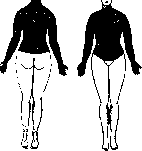
Figure 3. Loss of pain and temperature sensitivity.
lamicus to ascend further in the oral direction; it is clear that a process destroying the area of the posterior commissure should primarily affect these systems; but since the syringomyelic cavity also compresses the posterior horn and posterior columns, it is clear that the other types of sensitivity do not remain completely unaffected; pains and paresthesias are probably explained by those changes in the membranes that are so often encountered in syringomyelia. Thus, the features of the anesthesias in syringomyelia should be connected not with the nature but only with the topography of the pathological process, and therefore these features, however typical they may be for syringomyelia, do not yet contain anything pathognomonic: any other process with a similar localization (hematomyelia, central myelitides, intramedullary new growths) can lead to the development of exactly the same 'syringomyelic' dissociation. Here it should also be mentioned that there exists a rare variant of central gliomatosis when the posterior columns are predominantly affected (Oppenheim, Astvatsaturov). This is the so-called pseudotabes gliomatosa. And the clinical picture here becomes very similar to tabes dorsalis: areflexias, ataxias, etc., develop. The trophic disorders encountered in syringomyelia are extremely diverse. The mildest include cooling, cyanosis, hyperemia of the extremities; accidental minor wounds, scratches, etc., heal poorly. More severe disorders are not uncommon: the skin becomes dense, thick, rough, easily cracks, the fingers become thick, as if edematous, the subcutaneous tissue thickens; the hand often appears swollen, as if edematous ('succulent hand' Marinesco); sometimes vaso-motor changes are observed: erythromelalgia, urticaria factitia. Disorders of pigmentation have been observed. Sometimes disorders of nail trophics, shiny skin, develop. Such trophically altered hands extremely easily give rise to the development of persistently continuing panaritiums and phlegmons, which often lead to 48
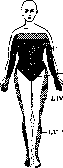
CVI-VII-VIIIDl
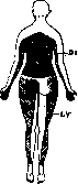
Fig. 4. Topography of tactile sensitivity disorder in that case.
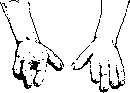
Figure 5. Panaritiums in syringomyelia.
leading to mutilation, to spontaneous amputation of entire phalanges (Fig. 5)—this form was in fact described by Morvan as an independent disease. Sometimes pathological adhesions form, sometimes blisters resembling pemphigus, sometimes necrotic skin ulcers on the hands, more rarely—perforating ulcers of the foot, spontaneous gangrene. The entire limb, whose soft tissues are thickened similar to elephantiasis, can increase in size also due to pathological thickening of the bones (cheiromegalia, podomegalia; unlike acromegaly, the process is usually asymmetric). Local anomalies of sweating, an- or hyperhidroses, affecting one half of the body or one half of the face, are common and are often an early symptom of the disease. Hypertrophies or pseudohypertrophies of muscles are common, as well as myosites with a tendency to marked hardening of muscle tissue up to the development of true myositis fibrosa or myositis ossificans. Hemiatrophia faciei has been observed. Bone atrophies are common, sometimes leading to so-called spontaneous fractures. Another very characteristic trophic symptom is syringomyelic arthropathy. In contrast to tabetic arthropathies, these affect more often the joints of the upper rather than the lower extremity: shoulder, elbow, wrist. The arthropathy can be markedly pronounced while the other syringomyelic symptoms are still very weak. Arthropathies often develop acutely, sometimes after disproportionately minor trauma. Effusion develops in the joint cavity, the articular surfaces of bones atrophy and are eroded, but at the same time hypertrophic changes in bones (exostoses) also develop. The process is usually painless, which is why patients continue to use the affected limb for a long time, despite the fact that movements are accompanied by creaking and cracking, and subluxations and dislocations form in the joint. These dislocations and the formation of a loose joint with abundant exudate are most characteristic of syringomyelic arthropathy of the shoulder, while the process in the elbow joint is accompanied by greater proliferation of exostoses; dislocations are also often observed in the wrist joint. In the spine, syringomyelic arthropathy is more often localized in the cervical and upper thoracic sections (in contrast to tabes, in which the lumbar vertebrae are most often affected) and runs less severely than tabetic. The affected joints often become suppurating (metastases from paronychia or phlegmon), sometimes with the formation of long-lasting non-healing fistulas.
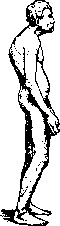
Furthermore, symptoms from the ocular branches of the cervical sympathetic nerve must be considered extremely typical. In a number of cases, anisocoria, narrowing of the palpebral fissure, enophthalmos (Claude Bernard-Horner syndrome) are observed. Mankovsky observed depigmentation of the corresponding iris in this syndrome, and sometimes changes in its stroma itself (rarefaction, the appearance of indentations in it), especially in patients in whom the disease began at a relatively young age. The origin of all these 'trophic' symptoms is probably connected with the fact that the process involves the cells of the lateral columns of the spinal cord, which give rise to preganglionic sympathetic fibers. Very often the process of central gliomatosis with the formation of syringomyelic cavities continues into the brainstem, as a result of which a picture of so-called syringobulbia develops. In such cases, a number of bulbar, mostly unilateral, symptoms are added to the general picture of S.: hemiatrophy of the tongue, paralysis of the soft palate or vocal cord, atrophy of m. sternocleidomastoidei; dyspnea, laryngeal crises, dysphagia, ageusia, salivation may develop; sometimes speech becomes monotonously scanned; pareses of the facial nerve are not common; on the contrary, disorders of sensitivity in the area of the trigeminal nerve are very often observed, often of a typically dissociated nature, in the form of circular zones surrounding the openings of the nose and mouth. At the same time, it must be borne in mind that anesthesia in the area of the trigeminal nerve can also develop with lower localizations due to the involvement of the spinal root of the trigeminal nerve. Another very common symptom is nystagmus. It is most often rotatory, sometimes rotatory-horizontal, more rarely—purely horizontal; true attacks of vestibular vertigo can also develop. Barre (Wagge) recently drew attention to the great importance of the vestibular syndrome for the diagnosis of syringobulbia. Sometimes in syringobulbia, polyuria and glycosuria were observed. Paralyses of the external eye muscles are rare; however, ptosis, paralyses of n. abducentis, disorders of pupillary reactions have been observed. Among other symptoms of the disease, the frequent affection of the optic nerve (stagnation papillae, neuritis, atrophy) deserves to be noted, apparently depending on complications from hydrocephalus. Also due to the latter should be attributed such symptoms as sometimes observed headaches, vomiting, weakening of intellect. As a rule, the psyche in S. remains normal. Occasionally, atypical hyperkineses were noted: cramps, spasmodic hiccup, blepharospasm. Sexual ability is usually preserved. The cerebrospinal fluid—with the exception of cases complicated by subarachnoid hemorrhages—shows no changes. In almost all cases—and this applies to the most typical symptoms of the disease—some signs of dysplastic structure can be found. Attention has long been drawn to the fact that these patients show a general 'coarse' build. Here special importance is attached to the tendency to develop kyphosis, resp. kyphoscoliosis, which occurs in 50-70% of all cases and is usually localized in the thoracic section of the spine (Fig. 6). Marie and Astier pointed to a characteristic boat-shaped depression of the chest (thorax en bateau), which can be observed without changes in the spine. Finzi attributed to the
signs of this dysplastic structure are too long compared to the growth of the arm, skeletal asymmetries of the face, narrow palate, irregularities of hair distribution, eccentric positioning of the pupils, frequently encountered status thymico-lymphaticus. Other authors noted anomalies in the structure of the skull (oxycephaly, underdevelopment of the supraorbital arches), anomalies of the bite, dislocation of the sternoclavicular joint, divergence of the straight abdominal muscles, shortening of ligam. nuchae, congenital ptosis, epicanthus, acromegalic structure of the facial skeleton, supernumerary nipples. Among these signs, particular importance belongs to the very frequent occurrence of spina bifida occulta in the lumbar-sacral or cervical regions of the spine in S. Sometimes in S., polydactyly was also observed, sometimes cervical ribs. It cannot be excluded that some signs from this list coincided with S. purely by chance, however the majority of them should apparently be considered as manifestations of that very anomaly in the raphe formation, which underlies S. Course of the disease as a rule is very slowly progressive. The disease usually begins in young or adult age. In most cases, the onset of the disease falls between the ages of 15 and 40 years. Individual cases of earlier and later onset have been observed. The disease lasts for many decades (40-50 years), rarely directly leading to death (septic processes following from phlegmons, sudden death from paralysis of respiration in syringobulbia), more often patients die from intercurrent diseases. Against the background of this slow progression, however, individual exacerbations are possible, which are probably connected with hemorrhages into the gliomatous tissue. In exceptional cases, the course of the process was much more unfavorable. Thus, the patient Gillen-Schmitt-Bert-rna died 9 months after the onset of the disease, which gave these authors the right to speak of an 'acute form' of S. However, since the process can remain latent for a long time and the first symptoms may only appear as a result of an accompanying hemorrhage, it is never possible to determine with certainty the time of the actual onset of the disease. Attempts have been made at a conditional classification of the separate clinical-anatomical types in which S. occurs. Thus, cervical (the most common), bulbar, sacral (the rarest) types were distinguished; the 'hemiplegic' type usually characterizes only a certain period of the disease, which later generalizes. Or types were divided according to the predominant prevalence of certain symptoms in the overall clinical picture. This resulted in: amyotrophic, spastic, osteoarthropathic, Morvan's type, pseudotabes gliomatosa and others. The diagnosis of S. is not always easy. Thus, rather similar conditions can be caused by neurosyphilis. In various types of scarring adhesive syphilitic meningitis, syphilitic myelitis and other processes, spinal cavities and cysts can develop, which clinically and anatomically somewhat resemble true S. (Oppenheim, Yafa, Crouzon and others); moreover, it should be borne in mind that even without the formation of central cavities, spinal syphilis can cause amyotrophies, mainly of the upper extremities, with sensory disturbances, sometimes dissociated, which sometimes present considerable difficulties for diagnosis. However, these syringomyelia-like syndromes of syphilitic origin in most cases are not pure, but are combined either with meningeal manifestations of syphilis or with tabetic symptoms; anatomically, here a combination with pachymeningitis, as well as with vascular processes and inflammatory processes, is very characteristic. If neurosyphilis leads to the formation of cysts and cavities, they differ from syringomyelic ones by their smaller size and distribution near blood vessels; spirochetes were found in the nervous tissue around these cavities; cavities can develop in all stages of syphilis, up to the very earliest (2 months after infection). The symptom complex of S. can also be caused by intramedullary gumma. In individual cases, a syringomyelic syndrome has been described in congenital syphilis as well. Specific therapy often gives remarkably good results in these conditions (Khait and Meerovich and others). Furthermore, there is much in common in the clinical picture between syringobulbia and basal syphilitic meningoencephalitis. In doubtful cases, examination of the cerebrospinal fluid helps in recognition. A special place belongs to the question of differential diagnosis between S. and tabes, which sometimes presents certain difficulties, since both processes are characterized by symptoms such as root-type anesthesias, anisocoria, trophic changes in bones and joints, hemiatrophy of the tongue, paresthesias, symptoms from the n. vago-accessorii and others. Moreover, one must always keep in mind the now well-studied and apparently not infrequent combination of 'tabes-syringomyelia' (Eisenlor, Egorov, Nonne, Oppenheim and many others). In these cases, syringomyelic symptoms during life were mostly hidden behind the symptoms of tabes, and the essence of the disease was discovered only at autopsy. The frequency of this combination led many authors to seek some causal connection here. Oppenheim thought that syphilis could be considered as a 'trigger'—provided that the spinal cord was already embryologically abnormal; other authors thought that the shrinkage of the atrophied posterior columns in tabes could mechanically lead to the formation of cavities in the spinal cord. However, Preobrazhensky, analyzing similar cases, came to the conclusion that here there is simply a coincidental occurrence, which is not rare only because syringomyelic cavities in the spinal cord are encountered relatively very frequently. The differential diagnosis of syringomyelia and tabes is further complicated by the fact that in so-called pseudotabes gliomatosa, clinical pictures develop that very much resemble tabetic ones. Diagnosis here can often be made only on the basis of indirect symptoms (presence or absence of syphilis, kyphosis, signs of dysraphic structure, condition of the cerebrospinal fluid, etc.). It is not always easy to distinguish from spinal cord tumor, especially from intramedullary tumor. It is important to remember that, unlike S., tumors are characterized by a more rapid course, more pronounced and earlier Brown-Séquard syndrome, rapid development of signs of transverse lesion, sometimes with a gradually shifting upward level, more intense and early signs of root irritation, early disturbance of sphincters. In unclear cases, examination of the cerebrospinal fluid or myelography can resolve the question. It is much easier to distinguish from tuberculous spondylitis (examination of the spine), from amyotrophic lateral sclerosis (in the latter, normal sensitivity and absence of trophic skin changes), from peripheral diseases. Finally, in areas endemic for leprosy, the question of differentiating the two diseases always arises, which can give far-reaching similarity, since in leprosy 'Morvan's hand', atrophic paralysis of the upper extremities, anesthesias, trophic skin disorders are also encountered. In differential diagnosis, the following is important to consider here: in leprosy, there are no spastic symptoms characteristic of S.; nerve trunks in S. are found to be normal on palpation, while leprotic nerve trunks appear spindle-shaped thickened; in leprosy, temperature elevations, loss of eyelashes and eyebrows, and characteristic tuberculous skin lesions are encountered; of course, the discovery of leprosy bacilli in the secretion of the nasal mucosa or in material obtained by biopsy is conclusive. The hypothesis that both diseases are essentially one and the same and that every S. has a leprotic origin has at present only historical interest. It is still difficult to say how widely diagnostic puncture of the syringomyelic cavity can be used for the purpose of obtaining fluid or for introducing lipiodol into the cavity with subsequent endomyelography (Pussepp, Yuzhilevsky). For a long time, the treatment of S. was considered completely hopeless and reduced to symptomatic measures (massage, electrification of the atrophying musculature, etc.), mostly without result. Recently, however, a shift has occurred in this question due to the introduction of two new methods of treatment: radiotherapy and surgical treatment. The first to propose treating S. with radiotherapy was Raymon (1905), who published cases with encouraging results. Since then, a number of individual cases have been described in which improvement in sensory disturbances, increase in range of motion, and increase in muscle strength were achieved. Only already developed muscle atrophies proved to be persistent. Recently, interest in this method of treatment has increased significantly. Good results were reported at special congresses (Eisenstein, Cherny, and Heinisman). Giese and Osinskaya saw good results in no less than 50% of all treated cases.
Sharapov (from the Blumenau clinic) also reports good results; among other things, he was able to observe that pain sensitivity recovered earlier than thermal sensitivity after treatment. Cherny and Heinisman point out that along with local symptoms, the general condition of patients usually improves; pains and paresthesias disappear; in one case, restoration of extinct tendon reflexes was even established; vasomotor-trophic disorders improved, fistulas and paronychia healed, and trophic ulcers scarred. The therapeutic effect is directly proportional to the freshness of the process and inversely proportional to the age of the patient. The levels of the spinal cord that are affected, as indicated by the results of neurological examination, should be treated. Regarding the mechanism of action of radiotherapy, it should be considered that here the effect is not on the syringomyelic cavity itself, but on the gliomatous process itself, and the gliomatous tissue is destroyed, freeing nerve cells and fibers from pressure. The surgical opening of the syringomyelic cavity was proposed by Elsberg (Elsberg, 1916), and subsequently by Pusse, who widely applied this method of treatment. According to Pusse's description, the operation is as follows: at the place where the cavity is predetermined clinically, against certain vertebrae, a 10-12 cm long incision is made along the midline of the skin and subcutaneous tissue; then, by conventional methods of typical laminectomy (see), the vertebral canal is exposed for a length of 2-3 vertebrae and 2-3 cm in width. After removing the fatty tissue separating the dura mater from the bony canal, the degree of tension of the spinal meninges is determined visually and by palpation. Then, with a thin narrow scalpel, an incision is made along the midline and along the length of the spinal cord, not penetrating deeper than just the dura mater, avoiding premature opening of the arachnoid membrane. The latter then appears protruding from under the dura mater incision as a delicate transparent membrane through which the spinal cord is visible. The arachnoid membrane is then opened with an identical longitudinal incision. After opening the membranes, the spinal cord in S. usually appears swollen and fluctuating upon palpation. Before opening the cavity, it is punctured and fluid is aspirated with a syringe. After obtaining the fluid, an incision is made along the length of the spinal cord, preferably 2-4 cm to the side of the midline, after which the cavity is emptied. The incision in the spinal cord is left unstitched, the membranes are sutured, and then the wound is closed in layers. Main indications: signs of compression of long conducting pathways, persistent trophic disorders, ununited fractures; as a contraindication, a tendency to significant spasms and mutilation was considered. As a postoperative complication, poor healing of the surgical wound, development of pareses, disturbance of sphincters, and appearance of pains between the shoulder blades were observed. The results of the operation were good in some cases, while in others it was ineffective. Peiper, in 44 cases, considers at least 12 improvements and at least 9 very significant improvements. The prognosis depends on the extent of the cavity, the degree of pressure inside the cavity, the duration of the process, and the intensity of gliosis. According to Sozon-Yaroshevich, the prognosis depends on the nature of the process itself: in the 'dry form', where there is little fluid inside the cavity, the prognosis is worse; in the 'hydropic form' with a cavity containing a large amount of fluid, the prognosis is better. There are reasons to believe that the communication of the syringomyelic cavity with the subarachnoid space may persist for a long time after the operation. In other cases, symptoms recurred, requiring reoperation. The operation gave very good results also in cases where there was pure hydromyelia. Pusse's operation is currently being actively discussed in the specialized literature. There are also opponents of this operation, who believe that the operation is completely useless, since nothing proves that the painful symptoms in S. depend specifically on the high pressure inside the syringomyelic cavity. Nevertheless, the fact of significant improvement in some cases is beyond doubt. Since, in contrast to radiotherapy, surgical treatment acts not on the gliomatous neoplasm, but on the cavity itself, it would be natural to base the indications for one or another type of treatment on what predominates in a given case: the solid gliomatous plug or the syringomyelic cavity. Unfortunately, in this respect, diagnosis is still poorly developed. Therefore, one has to join those authors who recommend the usual sequence of therapeutic measures: first conservative treatment (radiotherapy) and, in case of its failure and in the presence of appropriate indications, surgery. Questions of capacity for work and labor expertise in S. must be decided each time in a strictly individual manner: along with mildly progressing cases that allow for very good compensation for a long time, cases that quickly lead to complete disability are often encountered here. Each time, the nature of the work itself must be precisely taken into account: monotonous strain of muscles that are particularly prone to atrophy is always contraindicated in S. Furthermore, professions requiring well-preserved thermal sensitivity (possibility of burns) are always contraindicated. The question of the causal relationship of S. with a past trauma should also always be resolved through individual consideration. Here it should be remembered that although we have no grounds to consider S. as a disease with purely traumatic etiology, it is well known how often hematomyelia complicating the main process transforms the disease from a relatively mild suffering into a severe disease with loss of working capacity; on the other hand, there are many reasons to believe that in a number of cases trauma plays the role of a trigger causing the proliferation of embryologically defective glia,
SYRIN
gomyelia. Questions of capacity for work and labor expertise in S. must be decided each time in a strictly individual manner: along with mildly progressing cases that allow for very good compensation for a long time, cases that quickly lead to complete disability are often encountered here. Each time, the nature of the work itself must be precisely taken into account: monotonous strain of muscles that are particularly prone to atrophy is always contraindicated in S. Furthermore, professions requiring well-preserved thermal sensitivity (possibility of burns) are always contraindicated. The question of the causal relationship of S. with a past trauma should also always be resolved through individual consideration. Here it should be remembered that although we have no grounds to consider S. as a disease with purely traumatic etiology, it is well known how often hematomyelia complicating the main process transforms the disease from a relatively mild suffering into a severe disease with loss of working capacity; on the other hand, there are many reasons to believe that in a number of cases trauma plays the role of a trigger causing the proliferation of embryologically defective glia.
Related articles
Mentioned in
- Amyotrophic Lateral Sclerosis
- Anesthesia
- Asphyxia Localis Symmetrica
- Hemiatrophia
- Hemihypertrophy
- Locus Minoris Resistentiae
- Median Nerve
- Morvan's Disease
- Muscle Atrophy in Diseases of the Nervous System
- Mutilation
- Neuropathology
- Osteoarthropathy
- Pain Sensations
- Paraplegia
- Pemphigus
- Perforating Ulcer
- Remak's Paralysis
- Rice Bodies
- Scapula Alata
- SPINELLI
Cite this page
“Syringomyelia.” Soviet Medical Encyclopedia. English translation of Bolshaya Meditsinskaya Entsiklopediya, 1st ed. (Moscow, 1928–1936), ed. N. A. Semashko. https://sovietmedicalencyclopedia.pages.dev/article/syringomyelia/