Panaritium
Historical document, translated for reference. It reflects medical knowledge of the 1920s–30s and is not medical advice.
Summary
Panaritium refers to all non-specific purulent processes of the fingers. This article describes the causes, classification, symptoms, and treatment of various forms of panaritium, including skin, subcutaneous, bone, tendon, and joint involvement.
Encyclopedia article (1928–1936)
PANARITIUM (panaritium), all non-specific purulent processes of the fingers. No organ of the human body is subjected to such frequent injuries and diseases as the hand. According to Klyucharev's data, 43.4% of all injuries among manual workers affect the hands, with fingers accounting for 32.4%. The entry gates for infection here are visible and invisible damage to the skin coverings, through which lymphatic slits or subcutaneous cellular tissue are exposed. At the same time, the most dangerous are puncture wounds, splinters, as well as torn or bitten wounds. Based on existing observations, the possibility (Kanavel, Melchior) of bacteria penetrating inward through the numerous openings and ducts of sweat glands, which are abundantly supplied to the palmar surfaces of the fingers and hand, is not excluded. Even minor injuries to the fingers (abrasions, cuts, scratches, hangnails, pricks, etc.) with careless treatment and continued contact with infected material cause purulent processes, which result in workers being disabled. In a number of cases, P. must be considered a professional disease. Known are P. of slaughterhouse workers, sausage production workers, bakers, cooks, domestic workers, P. of fishing industry workers (pricks from bones), etc. For agriculture, P. is characteristic of persons caring for domestic animals (e.g., P. of milkmaids). Finally, everyone knows about malignant P. of anatomists, radiologists, surgeons, and medical personnel dealing with purulent diseases and purulent dressing material. As causative agents, staphylococci and streptococci are found predominantly. Much more rarely, pneumococcus, gonococcus, bacillus coli, diphtheria bacillus, Bact. proteus, pyocyaneus, etc., have been encountered. There is no unity of views in the classification of P. When divided according to anatomical principles, 5 groups are obtained: I. Skin P., II. Subcutaneous P. (p. subcutaneum), III. Bone P. (p. ossale), IV. Tendon P. (p. tendinosum, s. tendovaginitis), V. Joint P. (p. articulare).-The palmar surface of the fingers and hand is actually the 'working' grasping and holding surface, adapted for grasping and holding objects of very different densities, and here three facts of great practical importance should be noted: a) with the powerful development of the epidermal layer on the palmar surface, one cannot count on the independent breakthrough of pus to the surface of the skin, b) The presence in the subcutaneous layer of special 'spring' devices in the form of dense connective tissue strands directed almost perpendicularly from the papillary layer of the skin inward hinders the spread of infection in a plane parallel to the skin, and, conversely, promotes the advancement of bacteria inward, i.e., to the tendon and bone. This is precisely the peculiarity of the spread of P. c) Due to the scarcity of loose connective tissue, the individual cells of which are tightly compressed between connective tissue 'springs,' and the density of tissues in general, dangerous disorders of blood circulation for individual tissues can quickly occur. It should be borne in mind that for correct evaluation, the normal condition of the sensitivity of the fingers and hand of the patient is important, in particular pain and temperature sensitivity. Organic diseases of the spinal cord (syringomyelia, tabes dorsalis, etc.) can significantly affect the course and recognition of various forms of P. Recently, there has been a tendency (Molotkov, Turner) to single out a so-called panaritium neuriticum into a special group due to the assumed primary disease of trophic nerves in its basis. However, such a proposal is not yet sufficiently substantiated. I. In skin P., hangnails deserve attention as entry gates - small traumatic tears or elevations of the epidermis in the periungual skin fold. Cases of diverse infection of fingers, especially in persons in contact with pus (in surgeons, gynecologists, veterinarians before the widespread use of gloves), usually occurred due to the presence of hangnails. Recently, dangerous consequences of manicure infection (KbЫ) are also reported, and the requirement is put forward to use sterile instruments during manicures. Infection that has entered the skin of the finger may manifest in the form of a small blister, first filled with seropurulent, and then purely purulent or bloody fluid. Bacteria, most often streptococci, are located here directly under the detached epithelium near the nail bed and are often the beginning of severe general infection. Such dermatitis bullosa, accompanied by significant pain, sometimes increased t°, appearance of lymphangitis, requires rapid surgical intervention, expressed in careful excision of the entire blister and elimination of all 'pockets' where infection could subsequently hide. After this, for final healing, it is usually sufficient to apply antiseptic ointment dressings for several days. Similarly, in subepidermoidal abscess (abscessus subepidermoidalis), proceeding with the same clinical manifestations, the resulting pus separates the epidermis from the corium and accumulates here. Simple excision of the entire blister and dressing with indifferent ointment is sufficient, provided there is no narrow sinus in the center leading to a deeper purulent accumulation. Dermatitis, small abrasions and cracks around the nail bed develop particularly often in the confectionery industry, when sweet food masses (liquid sugar, jam, confectionery dough, etc.), sticking to the fingers and drying out, cause cracks and ulcers of the skin and nail bed. These minor injuries easily complicate with purulent processes, panaritiums, purulent dermatitis, which for a long time remove the worker from work. Constant inspection of workers' hands, gloves and other protective devices are necessary. In persistent cases - transfer to other work. Among skin P., purulent processes in the periungual fold (p. parunguale, s. paronychia, see Onychia) and in the subungual 'space' (p. subunguale) are most common. Clinical manifestations in p. parunguale, s. paronychia are expressed in severe, tearing pains in the fingertip, depriving the patient of rest and sleep, t° rises to 38°, the patient feels broken and cannot work. In view of this, as soon as signs of infection appear, measures should be taken to prevent its spread, which sometimes succeeds. Hartel recommends gray mercury ointment here. Also an effective means is the gentle separation of the skin periungual fold covering the nail from its root and the very careful introduction under it with the sharp tip of a scalpel as a spacer of a narrow strip of gauze soaked in a 1% solution of Argenti nitrici. If the infection has already penetrated under the root of the nail and p. subunguale has developed, the immediate removal of the detached part or the entire nail in one procedure is indicated (fig. 1). The most gentle dressings after this are strips of gauze soaked in liquid paraffin. Regeneration of the nail with its complete removal takes 3-4 months. II. Subcutaneous P. usually develops on the palmar surface of the fingertips and at the level of the phalangeo-metacarpal joints. Patho-anatomically, subcutaneous P. is revealed as a local acute inflammation (redness, swelling, sensation of heat and strong pulsating pains) with transition to purulent softening. Not only the dense tissue of the flexor side swells, but even more so the loose tissue on the back of the hand. Edema of the back of the hand can sometimes cause erroneous localization of the process. The inflammatory process can easily pass to the tendon sheaths, bones and cause lymphangitis and lymphadenitis. The symptoms of subcutaneous P. come down to pulsating pains disturbing sleep, moderate fever, general malaise, loss of appetite, insomnia and complete loss of working capacity.-TREATMENT. Beginning subcutaneous P. should be tried to be stopped with wet or alcohol compresses, which must be changed every 12 hours. Klapp considers the best means to be treatment by stagnant hyperemia by applying a bandage to the very finger, and even better - to the shoulder. In those cases where patients come with P. not at the beginning, but already when a purulent process is present, it is necessary to resort to surgical treatment. In this, it is important to consider three forms of damage: in the first, dry form, there is no pus, but a special dry necrosis, 'sequestrum' of soft tissues forms. An ordinary incision here is insufficient: it quickly sticks together and does not eliminate dangers in subsequent purulent melting of the 'sequestrum'. Therefore, it is necessary to join the authors (Klapp) who recommend excising the area with dry necrosis (fig. 2).-In the second, most common wet form of subcutaneous P., a simple incision is quite sufficient. A great mistake is to set indications for incision depending on the presence of fluctuation, which on the palmar surface of the fingers and hand can almost never be established with certainty. Therefore, incisions here often have a trial character. After incisions of subcutaneous P., tamponade or drainage is not required.-In the third, most malignant form, incision 4& is contraindicated.
These are particularly dangerous to life cases of infection when there is no clearly expressed local 'formation' of infection, when there is no pus, and when the infection immediately generalizes through the blood or lymphatic systems. Here, the necessary measures to save the patient are energetic local methods of active hyperemia, absolute immobilization of the entire limb with a Deso bandage with a load on the shoulder of the healthy side, and general therapeutic measures as in general infection (see Pyemia).-For deep inflammatory processes of the fingers and hand, a warm compress is not advisable as it increases exudation and raises hydrostatic pressure in the non-compliant tissues. For a compress, physiological saline solution or boiled water is desirable, as cases of necrosis of soft tissues have been recorded from all antiseptic agents, including Burrow's solution, especially when the skin integrity is compromised. - In the topical diagnosis of inflammatory foci of the fingers and hand, a button probe is convenient for palpating the skin, with which it is possible to determine the points of greatest tenderness with an accuracy of 1 mm. Palpation with a finger provides too large a surface of contact and obscures the picture. III. If in ordinary acute osteomyelitis of tubular bones the process begins with the bone marrow, then in bone panaritium it happens differently. The infection, which has most often spread from infected and inflamed subcutaneous tissue or introduced through some minor puncture wound, goes from the outside inward and initially affects the periosteum of the phalanx. In the development of the inflammatory process here, a necrotizing periostitis first occurs with inflammatory thrombosis of the vessels supplying the bone or their compression as a result of inflammatory 'swelling' of the tissues, and further bone necrosis is already a consequence of the death of the periosteal vessels. In addition, the harmful effect of bacterial toxins on the bone is also important here. For this reason, in bone panaritium, the formation of a sequestrum box cannot occur, as the periosteum dies here first. This form of panaritium is accompanied by extremely painful sensations, sleepless nights, and the entire clinical picture described above for subcutaneous panaritium. Even one sleepless night due to severe pain here is an absolute indication for surgery. For bone panaritium of the terminal phalanx, a cut in the shape of the Russian letter G is most convenient. Many authors transform it into a horseshoe shape (Fig. 2) in order to turn back from the bone in the form of a flap all the soft tissues. In the initial stages of bone panaritium, most often of the nail phalanges, it is easier, using hemostatic methods, to initially limit oneself to a cut to the bone and then wait for spontaneous completion of sequestration. In advanced cases with the presence of fistulas, it is necessary to make greater use of X-rays to thoroughly establish the boundaries of the entire area doomed to destruction. At the same time, it is important to take into account the pronounced anatomical isolation of the blood supply to the diaphyses and epiphyses of the phalanges, especially in growing individuals, thanks to which there is a full possibility, by cutting the bone at the metaphysis, to preserve the bases of the phalanges. With this method on the terminal phalanx, the attachment of the deep flexor can be preserved intact and it is possible to do without opening the interphalangeal joint and the distal dome-cuff of the tendon sheath. Thus, it is possible to completely avoid undesirable amputations of the fingers. Bone panaritium can sometimes be complicated by septicopyemia. IV. Of all forms of panaritium, purulent tenosynovitis presents the greatest danger due to possible extensive spread, significant loss of function, and the threat of general infection. It develops either from direct traumatic damage to the tendon sheath or from the entry of infection from nearby purulent-inflammatory foci, e.g., from soft tissues, bones, joints. In the latter case, infection is most often transmitted along lymphatic pathways, but it is also possible destruction of the wall of the tendon sheath under the influence of pus enzymes. For a clear understanding of the disease picture, an exact anatomical concept of the relationships of tendon sheaths (Figs. 3, 5 and 11) is necessary. The inability to predict in advance the architectural variant of tendon sheaths obliges the surgeon to observe the patient with great attention so as not to open uninfected sections during the operation (see Tenosynovitis). Particular importance is attached to the timeliness of surgical intervention. Any kind of incisions can be made, tamponaded or not tamponaded, but if days and sometimes even hours are missed, the death of the tendon is inevitable. The general condition in purulent tenosynovitis is in most cases severely disturbed due to the existing febrile state, sometimes with shaking chills. At the same time, patients experience extremely intense pain.-The diagnosis of purulent tenosynovitis is not always simple. In the medical history, minor puncture wounds along the course of tendon sheaths should be given great importance. Locally, it is possible to objectively establish an inflammatory painful swelling along the anatomical extent of the affected tendon, the forced position of the corresponding finger in a state of slight flexion, and pain on pressure (with a button probe) along the course of the tendon sheath. The latter symptom is especially important in areas corresponding to the upper or lower cuff of the tendon sheath, i.e., in areas remote from the portals of entry of infection. However, according to Sokolov's observations, the most reliable differential diagnostic sign of tenosynovitis should be considered the pain of passive extensions of the affected finger. This symptom should be elicited carefully, otherwise pain will also occur in ordinary phlegmon. If the disease is left to itself, all the described phenomena intensify until the wall of the tendon sheath ruptures with the outpouring of pus into the surrounding tissues. After this, the feeling of inflammatory tension decreases, but new phenomena appear, depending mainly on the developing phlegmon of the palmar surface of the hand or forearm. Patients with purulent tenosynovitis require hospital treatment. With a confirmed diagnosis, delay in incision, not only in days, but even in hours, can lead to the death of the tendon. The continuous median incision along the entire length of the tendon sheath and wrapping the tendon with tampons, still used by some surgeons, must be abandoned, as such methods contribute to the prolapse of the tendon into the subcutaneous tissue, its drying out, cooling, and subsequently necrosis or adhesion to the surrounding tissues; it must be covered with a piece of oilcloth or rubber, after which it is sometimes possible to save it (Gorodkov). In the surgical approach to purulent tenosynovitis, the following conditions must be met: 1) ether anesthesia; 2) Esmarch tourniquet on the shoulder for dehematization; 3) presence of an assistant. Failure to meet at least one of these conditions puts it at great risk
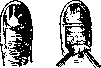

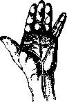
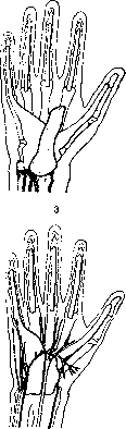
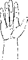
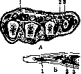
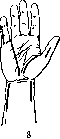
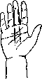
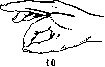

Figure 1. Incision for paronychia. Figure 2. Bivalved incision; left-outlining the arcuate incision. Figure 3. Tendon sheaths. Figure 4. Incisions along the lumbrical muscles in phlegmon of the midpalmar space and along the interdigital fold. Figure 5. A-transverse incision through the heads of the metacarpal bones: 1-fibrous barrier; 2-lumbrical muscle canal; 3-tendon sheaths; L-incision through the intermetacarpal space: 1-subcutaneous tissue; 2-fibrous barrier; 3-palmar cord of the lumbrical muscle. Figure 6. Location of incisions in phlegmon of tendon sheaths (according to Lesen). Figure 7. Blood vessel network. Figure 8. Location of incisions for opening tendon sheaths and extensions on the forearm. Figure 9. Incisions in tenosynovitis (the boundary of the synovial bursae is indicated by a dotted line). Figure 10. Incision for panaritium of the middle phalanx extending to the palm (the line of incision on the palm is indicated by a dotted line). Figure 11. Cross-section of tendon sheaths: 2-vagina fibrosa; 2-vagina mucosa; 3-tendon; 4-mesotenon.
surrounding tissues. It is much more advisable to make separate lateral incisions on each phalanx (Bardenheuer, Bier, Klapp, etc.), thanks to which the tendons remain covered by living tissues, i.e. continue to remain in the moist chamber of the tendon sheath and at body temperature. In addition, the indicated incisions spare the transverse palmar grooves of the fingers, corresponding to the flexion lines of the interphalangeal joints, and thereby reduce the possibility of the development of function-limiting scars. With lateral incisions, it is necessary to preserve the major blood vessels and nerves intact, which can be seen during artificial exsanguination, otherwise there is a threat of gangrene of the finger (Fig. 7). If for any reason the tendon is exposed as a result of the operation. As long as during the incision on the bottom of the relatively deep and bloodless wound the tendon does not shine with its pearly luster, the surgeon cannot be sure that he has opened the tendon, and cannot be confident in the diagnosis of tenosynovitis (Sokolov). On the hand, in cases of tenosynovitis and deep phlegmons, wide incisions with ligation of the arcus volaris superficialis are necessary (Figures 4, 9 and 10). With lesions of the ulnar or radial synovial bursae of the hand, incisions on the forearm corresponding to the location of these bursae are mandatory (Figures 6 and 8). In tenosynovitis of the thumb, it is necessary to preserve the motor branch (ramus muscularis) of the n. medianus to the thenar muscles (m. opponens!). The function of the m. opponens is considered even more important than the function of the m. flexoris pollicis longi. The forbidden zone for the knife when protecting the named nerve branch extends one diameter of the thumb distal to the skin projection of the ligamentum carpi transversum. Incisions in tenosynovitis may adhere. To prevent this, many methods have been proposed, up to and including special wooden and metal (Tiegel) spreaders. Based on his observations, Sokolov believes that in purulent tenosynovitis the main thing lies in the timeliness and urgency of surgical intervention. Therefore, strips of paraffinized gauze or rubber strips placed during the operation as spreaders, but without contact with the tendon, do no harm. It is more convenient to place these strips in such a way that between them and the tendon there is the 'edge' of the opened tendon sheath. Such spreaders should not be changed until the temperature has stably returned to normal, and should not be introduced later. The basic principle of purulent surgery - not to proceed with restoring function until the danger to life is eliminated - retains its significance even in tenosynovitis. In restoring function, hot water or dry-air baths are important. For water baths, physiol. solution is best, as plain water promotes undesirable swelling of the tendons. Active movements of the fingers should begin as early as possible, but it is impossible to set specific deadlines for all cases. In the long struggle to restore function in tendon P., the most important thing is the active self-activity of the patient. One bath or one session of passive gymnastics a day has negligible importance compared to the active movements that the patient must perform himself. The patient's active work should continue all the time while he is awake. For this, after the temperature has stabilized, a very soft white rubber ball is screwed on, as shown in Fig. 12. With proper gripping of the ball by the fingers, the desired average physiological position of the fingers is achieved and the continuity of the active work of the entire neuromuscular-tendon-articular apparatus is ensured. If the patient is not trained in this direction, the task of restoring tendon function may be completely unachievable. Subsequently after wound healing, persistent application of massage, passive gymnastics, active performance of tasks, and exercise in special mechano-therapy apparatuses is necessary for several months.-In purulent lesions of the flexor surface of the hand and forearm, the subsequent scar-smoothing process can cause the fingers and hand to be drawn into a position of hyperflexion, which in persistent contractures is unfavorable for function. To prevent this, special splints are used that hold the hand in the most physiological position of slight extension. It is simpler to use a plaster splint at an angle of 60°, to which the hand is bandaged, as shown in Fig. 12. As a result of the use of lateral incisions and modern postoperative treatment methods, the function of tendons after purulent tenosynovitis can currently be preserved in 34% of cases (Sokolov). According to European and American data, the number of preserved and functioning tendons can be increased under favorable conditions on average to 50%, and in very early cases even up to 90% (Klapp). From what has been said, it is clear what enormous importance, along with preventive measures, the quality of the treatment itself has. A timely incision shortens the treatment period. Moreover: a properly performed incision and exposure of the tendon save the working hand and thereby save the victim from disability. Proceeding from the political significance of reducing morbidity and restoring the working capacity of workers, the preservation of tendons in purulent tenosynovitis has acquired
surrounding tissues. It is much more advisable to make separate lateral incisions on each phalanx (Bardenheuer, Bier, Klapp, etc.), thanks to which the tendons remain covered by living tissues, i.e. continue to remain in the moist chamber of the tendon sheath and at body temperature. In addition, the indicated incisions spare the transverse palmar grooves of the fingers, corresponding to the flexion lines of the interphalangeal joints, and thereby reduce the possibility of the development of function-limiting scars. With lateral incisions, it is necessary to preserve the major blood vessels and nerves intact, which can be seen during artificial exsanguination, otherwise there is a threat of gangrene of the finger (Fig. 7). If for any reason the tendon is exposed as a result of the operation. As long as during the incision on the bottom of the relatively deep and bloodless wound the tendon does not shine with its pearly luster, the surgeon cannot be sure that he has opened the tendon, and cannot be confident in the diagnosis of tenosynovitis (Sokolov). On the hand, in cases of tenosynovitis and deep phlegmons, wide incisions with ligation of the arcus volaris superficialis are necessary (Figures 4, 9 and 10). With lesions of the ulnar or radial synovial bursae of the hand, incisions on the forearm corresponding to the location of these bursae are mandatory (Figures 6 and 8). In tenosynovitis of the thumb, it is necessary to preserve the motor branch (ramus muscularis) of the n. medianus to the thenar muscles (m. opponens!). The function of the m. opponens is considered even more important than the function of the m. flexoris pollicis longi. The forbidden zone for the knife when protecting the named nerve branch extends one diameter of the thumb distal to the skin projection of the ligamentum carpi transversum. Incisions in tenosynovitis may adhere. To prevent this, many methods have been proposed, up to and including special wooden and metal (Tiegel) spreaders. Based on his observations, Sokolov believes that in purulent tenosynovitis the main thing lies in the timeliness and urgency of surgical intervention. Therefore, strips of paraffinized gauze or rubber strips placed during the operation as spreaders, but without contact with the tendon, do no harm. It is more convenient to place these strips in such a way that between them and the tendon there is the 'edge' of the opened tendon sheath. Such spreaders should not be changed until the temperature has stably returned to normal, and should not be introduced later. The basic principle of purulent surgery - not to proceed with restoring function until the danger to life is eliminated - retains its significance even in tenosynovitis. In restoring function, hot water or dry-air baths are important. For water baths, physiol. solution is best, as plain water promotes undesirable swelling of the tendons. Active movements of the fingers should begin as early as possible, but it is impossible to set specific deadlines for all cases. In the long struggle to restore function in tendon P., the most important thing is the active self-activity of the patient. One bath or one session of passive gymnastics a day has negligible importance compared to the active movements that the patient must perform himself. The patient's active work should continue all the time while he is awake. For this, after the temperature has stabilized, a very soft white rubber ball is screwed on, as shown in Fig. 12. With proper gripping of the ball by the fingers, the desired average physiological position of the fingers is achieved and the continuity of the active work of the entire neuromuscular-tendon-articular apparatus is ensured. If the patient is not trained in this direction, the task of restoring tendon function may be completely unachievable. Subsequently after wound healing, persistent application of massage, passive gymnastics, active performance of tasks, and exercise in special mechano-therapy apparatuses is necessary for several months.-In purulent lesions of the flexor surface of the hand and forearm, the subsequent scar-smoothing process can cause the fingers and hand to be drawn into a position of hyperflexion, which in persistent contractures is unfavorable for function. To prevent this, special splints are used that hold the hand in the most physiological position of slight extension. It is simpler to use a plaster splint at an angle of 60°, to which the hand is bandaged, as shown in Fig. 12. As a result of the use of lateral incisions and modern postoperative treatment methods, the function of tendons after purulent tenosynovitis can currently be preserved in 34% of cases (Sokolov). According to European and American data, the number of preserved and functioning tendons can be increased under favorable conditions on average to 50%, and in very early cases even up to 90% (Klapp). From what has been said, it is clear what enormous importance, along with preventive measures, the quality of the treatment itself has. A timely incision shortens the treatment period. Moreover: a properly performed incision and exposure of the tendon save the working hand and thereby save the victim from disability. Proceeding from the political significance of reducing morbidity and restoring the working capacity of workers, the preservation of tendons in purulent tenosynovitis has acquired
surrounding tissues. It is much more advisable to make separate lateral incisions on each phalanx (Bardenheuer, Bier, Klapp, etc.), thanks to which the tendons remain covered by living tissues, i.e. continue to remain in the moist chamber of the tendon sheath and at body temperature. In addition, the indicated incisions spare the transverse palmar grooves of the fingers, corresponding to the flexion lines of the interphalangeal joints, and thereby reduce the possibility of the development of function-limiting scars. With lateral incisions, it is necessary to preserve the major blood vessels and nerves intact, which can be seen during artificial exsanguination, otherwise there is a threat of gangrene of the finger (Fig. 7). If for any reason the tendon is exposed as a result of the operation. As long as during the incision on the bottom of the relatively deep and bloodless wound the tendon does not shine with its pearly luster, the surgeon cannot be sure that he has opened the tendon, and cannot be confident in the diagnosis of tenosynovitis (Sokolov). On the hand, in cases of tenosynovitis and deep phlegmons, wide incisions with ligation of the arcus volaris superficialis are necessary (Figures 4, 9 and 10). With lesions of the ulnar or radial synovial bursae of the hand, incisions on the forearm corresponding to the location of these bursae are mandatory (Figures 6 and 8). In tenosynovitis of the thumb, it is necessary to preserve the motor branch (ramus muscularis) of the n. medianus to the thenar muscles (m. opponens!). The function of the m. opponens is considered even more important than the function of the m. flexoris pollicis longi. The forbidden zone for the knife when protecting the named nerve branch extends one diameter of the thumb distal to the skin projection of the ligamentum carpi transversum. Incisions in tenosynovitis may adhere. To prevent this, many methods have been proposed, up to and including special wooden and metal (Tiegel) spreaders. Based on his observations, Sokolov believes that in purulent tenosynovitis the main thing lies in the timeliness and urgency of surgical intervention. Therefore, strips of paraffinized gauze or rubber strips placed during the operation as spreaders, but without contact with the tendon, do no harm. It is more convenient to place these strips in such a way that between them and the tendon there is the 'edge' of the opened tendon sheath. Such spreaders should not be changed until the temperature has stably returned to normal, and should not be introduced later. The basic principle of purulent surgery - not to proceed with restoring function until the danger to life is eliminated - retains its significance even in tenosynovitis. In restoring function, hot water or dry-air baths are important. For water baths, physiol. solution is best, as plain water promotes undesirable swelling of the tendons. Active movements of the fingers should begin as early as possible, but it is impossible to set specific deadlines for all cases. In the long struggle to restore function in tendon P., the most important thing is the active self-activity of the patient. One bath or one session of passive gymnastics a day has negligible importance compared to the active movements that the patient must perform himself. The patient's active work should continue all the time while he is awake. For this, after the temperature has stabilized, a very soft white rubber ball is screwed on, as shown in Fig. 12. With proper gripping of the ball by the fingers, the desired average physiological position of the fingers is achieved and the continuity of the active work of the entire neuromuscular-tendon-articular apparatus is ensured. If the patient is not trained in this direction, the task of restoring tendon function may be completely unachievable. Subsequently after wound healing, persistent application of massage, passive gymnastics, active performance of tasks, and exercise in special mechano-therapy apparatuses is necessary for several months.-In purulent lesions of the flexor surface of the hand and forearm, the subsequent scar-smoothing process can cause the fingers and hand to be drawn into a position of hyperflexion, which in persistent contractures is unfavorable for function. To prevent this, special splints are used that hold the hand in the most physiological position of slight extension. It is simpler to use a plaster splint at an angle of 60°, to which the hand is bandaged, as shown in Fig. 12. As a result of the use of lateral incisions and modern postoperative treatment methods, the function of tendons after purulent tenosynovitis can currently be preserved in 34% of cases (Sokolov). According to European and American data, the number of preserved and functioning tendons can be increased under favorable conditions on average to 50%, and in very early cases even up to 90% (Klapp). From what has been said, it is clear what enormous importance, along with preventive measures, the quality of the treatment itself has. A timely incision shortens the treatment period. Moreover: a properly performed incision and exposure of the tendon save the working hand and thereby save the victim from disability. Proceeding from the political significance of reducing morbidity and restoring the working capacity of workers, the preservation of tendons in purulent tenosynovitis has acquired
surrounding tissues. It is much more advisable to make separate lateral incisions on each phalanx (Bardenheuer, Bier, Klapp, etc.), thanks to which the tendons remain covered by living tissues, i.e. continue to remain in the moist chamber of the tendon sheath and at body temperature. In addition, the indicated incisions spare the transverse palmar grooves of the fingers, corresponding to the flexion lines of the interphalangeal joints, and thereby reduce the possibility of the development of function-limiting scars. With lateral incisions, it is necessary to preserve the major blood vessels and nerves intact, which can be seen during artificial exsanguination, otherwise there is a threat of gangrene of the finger (Fig. 7). If for any reason the tendon is exposed as a result of the operation. As long as during the incision on the bottom of the relatively deep and bloodless wound the tendon does not shine with its pearly luster, the surgeon cannot be sure that he has opened the tendon, and cannot be confident in the diagnosis of tenosynovitis (Sokolov). On the hand, in cases of tenosynovitis and deep phlegmons, wide incisions with ligation of the arcus volaris superficialis are necessary (Figures 4, 9 and 10). With lesions of the ulnar or radial synovial bursae of the hand, incisions on the forearm corresponding to the location of these bursae are mandatory (Figures 6 and 8). In tenosynovitis of the thumb, it is necessary to preserve the motor branch (ramus muscularis) of the n. medianus to the thenar muscles (m. opponens!). The function of the m. opponens is considered even more important than the function of the m. flexoris pollicis longi. The forbidden zone for the knife when protecting the named nerve branch extends one diameter of the thumb distal to the skin projection of the ligamentum carpi transversum. Incisions in tenosynovitis may adhere. To prevent this, many methods have been proposed, up to and including special wooden and metal (Tiegel) spreaders. Based on his observations, Sokolov believes that in purulent tenosynovitis the main thing lies in the timeliness and urgency of surgical intervention. Therefore, strips of paraffinized gauze or rubber strips placed during the operation as spreaders, but without contact with the tendon, do no harm. It is more convenient to place these strips in such a way that between them and the tendon there is the 'edge' of the opened tendon sheath. Such spreaders should not be changed until the temperature has stably returned to normal, and should not be introduced later. The basic principle of purulent surgery - not to proceed with restoring function until the danger to life is eliminated - retains its significance even in tenosynovitis. In restoring function, hot water or dry-air baths are important. For water baths, physiol. solution is best, as plain water promotes undesirable swelling of the tendons. Active movements of the fingers should begin as early as possible, but it is impossible to set specific deadlines for all cases. In the long struggle to restore function in tendon P., the most important thing is the active self-activity of the patient. One bath or one session of passive gymnastics a day has negligible importance compared to the active movements that the patient must perform himself. The patient's active work should continue all the time while he is awake. For this, after the temperature has stabilized, a very soft white rubber ball is screwed on, as shown in Fig. 12. With proper gripping of the ball by the fingers, the desired average physiological position of the fingers is achieved and the continuity of the active work of the entire neuromuscular-tendon-articular apparatus is ensured. If the patient is not trained in this direction, the task of restoring tendon function may be completely unachievable. Subsequently after wound healing, persistent application of massage, passive gymnastics, active performance of tasks, and exercise in special mechano-therapy apparatuses is necessary for several months.-In purulent lesions of the flexor surface of the hand and forearm, the subsequent scar-smoothing process can cause the fingers and hand to be drawn into a position of hyperflexion, which in persistent contractures is unfavorable for function. To prevent this, special splints are used that hold the hand in the most physiological position of slight extension. It is simpler to use a plaster splint at an angle of 60°, to which the hand is bandaged, as shown in Fig. 12. As a result of the use of lateral incisions and modern postoperative treatment methods, the function of tendons after purulent tenosynovitis can currently be preserved in 34% of cases (Sokolov). According to European and American data, the number of preserved and functioning tendons can be increased under favorable conditions on average to 50%, and in very early cases even up to 90% (Klapp). From what has been said, it is clear what enormous importance, along with preventive measures, the quality of the treatment itself has. A timely incision shortens the treatment period. Moreover: a properly performed incision and exposure of the tendon save the working hand and thereby save the victim from disability. Proceeding from the political significance of reducing morbidity and restoring the working capacity of workers, the preservation of tendons in purulent tenosynovitis has acquired
surrounding tissues. It is much more advisable to make separate lateral incisions on each phalanx (Bardenheuer, Bier, Klapp, etc.), thanks to which the tendons remain covered by living tissues, i.e. continue to remain in the moist chamber of the tendon sheath and at body temperature. In addition, the indicated incisions spare the transverse palmar grooves of the fingers, corresponding to the flexion lines of the interphalangeal joints, and thereby reduce the possibility of the development of function-limiting scars. With lateral incisions, it is necessary to preserve the major blood vessels and nerves intact, which can be seen during artificial exsanguination, otherwise there is a threat of gangrene of the finger (Fig. 7). If for any reason the tendon is exposed as a result of the operation. As long as during the incision on the bottom of the relatively deep and bloodless wound the tendon does not shine with its pearly luster, the surgeon cannot be sure that he has opened the tendon, and cannot be confident in the diagnosis of tenosynovitis (Sokolov). On the hand, in cases of tenosynovitis and deep phlegmons, wide incisions with ligation of the arcus volaris superficialis are necessary (Figures 4, 9 and 10). With lesions of the ulnar or radial synovial bursae of the hand, incisions on the forearm corresponding to the location of these bursae are mandatory (Figures 6 and 8). In tenosynovitis of the thumb, it is necessary to preserve the motor branch (ramus muscularis) of the n. medianus to the thenar muscles (m. opponens!). The function of the m. opponens is considered even more important than the function of the m. flexoris pollicis longi. The forbidden zone for the knife when protecting the named nerve branch extends one diameter of the thumb distal to the skin projection of the ligamentum carpi transversum. Incisions in tenosynovitis may adhere. To prevent this, many methods have been proposed, up to and including special wooden and metal (Tiegel) spreaders. Based on his observations, Sokolov believes that in purulent tenosynovitis the main thing lies in the timeliness and urgency of surgical intervention. Therefore, strips of paraffinized gauze or rubber strips placed during the operation as spreaders, but without contact with the tendon, do no harm. It is more convenient to place these strips in such a way that between them and the tendon there is the 'edge' of the opened tendon sheath. Such spreaders should not be changed until the temperature has stably returned to normal, and should not be introduced later. The basic principle of purulent surgery - not to proceed with restoring function until the danger to life is eliminated - retains its significance even in tenosynovitis. In restoring function, hot water or dry-air baths are important. For water baths, physiol. solution is best, as plain water promotes undesirable swelling of the tendons. Active movements of the fingers should begin as early as possible, but it is impossible to set specific deadlines for all cases. In the long struggle to restore function in tendon P., the most important thing is the active self-activity of the patient. One bath or one session of passive gymnastics a day has negligible importance compared to the active movements that the patient must perform himself. The patient's active work should continue all the time while he is awake. For this, after the temperature has stabilized, a very soft white rubber ball is screwed on, as shown in Fig. 12. With proper gripping of the ball by the fingers, the desired average physiological position of the fingers is achieved and the continuity of the active work of the entire neuromuscular-tendon-articular apparatus is ensured. If the patient is not trained in this direction, the task of restoring tendon function may be completely unachievable. Subsequently after wound healing, persistent application of massage, passive gymnastics, active performance of tasks, and exercise in special mechano-therapy apparatuses is necessary for several months.-In purulent lesions of the flexor surface of the hand and forearm, the subsequent scar-smoothing process can cause the fingers and hand to be drawn into a position of hyperflexion, which in persistent contractures is unfavorable for function. To prevent this, special splints are used that hold the hand in the most physiological position of slight extension. It is simpler to use a plaster splint at an angle of 60°, to which the hand is bandaged, as shown in Fig. 12. As a result of the use of lateral incisions and modern postoperative treatment methods, the function of tendons after purulent tenosynovitis can currently be preserved in 34% of cases (Sokolov). According to European and American data, the number of preserved and functioning tendons can be increased under favorable conditions on average to 50%, and in very early cases even up to 90% (Klapp). From what has been said, it is clear what enormous importance, along with preventive measures, the quality of the treatment itself has. A timely incision shortens the treatment period. Moreover: a properly performed incision and exposure of the tendon save the working hand and thereby save the victim from disability. Proceeding from the political significance of reducing morbidity and restoring the working capacity of workers, the preservation of tendons in purulent tenosynovitis has acquired
surrounding tissues. It is much more advisable to make separate lateral incisions on each phalanx (Bardenheuer, Bier, Klapp, etc.), thanks to which the tendons remain covered by living tissues, i.e. continue to remain in the moist chamber of the tendon sheath and at body temperature. In addition, the indicated incisions spare the transverse palmar grooves of the fingers, corresponding to the flexion lines of the interphalangeal joints, and thereby reduce the possibility of the development of function-limiting scars. With lateral incisions, it is necessary to preserve the major blood vessels and nerves intact, which can be seen during artificial exsanguination, otherwise there is a threat of gangrene of the finger (Fig. 7). If for any reason the tendon is exposed as a result of the operation. As long as during the incision on the bottom of the relatively deep and bloodless wound the tendon does not shine with its pearly luster, the surgeon cannot be sure that he has opened the tendon, and cannot be confident in the diagnosis of tenosynovitis (Sokolov). On the hand, in cases of tenosynovitis and deep phlegmons, wide incisions with ligation of the arcus volaris superficialis are necessary (Figures 4, 9 and 10). With lesions of the ulnar or radial synovial bursae of the hand, incisions on the forearm corresponding to the location of these bursae are mandatory (Figures 6 and 8). In tenosynovitis of the thumb, it is necessary to preserve the motor branch (ramus muscularis) of the n. medianus to the thenar muscles (m. opponens!). The function of the m. opponens is considered even more important than the function of the m. flexoris pollicis longi. The forbidden zone for the knife when protecting the named nerve branch extends one diameter of the thumb distal to the skin projection of the ligamentum carpi transversum. Incisions in tenosynovitis may adhere. To prevent this, many methods have been proposed, up to and including special wooden and metal (Tiegel) spreaders. Based on his observations, Sokolov believes that in purulent tenosynovitis the main thing lies in the timeliness and urgency of surgical intervention. Therefore, strips of paraffinized gauze or rubber strips placed during the operation as spreaders, but without contact with the tendon, do no harm. It is more convenient to place these strips in such a way that between them and the tendon there is the 'edge' of the opened tendon sheath. Such spreaders should not be changed until the temperature has stably returned to normal, and should not be introduced later. The basic principle of purulent surgery - not to proceed with restoring function until the danger to life is eliminated - retains its significance even in tenosynovitis. In restoring function, hot water or dry-air baths are important. For water baths, physiol. solution is best, as plain water promotes undesirable swelling of the tendons. Active movements of the fingers should begin as early as possible, but it is impossible to set specific deadlines for all cases. In the long struggle to restore function in tendon P., the most important thing is the active self-activity of the patient. One bath or one session of passive gymnastics a day has negligible importance compared to the active movements that the patient must perform himself. The patient's active work should continue all the time while he is awake. For this, after the temperature has stabilized, a very soft white rubber ball is screwed on, as shown in Fig. 12. With proper gripping of the ball by the fingers, the desired average physiological position of the fingers is achieved and the continuity of the active work of the entire neuromuscular-tendon-articular apparatus is ensured. If the patient is not trained in this direction, the task of restoring tendon function may be completely unachievable. Subsequently after wound healing, persistent application of massage, passive gymnastics, active performance of tasks, and exercise in special mechano-therapy apparatuses is necessary for several months.-In purulent lesions of the flexor surface of the hand and forearm, the subsequent scar-smoothing process can cause the fingers and hand to be drawn into a position of hyperflexion, which in persistent contractures is unfavorable for function. To prevent this, special splints are used that hold the hand in the most physiological position of slight extension. It is simpler to use a plaster splint at an angle of 60°, to which the hand is bandaged, as shown in Fig. 12. As a result of the use of lateral incisions and modern postoperative treatment methods, the function of tendons after purulent tenosynovitis can currently be preserved in 34% of cases (Sokolov). According to European and American data, the number of preserved and functioning tendons can be increased under favorable conditions on average to 50%, and in very early cases even up to 90% (Klapp). From what has been said, it is clear what enormous importance, along with preventive measures, the quality of the treatment itself has. A timely incision shortens the treatment period. Moreover: a properly performed incision and exposure of the tendon save the working hand and thereby save the victim from disability. Proceeding from the political significance of reducing morbidity and restoring the working capacity of workers, the preservation of tendons in purulent tenosynovitis has acquired
holds particular importance. For this, it is necessary to have precise knowledge of the modern methods of treating this disease (panaritium) by physicians at health stations, outpatient clinics, dispensaries, etc. V. Joint P. (panaritium articulare, s. arthritis interphalangea). The greatest danger of infection penetrating into the joint exists when the back of the fingers and hand are wounded, where the thickness of soft tissues from the skin surface to the joint cavity is only 2 mm. Often the joint and bone become infected from the inflamed subcutaneous tissue in the presence of subcutaneous P. Clinical manifestations in joint P. consist of severe swelling of the joint, pain, and general symptoms described above for other forms of P. Purulent joint involvement is recognized based on limited ring-shaped tenderness of the corresponding joint, as well as crepitus in lateral movements of an abnormally lax joint. In early cases of purulent interphalangeal synovitis, aspiration of pus and instillation of several drops of rivanol (1:500) should be attempted. After arthrotomy with two lateral incisions along the edges of the extensor tendon, great benefit can be obtained from Virchow's hyperemia. In pronounced interphalangeal arthritis, if the tendon is preserved, early resection followed by active work by the patient sometimes allows preservation of some joint function. The resection performed as a rule requires immobilization of the finger with a plaster cast with the expectation of ankylosis, for which it is necessary to place the finger in the corresponding average physiological position. After resection of interphalangeal joints, the French school of surgeons, using a dressing with Peru balsam, leaves the plaster in place for at least three weeks (Iselin). In persistent contractures or ankylosis of interphalangeal joints in the near future (not earlier than 3 months), operative mobilization of the joints is possible. All operations in suppurative processes of the fingers and hand must be completely painless. Local anesthesia by freezing with a stream of chloroethyl does not achieve the goal in cases of high pain sensitivity of the fingers. The scope of application of local anesthesia is limited to lesions of the terminal phalanx. All other interventions are performed under general chloroethyl or ether anesthesia. 689 With simultaneous involvement of bones, joints, and tendon sheaths, i.e., in so-called pandactylitis, short amputation at the base of the phalanx with preservation of the phalanx base is indicated. Operations of the exarticulation type should be fundamentally abandoned and should not find application in hand surgery. On the thumb, a persistent struggle must be waged for every millimeter, as the loss of the thumb in a person's work process is almost equivalent to the loss of an eye. It should be remembered that immobilization of the fingers and hand is performed in a strictly defined position: in the wrist joint, it is desirable to have the hand in extension at an angle of 60° (Fig. 12). The finger joints are in flexion at an angle of 60°. The thumb is in adduction against the III finger, semi-flexed in both joints, and slightly opposed. The indicated positions are most favorable in case of possible persistent contractures or ankyloses of the fingers and hand. In treating P., starting from the first indications for incision, the attending physician must remember that he is dealing with a finger, i.e., a working organ, the violation of whose integrity depends not only on loss of working days during treatment, but also on loss of capacity for work (disability) after treatment completion. Both of these, with proper organization of medical care, should be minimized in the USSR. Prevention. Work processes involving the possibility of frequent injuries and work in a wet, infected environment require special protection from both the danger of injury and further infection of an already existing wound. Mechanization of production, especially in the food industry and livestock farming (e.g., electric milking, mechanical fish cleaning, etc.), by eliminating work with hand tools, keeps the fingers away from direct contact with infected material, thereby reducing the possibility of their injury. Protective devices-gloves, finger cots, etc.-should protect the hand and fingers from secondary infection of the wound in cases where the initial injury is so insignificant that the injured person continues to work. The task of the physician at a health station studying minor industrial injuries is to prevent complications through properly implemented preventive measures. Preventive measures should follow two main directions: 1. In addition to minimizing traumatic injuries, it is necessary to ensure that every injury to the finger, even the most superficial, is treated immediately on the spot by appropriate means (Jodi's method, bromferon, etc.). 2. After wound care, the finger is placed in a resting state and the patient is advised to stop work for a period of 1 to 3-4 days, depending on the case. With such measures, it is often possible to prevent the appearance of P. and return the patient to work capacity as soon as possible.
Related articles
Mentioned in
Cite this page
“Panaritium.” Soviet Medical Encyclopedia. English translation of Bolshaya Meditsinskaya Entsiklopediya, 1st ed. (Moscow, 1928–1936), ed. N. A. Semashko. https://sovietmedicalencyclopedia.pages.dev/article/panaritium/