Palpation
Historical document, translated for reference. It reflects medical knowledge of the 1920s–30s and is not medical advice.
Summary
Palpation is a method of examining the body or organs through touch to detect certain phenomena and study physical properties and topographical relationships. This article details various palpation techniques for different body parts including the heart, pulse, lungs, and abdomen.
Encyclopedia article (1928–1936)
PALPATION (from Latin palpatio - feeling, probing), a method of examining the body or organs by means of touch for the purpose of detecting certain phenomena in the body, as well as studying the physical properties of organs and their topographical relationships. Palpation represents one of the methods of physical examination, widely used in daily medical practice. Palpation in the broad sense was known even in ancient times. It is mentioned in the works of Hippocrates. But physicians used this method mainly for studying the physical properties of superficially located organs (e.g., skin, joints, bones) or pathological formations (tumors), as well as for studying the properties of the pulse. The use of palpation to detect certain physical phenomena in internal organs began only in relatively recent times. For example, the study of vocal fremitus and the apical cardiac impulse entered clinical practice approximately in the middle of the 19th century, since the time of Laennec, Piorry, Skoda, and others, while systematic palpation of the abdominal cavity only began at the end of the 19th century, mainly after the publication of works by Glenard, Obraztsov, Gaussman, Strazhesko, and others. Depending on the purpose pursued, the organ or system being examined, palpation is conducted differently, but always according to certain rules, the non-observance of which makes the results of palpation unclear and sometimes erroneous. For example, the skin or muscles are probed by taking them in a fold, with the aim of determining their thickness, elasticity, resilience, etc.; the pulse is felt by touching the artery with the fingers at the site of its passage to determine the properties of the arterial wall, the character and quality of the pulse; the chest is palpated by placing the hand flat on it and having the patient produce short, sharp sounds to determine the nature of the chest's fremitus (see Vocal Fremitus); the hand is placed flat on the abdomen and various movements are made with it during superficial, orienting palpation, or the hand is pressed in during deep palpation, with the fingers bent in a certain way, using their approximation to the posterior abdominal wall during expiration and sliding along it (sliding palpation) (see Abdomen); one or several fingers are inserted into the vagina or rectum during gynecological examination (see Obstetrical Examination, Gynecological Examination), etc. But despite the diversity of technique in all applications of palpation, the basis of the method is a certain sensation in the examiner from the touch or contact of his fingers or hand with the object being palpated and from the movement of the latter (Gaussman). Palpation of the heart is performed to determine the location of the apical cardiac impulse, study its properties, as well as to find certain oscillations and tremors in the precordial area (purr-like sound, short impulses in gallop rhythm, pericardial friction rub), observed in diseases of the heart valves, myocardium, or pericardium. Some clinicians (Bard) attach greater importance to palpation of the heart in diagnosing its diseases than to percussion or even auscultation. Palpation of the heart is performed with the patient in the vertical or recumbent position. The doctor, sitting to the left of the patient, places two or three fingers of his hand or even the entire palm on the precordial area, first trying to determine the location of the apical cardiac impulse, its properties, extent, displacement during deep breathing or when changing the patient's position, to recognize whether it is positive or negative (not coinciding with the heart's systole), and secondly to palpate the normal heart tones. The development of subcutaneous fat, muscles, and in women, the large mammary gland, often interfere with palpation; to facilitate it, the other free hand pushes away as much as possible a thick layer of tissue in the area of the heart, and after this, palpation is performed. When palpating the apical impulse, its location, frequency of beating, regularity of rhythm, as well as strength are determined. In obese subjects due to the great thickness of the chest wall, and in emphysematous subjects due to the lungs being located in front of the heart, the impulse is either not palpated at all or is markedly weakened. The impulse weakens with fluid accumulation in the pericardial sac, acute myocarditis, decreased energy of heart contraction in the stage of asystole, and intensifies during excitement of the heart or its hypertrophy. In some cases, the cardiac impulse acquires the following features: it may become dome-shaped (choc en dôme), producing the impression of a bulging upward during systole under the palpating fingers, which is observed with significant hypertrophy of the left heart, mainly with aortic valve insufficiency; sometimes additional impulses are added to the apical impulse, before it in presystole or after it in protodiastole, during which additional tones are also heard (gallop rhythm). With aortic insufficiency in the stage of left heart decompensation, the protodiastolic impulse may be so pronounced that the apical cardiac impulse becomes double (Strazhesko). Finally, in rare cases, during the impulse, a shaking of the entire precordial area may be observed, which occurs with early extrasystoles, when the heart is not yet sufficiently filled with blood, or with the coincidence of atrial systole with ventricular systole in transverse dissociation in the heart (Strazhesko). Normally, only in lean people, especially with excited heart activity, short vibrations corresponding to the normal heart tones are palpated in the precordial area. With intensification of the heart tones (I tone with mitral stenosis and II with sclerosis of the aortic or pulmonary valves, as well as with increased arterial pressure), short impulses corresponding to the tones are palpated in the precordial area. With inflammation of the pericardial sac and rough deposits on the pericardium, a pericardial friction rub is palpated in the middle of systole and middle of diastole, especially when the patient is sitting or leaning forward. A special tremor (fremissement) is palpated with narrowing or widening of the heart openings. It resembles a cat's purr (cataire) and depends on the sound vibrations produced by the blood flow at the site of narrowing or widening of the vessel, or violation of the heart valves. This tremor is most often found at the apex with mitral stenosis, in the second intercostal space on the right - with aortic stenosis and on the left - with pulmonary artery stenosis; but it can also be found at the xiphoid process with tricuspid stenosis and aortic insufficiency. Palpation of the pulse has always played a prominent role in the recognition of diseases. Having begun to use it 2,000 years before Christ (China), physicians widely use it to this day. Palpation of the pulse makes it possible to judge the rhythm and strength of the heart, arterial pressure, the state of peripheral arteries, sometimes the condition of heart valves and the febrile state. Any artery can be palpated, but it is more convenient to palpate superficially located arteries, such as the radial artery, temporal artery, or carotid artery. In practice, the radial artery is most often palpated. It is useful to palpate the radial arteries on both hands simultaneously or successively to avoid errors in conclusion in case of anomaly in the development or location of the radial artery, as well as to establish the difference in pulse properties in the radial arteries (aortic aneurysm, brachial artery stenosis). The radial artery is palpated between the styloid process of the radius (resp. the tendon of the long supinator) and the tendon of the radial flexor muscle, placing the ends of three fingers - index, middle, and ring - in the radial groove. During palpation, various pressures are applied to the artery with one finger and then another, as well as by pressing the artery against the underlying bone, rolling over it, shifting in a direction transverse to the axis of the artery, with the aim of clarifying the physical properties of the arterial wall and their tension. Palpation of arteries makes it possible to judge the physical properties of the arterial wall (elasticity, rigidity, nodularity, etc.), the frequency, rhythm, speed of the pulse, the strength of the pulse and its tension (see Pulse), as well as the influence of the phases of respiration on the activity of the heart and the properties of the pulse. Palpation of the lungs and pleura through the chest is used to determine various rales, pleural friction rub, splashing noise in the presence of fluid and gas in the pleura, as well as to determine the fremitus in the chest area, changes in the resistance of its walls, determination of abnormal pulsations, etc. Palpation makes it possible to determine in the chest area superficial purulent accumulations (abscess, phlegmon), as well as ruptures under the skin of purulent pleurisy. In some cases, palpation reveals the pulsation of the lung, depending on the transmission to the lung of the movements of a pulsating tumor (aneurysm, a tumor of the lung in contact with large vessels, glands, etc.). With relaxation of the soft parts of the chest and accumulation of fluid in the pleura, especially on the left, the movements of the heart can be transmitted through the fluid to the chest, which is detected by chest palpation (see Pleurisy). Palpation of the chest is also used to determine changes in the nature of vocal fremitus, which is of very great importance for recognizing various diseases of the lungs and pleura (see Vocal Fremitus).
Palpation of the abdominal cavity, along with X-rays, is the main method of physical examination in diagnosing diseases of its organs. This method, the importance of which for clinical practice was first appreciated by French physicians (Glenarcl), was developed mainly by Russian therapists (Obraztsov, Gausman, Strazhesko and others). The merit of Glenar is, on the one hand, the proposal of the method of abdominal palpation, on the other, the attempt to introduce systematic examinations of the abdominal cavity into clinical practice, and finally, the indication that not only solid organs of the abdominal cavity but also various parts of the intestine can be palpated. However, Glenar, having established that the cecum, transverse colon, and sigmoid colon can sometimes be palpated, still considered that their 'palpability' served as an indication on their pathological condition. Only Obraztsov, developing independently of Glenar the technique for examining the gastrointestinal tract, became convinced that the 'palpability' of various parts of the stomach and intestines is observed both under physiological conditions, and for the first time gave an exact and detailed description of the physical properties of each of the palpable parts in their normal state. This circumstance first laid the foundation for palpation to enter clinical practice along with other physical methods, secondly made it possible to study topographical relationships in the abdominal cavity in a living person even in the pre-X-ray era, and thirdly made it possible to draw extremely valuable conclusions for the diagnosis of diseases of the abdominal cavity from comparing the physical properties of organs and their topographical relationships in normal and various pathological conditions. Subsequently, Obraztsov himself and his students (Strazhesko, Rutkevich, Mikhailov and others), and especially the diligent follower of Obraztsov, Gausman, developed in detail the technique of abdominal palpation, studied which organs and their parts can be palpated and under what circumstances, and also described in detail the normal palpatory properties of organs and their properties in the most diverse pathological processes in them, and thus, together with Western European, mainly French clinicians, finally consolidated the position of abdominal palpation as a method without which it is quite impossible to do in clinical practice. However, mastering the method of palpation and using it for diagnostic purposes is a difficult task requiring study of the method and exercise for many years under experienced guidance and with control by X-ray, laparotomy, and section. This circumstance can explain the fact that even in our time many, even experienced clinicians, do not possess this valuable method to a sufficient degree. The technique and setting among various clinicians who master the palpation method are not identical in all details and differs in the position of the examining hands, in the position of the patient during the examination, and finally in the position of the doctor in relation to the patient. The method of palpation described below belongs to the Russian school (Obraztsov-Gausman-Strazhesko). When proceeding with palpation, one must first ensure that the abdominal cavity is most accessible for palpation, i.e., that the musculature of the abdominal press is relaxed in the subject and that the examiner does not cause its tension with his touches and techniques. For this purpose, the patient, relaxing all his musculature, should lie quietly on a comfortable, not too soft bed or couch with legs extended and hands folded on the chest and breathe calmly and deeply, using diaphragmatic breathing, with his head resting on a small, not too soft pillow. The doctor should sit on the right side of the bed, facing the patient, on a firm stool or chair, the height of which should be level with the patient's bed. The room in which the examination is conducted should be warm, and the patient's abdomen should be completely uncovered. The doctor's hands should be warm and dry. The examination should be done carefully and gently, without causing pain if possible, since any touch with cold hands or a rough, painful examination causes reflex contraction of the abdominal press musculature, which makes it difficult to palpate the organs of the abdominal cavity. In persons with a distended abdomen, it is sometimes necessary to first empty the intestine by prescribing a laxative or enema, and to achieve complete relaxation of the abdominal press musculature, it is necessary to conduct the examination in a warm bath. Such is the setting for the examination of abdominal palpation in the lying position. However, one should not limit oneself to examination only in the lying position, since some organs or their parts, descending according to the laws of gravity when the subject stands up, become more accessible to palpation (left lobe of the liver, lesser curvature of the stomach, spleen, kidneys, cecum, tumor). In the standing position, the epigastric region and the lateral parts of the abdominal cavity (flanks) are examined (Fig. 1). The goal pursued in palpation is, on the one hand, to confirm the presence of normal topographical relationships and the normal physical condition of organs, on the other hand, in case of development of any pathological process that changes the morphological state of organs and their topographical relationships or perverts their function, to detect it and then form an idea of its localization, nature, and character. For this, two kinds of palpation are used—superficial and deep, which allows one to become acquainted with the physical, and sometimes functional, state of organs and determine their position in the abdominal cavity, i.e., to clarify the topography of the abdominal cavity (topographical palpation).-Superficial orienting palpation is performed as follows: the doctor, taking the described position, places his right hand flat on the patient's abdomen or slightly bending the fingers and step by step, carefully, without striving particularly to penetrate deeply, palpatorily examines all areas of the abdomen, paying first attention to the tension of the abdominal press, its tenderness, and the localization of the latter. In case of significant increase in parenchyma
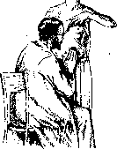
FIG. 1.
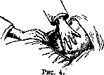
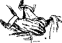
Palpation of the organs of the abdomen, such as the stomach or loops of intestines, as well as the appearance of large tumors, even superficial palpation provides much data for diagnosis. However, detailed and comprehensive familiarization with the state of the abdominal cavity and its organs, as well as their topography, can only be achieved through deep systematic palpation, and for palpating the stomach and intestines, it is necessary to apply the methodical deep sliding palpation according to the method of Glenar-Obraztsov-Gaussman. When proceeding with deep palpation, one must always remember the anatomical relations of the abdominal cavity, the shape, physical properties of the organs and their supporting apparatus, and deviations in topographical relations depending on the patient's constitution, nutritional status, and relaxation of the abdominal muscles—'one must think while palpating and palpate while thinking' (Boas). When palpating organs, one must use their respiratory excursions and examine them according to a strictly defined plan and order, starting with organs more accessible to palpation and moving to less accessible ones. The most acceptable sequence is: S-Romanum, cecum with appendix, terminal part of the ileum (pars caecalis ilei or ileum terminate), stomach with its parts, transverse colon, liver, spleen, duodenum, pancreas, and kidneys. Palpation of organs must be performed according to certain rules, from the strict observance of which its success depends. When palpating the edge of an organ, one should place the ends of the folded fingers of the right hand along this edge, press the abdominal wall slightly, and hold the fingers motionless, causing the subject to breathe deeply with the diaphragm, then the moving organ during breathing slips out from under the fingers and approaches them again, which makes it possible to palpate it and form an idea of its physical properties. In deep palpation, 'based on the principle that one penetrates deeply with the fingertips, carefully moving step by step, as if creeping, one uses the relaxation of the abdominal wall occurring with each exhalation in order to gradually reach the posterior wall or a deeply lying organ. Upon reaching sufficient depth according to the Glenar-Obraztsov principle, the fingertips slide in a direction transverse to the axis of the organ being examined, also using the moment of exhalation for this purpose. The fingers pass across the organ being examined and press it slightly against the posterior abdominal wall, fixing it there. Depending on the direction of the organs, the sliding movements go from top to bottom (stomach, transverse colon) or from inside to outside (cecum, sigmoid flexure), passing into a more or less oblique direction as these organs deviate from the horizontal or vertical course. Thus, sliding movements are performed in the frontal plane of the abdomen in different directions, starting at some distance from one side of the body being palpated and ending when the fingers have moved to the other side; at the same time, the sliding movements are not performed on the skin, but together with it' (Gaussman). Palpation is usually performed with the right hand alone or with the so-called double hand, when the right hand palpates while the left, placed on top, presses on it, or finally with both hands simultaneously (bimanual palpation). If palpating with one hand, the other is used either to press on the abdominal wall away from the field of palpation in order to perceive resistance of the abdominal wall in that place, and consequently relaxation of the abdominal muscles in the area being palpated, or to bring the organ being examined closer to the palpating hand, or finally for palpating the organ between two hands. Palpation of the intestine. Palpation of the S-Romanum is performed from the right, top and inside to the left, down and outside, perpendicular to the axis of the intestine, which in the average is obliquely located in the right iliac fossa on the border between the middle and outer thirds of the line umbilico-iliacae (the line connecting the navel with the anterior superior iliac spine). Palpation is performed with the four fingers folded together and slightly bent or with the ulnar edge of the little finger of the right hand. Having inserted the fingers inside from the presumed position of the intestine and reaching the posterior wall of the abdominal cavity with them, slide along it in the indicated direction, i.e., outward and downward. During this movement, the intestine, being pressed against the posterior wall, first slides along it, but then (since its mesentery has a certain width and is stretched) the intestine slips out from under the fingers as the hand continues to move, and at this moment the palpating fingers go around the intestine almost along its entire periphery, i.e., palpate the intestine (Fig. 2). Using the described technique, it is possible to palpate the S-Romanum in 90-95 out of 100 people. Only in cases of excessive abdominal distension and in obese individuals is the S-Romanum not palpable. If we do not find the S-Romanum in its usual place, this means that due to its long mesentery and excessive mobility, it is located somewhere else in the abdomen, most often closer to the navel and to the right. After performing deep palpation of the lower umbilical and suprapubic area according to the rules, we soon find it. In normal conditions, the S-Romanum is palpated over a length of 20-25 cm in the form of a smooth, moderately dense cylinder, as thick as a thumb, painless on palpation, not rumbling, peristalsizing very slowly and rarely. It can be displaced to one side or the other within 3-5 cm. When palpating the cecum, the technique is the same, only the direction in which palpation is performed is different. Since the cecum in the average lies on the border between the middle and outer thirds of lin. umbilico-iliacae (5 cm from the iliac spine), palpation is performed along this line or parallel to it (Fig. 3). On palpation, not only the cecal sac is found, but also a part of the ascending colon, about 10-12 cm, i.e., that part of the large intestine which in clinical practice has received the name 'typhlon'. The cecum in normal conditions is palpated in 80-85% in the form of a moderately tense, somewhat expanding downward cylinder with a rounded bottom, 2-3 cm in diameter, which gives a rumbling sound when pressed on it. Palpation of the intestine does not cause pain and allows one to ascertain some passive mobility of the intestine within 2-3 cm. The lower edge of the cecal sac is located in men 1/2 cm above the interiliac line, in women 1-1/2 cm below it. During further palpatory examination of the right iliac region, it is possible in 80-85% to palpate over a length of 15-20 cm that segment of the ileum which rises from below and left from the pelvis to connect with the large intestine—ileum terminate. The direction of this segment is mostly from below and left upward and to the right, so palpation is performed almost parallel to lin. umbilico-iliaca, but below it. The terminal segment of the ileum is palpated in the depth of the right iliac fossa in the form of a soft, easily peristaltizing, passively mobile cylinder, as thick as a little finger or a thick pencil, which gives a clear rumbling sound when slipping out from under the fingers. Having found the terminal segment of the ileum, one can attempt to find the vermiform appendix above or below it. The finding of the appendix is facilitated if, following Gaussman's advice, one first palpates the m. psoatis, the finding of which is facilitated by the subject slightly raising the right leg extended, and palpate the appendix on the contracted belly of the muscle. The appendix is palpated in 20-25% of all cases in the form of a thin cylinder, as thick as a goose feather, painless, which does not change its consistency under the hands and does not rumble. However, after palpating this cylinder above or below the ileum, one cannot yet be sure that one is palpating the vermiform appendix, since a duplication of the mesentery and a lymphatic bundle can imitate the appendix. In an inflammatory condition of the appendix, due to its thickening, distortion, fixation, and hardening, confidence in the palpatory finding of the appendix is significantly greater. Palpation of the cecum, terminal segment of the ileum, and vermiform appendix is performed with the right hand with the four fingers folded together and slightly bent at their joints. When the abdominal muscles are tense, in order to cause their relaxation in the area of palpation, it is useful to press with the radial edge of the left hand in the umbilical region (Fig. 4). Palpation of the transverse colon is performed with the right hand alone with the four fingers folded together and slightly bent, or with both hands (bilateral palpation). Since the position of the transverse colon is not constant, in order to know where to look for it, it is useful before its palpation to determine by means of 'Obraztsov's percussive palpation' the position', 'Figure 5', 'Figure 6'.

Palpation of the transverse colon is performed by placing the right hand on the right hypochondrium and the left hand on the left hypochondrium, with the fingers directed toward the midline. The patient is asked to take a deep breath, and during exhalation, the hands are pressed toward each other, sliding along the colon. This method allows the examiner to feel the entire length of the transverse colon, which can normally be palpated as a soft, mobile, slightly tubular structure. In pathological conditions, such as tumors or inflammatory processes, the transverse colon may become thickened, fixed, or painful on palpation. The technique requires careful attention to the patient's respiratory movements and the ability to adjust the pressure according to the resistance felt. The transverse colon is usually not palpable in its entirety in healthy individuals due to its mobility and position beneath the greater omentum, but parts of it may be palpable, especially in thin individuals. The normal width of the transverse colon is about 4-6 cm, and it should not be tender on palpation. If tenderness is present, it may indicate pathological processes such as colitis, tumors, or adhesions. The palpation should be performed systematically, starting from the hepatic flexure and moving toward the splenic flexure, paying attention to any irregularities in the contour, consistency, or mobility of the colon. The transverse colon may be displaced downward in cases of ptosis or upward in cases of abdominal distension. The technique of bimanual palpation allows the examiner to better appreciate the depth and mobility of the colon by using both hands to create a 'sandwich' effect, with one hand on each side of the abdominal wall. This is particularly useful for detecting deep-seated lesions or for assessing the mobility of the colon in relation to surrounding structures. The transverse colon should be palpated in relation to other abdominal organs, such as the liver and spleen, to determine if there is any displacement or compression. In cases of suspected pathological conditions, the palpation of the transverse colon should be combined with other diagnostic methods, such as percussion and auscultation, to obtain a comprehensive assessment of the abdominal cavity. The normal transverse colon should not produce any abnormal sounds on auscultation, and its peristaltic movements should be regular and not exaggerated. If the transverse colon is palpable as a tense, board-like structure, it may indicate the presence of fecal impaction or a pathological mass. The technique of palpation should be adjusted according to the patient's body habitus, as obese individuals may require deeper palpation, while thin individuals may require more gentle pressure. The transverse colon may be difficult to palpate in cases of excessive gas in the intestines or in the presence of ascites. In such cases, the examiner may need to use alternative positions, such as having the patient lie on their side, to facilitate palpation. The transverse colon should be palpated in a systematic manner, following the anatomical course from right to left, and any abnormalities should be documented with respect to their location, size, consistency, and mobility. The normal transverse colon should not be fixed to the abdominal wall, and its mobility should be assessed by moving it gently with the fingertips. If the transverse colon is fixed, it may indicate the presence of adhesions or inflammatory processes involving the peritoneum. The palpation of the transverse colon should be performed with clean hands and with the patient's consent, and the examiner should explain the procedure to the patient to minimize discomfort and anxiety. The transverse colon may be palpated in different positions, such as supine, lateral, or knee-chest, to optimize the examination and to detect any positional changes in the colon. The normal transverse colon should not be tender on deep palpation, and if tenderness is present, it may indicate pathological conditions such as colitis, diverticulitis, or tumors. The palpation of the transverse colon should be performed as part of a complete abdominal examination, which includes inspection, auscultation, percussion, and palpation of all abdominal organs. The transverse colon may be palpable in its entirety in cases of abdominal distension or in thin individuals, and its normal contour should be smooth and regular. If the transverse colon is palpable as a nodular or irregular structure, it may indicate the presence of pathological conditions such as tumors or inflammatory polyps. The technique of palpation should be adjusted according to the patient's symptoms and the suspected diagnosis, and the examiner should be prepared to modify the technique as needed to obtain the most accurate information. The transverse colon should be palpated in relation to the surrounding structures, such as the greater omentum and the anterior abdominal wall, to determine if there is any displacement or compression. The normal transverse colon should not be palpable as a separate structure in most individuals, but parts of it may be palpable, especially in thin individuals. The palpation of the transverse colon should be performed systematically, following the anatomical course from right to left, and any abnormalities should be documented with respect to their location, size, consistency, and mobility. The normal transverse colon should not be fixed to the abdominal wall, and its mobility should be assessed by moving it gently with the fingertips. If the transverse colon is fixed, it may indicate the presence of adhesions or inflammatory processes involving the peritoneum. The palpation of the transverse colon should be performed with clean hands and with the patient's consent, and the examiner should explain the procedure to the patient to minimize discomfort and anxiety. The transverse colon may be palpated in different positions, such as supine, lateral, or knee-chest, to optimize the examination and to detect any positional changes in the colon. The normal transverse colon should not be tender on deep palpation, and if tenderness is present, it may indicate pathological conditions such as colitis, diverticulitis, or tumors. The palpation of the transverse colon should be performed as part of a complete abdominal examination, which includes inspection, auscultation, percussion, and palpation of all abdominal organs. The transverse colon may be palpable in its entirety in cases of abdominal distension or in thin individuals, and its normal contour should be smooth and regular. If the transverse colon is palpable as a nodular or irregular structure, it may indicate the presence of pathological conditions such as tumors or inflammatory polyps. The technique of palpation should be adjusted according to the patient's symptoms and the suspected diagnosis, and the examiner should be prepared to modify the technique as needed to obtain the most accurate information. The transverse colon should be palpated in relation to the surrounding structures, such as the greater omentum and the anterior abdominal wall, to determine if there is any displacement or compression.
Palpation of the liver is an essential part of the abdominal examination, providing valuable information about its size, consistency, and surface. The normal liver is not palpable in most individuals, but in thin people or during deep inspiration, the lower edge may be felt just below the costal margin. To palpate the liver, the examiner places the right hand flat on the right upper quadrant, with the fingers pointing toward the costal margin. The patient is asked to take a deep breath, and during inspiration, the diaphragm descends, bringing the liver downward. If the liver is enlarged, it may be felt as it moves downward beneath the fingertips. The liver should be palpated systematically, starting from the right hypochondrium and moving toward the epigastrium. The normal liver edge is smooth, firm, and not tender. If the liver is palpable as a hard, nodular structure, it may indicate cirrhosis or tumor. If it is tender, it may suggest hepatitis or other inflammatory conditions. The liver should be assessed for its consistency, which can be described as soft, firm, or hard. A soft liver may indicate fatty infiltration, while a hard liver may suggest cirrhosis or tumor. The surface of the liver should be smooth in normal conditions, but irregularities may indicate pathological processes such as tumors or abscesses. The liver should also be assessed for its mobility, which can be evaluated by moving it gently with the fingertips. A fixed liver may indicate adhesions or invasion by tumor. The palpation of the liver should be performed with the patient in supine position, with the knees slightly flexed to relax the abdominal muscles. The examiner should use the fingertips of both hands to palpate the liver, with one hand on each side of the abdominal wall. This technique allows the examiner to better appreciate the depth and mobility of the liver. The liver should be palpated in relation to the surrounding structures, such as the gallbladder and the right kidney, to determine if there is any displacement or compression. The normal liver should not be palpable as a separate structure in most individuals, but its lower edge may be felt during deep inspiration in thin people. The palpation of the liver should be performed systematically, following the anatomical course from right to left, and any abnormalities should be documented with respect to their location, size, consistency, and mobility. The normal liver should not be fixed to the abdominal wall, and its mobility should be assessed by moving it gently with the fingertips. If the liver is fixed, it may indicate the presence of adhesions or inflammatory processes involving the peritoneum. The palpation of the liver should be performed with clean hands and with the patient's consent, and the examiner should explain the procedure to the patient to minimize discomfort and anxiety. The liver may be palpated in different positions, such as supine, lateral, or knee-chest, to optimize the examination and to detect any positional changes in the liver. The normal liver should not be tender on deep palpation, and if tenderness is present, it may indicate pathological conditions such as hepatitis, abscess, or tumor. The palpation of the liver should be performed as part of a complete abdominal examination, which includes inspection, auscultation, percussion, and palpation of all abdominal organs. The liver may be palpable in its entirety in cases of hepatomegaly, and its normal contour should be smooth and regular. If the liver is palpable as a nodular or irregular structure, it may indicate the presence of pathological conditions such as cirrhosis or tumor. The technique of palpation should be adjusted according to the patient's symptoms and the suspected diagnosis, and the examiner should be prepared to modify the technique as needed to obtain the most accurate information. The liver should be palpated in relation to the surrounding structures, such as the gallbladder and the right kidney, to determine if there is any displacement or compression. The normal liver should not be palpable as a separate structure in most individuals, but its lower edge may be felt during deep inspiration in thin people. The palpation of the liver should be performed systematically, following the anatomical course from right to left, and any abnormalities should be documented with respect to their location, size, consistency, and mobility. The normal liver should not be fixed to the abdominal wall, and its mobility should be assessed by moving it gently with the fingertips. If the liver is fixed, it may indicate the presence of adhesions or inflammatory processes involving the peritoneum. The palpation of the liver should be performed with clean hands and with the patient's consent, and the examiner should explain the procedure to the patient to minimize discomfort and anxiety. The liver may be palpated in different positions, such as supine, lateral, or knee-chest, to optimize the examination and to detect any positional changes in the liver. The normal liver should not be tender on deep palpation, and if tenderness is present, it may indicate pathological conditions such as hepatitis, abscess, or tumor. The palpation of the liver should be performed as part of a complete abdominal examination, which includes inspection, auscultation, percussion, and palpation of all abdominal organs. The liver may be palpable in its entirety in cases of hepatomegaly, and its normal contour should be smooth and regular. If the liver is palpable as a nodular or irregular structure, it may indicate the presence of pathological conditions such as cirrhosis or tumor. The technique of palpation should be adjusted according to the patient's symptoms and the suspected diagnosis, and the examiner should be prepared to modify the technique as needed to obtain the most accurate information. The liver should be palpated in relation to the surrounding structures, such as the gallbladder and the right kidney, to determine if there is any displacement or compression.
of the lower border of the stomach and to conduct the research, moving downward by 2-3 cm. Palpation is performed as follows: placing the right hand, resp. both hands, with bent fingers (fig. 5 and 6) along the white line and pushing the skin slightly upward, gradually immerse the hand, taking advantage of the relaxation of the abdominal press during exhalation, until contact is made with the posterior wall of the abdomen. Upon reaching the posterior wall, slide downward along it, and in case the intestine is palpable, find it in the form of an arc-shaped and transverse cylinder of moderate density, 2-21/2 cm thick, easily movable up and down, but not rumbling and painless. If the intestine is not found at the specified location, then using the same technique, the abdominal cavity is examined below and in the lateral flank areas, appropriately changing the position of the palpating hands. The transverse colon is palpable in 60-70% of all cases under normal conditions. Besides the indicated segments of the intestines, in rare cases it is possible to palpate the horizontal parts of the duodenum and the flexures of the colon, as well as any loop of small intestine that has accidentally fallen into the iliac fossae. Generally, however, the small intestines, being located very deeply and excessively mobile and having thin walls, are not amenable to palpation, since due to these circumstances they cannot be pressed against the posterior wall of the abdominal cavity, without which it is impossible to palpate a segment of the intestine in a normal state. Digital palpation of the rectum is performed with the patient in the knee-elbow position, after preliminary cleansing of the rectum with an enema; a greased index finger is inserted into it and is slowly and carefully advanced to the maximum possible depth. In cases of extreme sensitivity of the patient, with fissures and inflammatory processes, it is necessary to anesthetize the sphincter part and the ampulla of the rectum by inserting a tampon soaked in a 1-2% solution of cocaine before introducing the finger. After passing the sphincter, the finger encounters in front in the male the prostate, and in the female the vaginal part of the uterus; along it the finger should be advanced upward, bypass the sacrococcygeal fold and, if possible, reach the terminal fold (plica terminalis recti), which covers the entrance to the rectosigmoid and is located 11-13 cm above the anus. After examining the anterior wall with the finger, the finger is turned backward and the posterior sacral and then the lateral walls are palpated, everywhere forming an impression based on palpation of the condition of the mucous membrane (ulcers, papillomas, polyps, varicose nodes, swelling and swelling of the mucous membrane, scar constrictions, neoplasms, etc.), as well as the condition of the cellular tissue surrounding the rectum, Douglas' space, 5C2-. the prostate, the uterus with its appendages, and the pelvic bones. Palpation of the stomach-see Stomach-palpation. Palpation of the liver and gallbladder is performed both with the subject standing and lying on his back. In some cases, palpation of the liver is facilitated by the patient taking a diagonal position on the left side-at this time the liver, due to gravity, protrudes from under the ribs; then it is easier to palpate its lower-anterior edge. Palpation of the liver and gallbladder is performed according to the general rules of Palpation, with the greatest attention being paid to the lower-anterior edge of the liver, the properties of which determine the physical condition of the liver itself, its position and shape. In many cases (especially with prolapse or enlargement of the organ), besides the edge of the liver, which can often be followed from the left hypochondrium to the right by palpation, it is also possible to palpate the upper-anterior and lower-posterior surfaces. The researcher sits to the right next to the bed on a chair or stool facing the subject, places the palm and 4 fingers of the left hand on the right lumbar region, and with the thumb of the left hand presses from the side and in front on the costal arch, which facilitates the approach of the liver to the palpating right hand, and hinders the displacement of the thoracic
The palm of the right hand is placed flat with slightly bent fingers on the abdomen of the patient, immediately below the costal arch, along the sides of the nipple line, and slight depression of the abdominal wall is made with the fingertips (fig. 7). After this hand position is established, the patient is asked to take a deep breath, and the liver, descending, first approaches the fingers, then bypasses them, and finally slips out from under them, i.e., it is palpated. The examiner's hand remains stationary throughout; the maneuver is repeated several times. Since the position of the liver edge may vary depending on various circumstances, it is useful to first determine the position of the lower edge of the liver by percussion in order to know where to place the fingers of the palpating hand. The edge of the normal liver, palpated at the end of a deep breath 1-2 cm below the costal arch, feels soft, sharp, easily turnable, and not tender. According to Obraztsov, the normal liver is palpable in 88% of cases. In cases of significant abdominal distension, it is helpful to perform the examination fasting, after administering a laxative, and in cases of large fluid accumulations in the abdominal cavity, it is necessary to first drain the fluid by paracentesis. The gallbladder, because it is soft and protrudes very little from under the edge of the liver, is not palpable under normal conditions. However, when the bladder is enlarged (hydrops, filling with stones, cancer, etc.), it becomes accessible to palpation. Palpation of the bladder is performed in the same position as palpation of the liver. After finding the edge of the liver, immediately below it, at the outer edge of the right rectus muscle, palpation of the gallbladder is performed according to the rules for palpating the liver itself. Palpatorily, it presents as a pear-shaped body of varying size, density, and tenderness, depending on the nature of the pathological process in it or in the surrounding organs (e.g., a soft elastic bladder in obstruction of the common bile duct - a sign of Terrier-Courvoisier, a dense nodular bladder in neoplasms in its wall or overfilling with stones, and in inflammation of the wall, etc.). An enlarged bladder is mobile during respiration and performs lateral pendulum-like movements. Mobility of the bladder is lost in inflammation of the peritoneum covering it-pericholecystitis. The described technique for palpation of the liver and gallbladder appears to be the simplest, most convenient, and gives the best results. The difficulty of liver palpation and at the same time the awareness that only it can provide valuable diagnostic data led to seeking the best method of palpation. Various techniques have been proposed, which mainly come down to different positions of the examiner's hands (Glenar's method-procede du pouce, Mathieu's method, Chauffard's method, Chiray's method for the gallbladder), or to changing the examiner's position relative to the patient [e.g., examining the liver and bladder by embracing the patient bent forward from behind-Chiray's method, palpating the edge of the liver with two hands, with their fingertips simultaneously touching, placing one above and the other below-Gilbert's method, etc.]. Palpation of the liver and gallbladder by all these methods has no advantages. The issue is not in the variety of techniques, but in the experience of the examiner and the systematic conduct of a plan for examining the abdominal cavity as a whole. Palpation of the spleen is performed with the patient lying on their back or in the right lateral diagonal position. The examiner places the left hand flat on the left half of the chest in the area of the VII and X ribs and presses slightly on it, thereby fixing the left half of the chest and increasing the respiratory excursions of the left dome of the diaphragm. The right hand with slightly bent fingers is placed flat immediately below the costal margin along the line representing the continuation of the X rib, and slightly depresses the abdominal wall, after which the patient is asked to take a deep breath; the edge of the spleen approaches the fingers, bypasses them, and slips out, i.e., it is palpated. This maneuver is performed several times, with the palpating hand remaining stationary throughout. If the edge of the spleen is not found immediately below the costal arch, especially when a vague resistance is felt as if from some body in this location, the fingers of the right hand are moved 2-3 cm lower or slightly to the side, and the patient is asked to take deep breaths. Sometimes palpation is facilitated by the left hand, placed under the patient, pressing from behind on the lower ribs. A normal, non-enlarged spleen is not palpable; it can only be palpated in cases of significant enteroptosis. If the spleen is palpable at all, this means it is enlarged. After palpating the spleen, an attempt is made to determine its consistency, tenderness, and the condition of its edge and surface. Palpation of the pancreas is extremely difficult due to the deep position and soft consistency of the organ. Only the patient's emaciation, relaxation of the abdominal press, and ptosis of the viscera allow palpation of the normal gland in 4-5% of women and 1-2% of men. A hardened pancreatic gland in cirrhosis, neoplasm, or with a cyst in it is palpated much more easily. Palpation of the pancreatic gland should be performed in the morning fasting, after administering a laxative, and with an empty stomach. It is necessary to first palpate the greater curvature of the stomach, determine the position of the pylorus, and palpate the right flexure of the transverse colon. It is desirable to also palpate the lower horizontal part of the duodenum. Then the location where to look for the head of the pancreas by palpation will be determined; it is still easier to palpate than the body of the gland due to its larger size and more frequent hardening. Palpation is performed according to the rules of deep sliding palpation, usually above the right part of the greater curvature of the stomach. Conclusions regarding the palpability of the gland must be made with extreme caution-one can easily mistake part of the stomach, part of the transverse colon, a packet of lymph nodes, glands, etc., for the gland.

Figure 8.
(of the right kidney) or the spleen (for the left kidney). When palpating the right kidney, the examiner places the right hand with slightly bent fingers on the patient's abdomen along the outer edge of the rectus muscle, so that the fingertips are 2-3 cm below the costal margin, and places the left hand under the lumbar region (Fig. 8). At each expiration, the physician tries to push the fingertips of the right hand deeper until they touch the posterior wall of the abdominal cavity and through it to the left hand. Then, with lifting movements of the left hand through the thickness of the lumbar muscles, the kidney lying on them is raised and brought under the fingertips of the right hand; at this time the patient should take a shallow breath. If the kidney is palpable, it is either entirely or only its lower rounded pole comes under the fingertips of the right hand, which grasps it, increasing the pressure backward. Then, without reducing the pressure and without moving the hands apart, the fingertips of the right hand are made to slide downward; the kidney, being fixed, attempts to slip downward when the right hand tries to displace it, and at this moment the examiner forms a final impression of its size, shape, consistency, and degree of mobility. If the kidney is highly mobile or wandering, it should be grasped with the right hand and the limits of its mobility established by displacing it sideways, upward, and downward. It is also useful for determining the nature of kidney enlargement to apply the method of ballottement proposed by Guyon. After palpating the kidney between the two hands, a series of thrusts is applied to the lumbar region by jerky flexions of the fingers of the hand placed behind, which are transmitted through the kidney to the other hand; this allows for better judgment of its tenderness, consistency, contents of a cystic tumor of the kidney, etc. The palpation of abdominal tumors, strictly speaking, gave rise to the detailed and systematic development of the methodical P., as this method is at present perhaps almost the only one for their diagnosis. By means of palpation, the presence of a tumor is detected, its belonging to the abdominal cavity and relation to neighboring organs is determined, the nature of the tumor is established, and an impression is formed of the possibility of its surgical removal. With the introduction of X-ray examination into clinical practice, palpation of a tumor is often performed under X-ray control. After detecting a tumor, one should first establish its localization, i.e., whether it is located in the abdominal wall itself, inside the abdominal cavity, or behind the peritoneum; having established the presence of a tumor in the abdominal cavity, it is necessary to accurately determine its belonging to one or another organ and its relation to neighboring organs, its mobility, nature, as well as whether there is an inflammatory process in the peritoneum around it. Tumors of the abdominal wall, in contrast to intra-abdominal and retroperitoneal tumors, are located more superficially, are easily detected by inspection, are clearly palpable, and when the abdominal muscles are tensed, they become fixed and less palpable, but when the muscles contract, they do not completely disappear from the field of P., as happens with intra-abdominal tumors; during respiratory excursions they move in the anteroposterior direction with protrusion of the abdominal wall during inspiration and its retraction during expiration. Tumors located behind the peritoneum are characterized by rather close contact with the posterior wall of the abdominal cavity, are little mobile during respiration and less mobile during P., and mainly they are always covered by the intestines or stomach. An exception in terms of mobility are small kidney tumors and tumors of the tail of the pancreas, which, despite their retroperitoneal location, are often sufficiently mobile. Even greater respiratory and passive mobility is characteristic of tumors located intra-abdominally; the closer they are located to the diaphragm, the greater their mobility from top to bottom during inspiration. Depending on the width or length of the covering ligaments of the organ to which the tumor belongs, its passive mobility is found. Here, however, it should be noted that sometimes tumors of normally firmly fixed parts of the digestive tract acquire greater mobility due to congenital excessive length of the mesentery and ligaments or stretching of the supporting apparatus during tumor growth; for example, tumors of the pylorus of the stomach or tumors of the cecum often have great mobility. Intra-abdominal tumors lose both respiratory and passive mobility if inflammation of the peritoneum develops around them, after which dense adhesions of the tumor with the surrounding organs are observed. The detection of a tumor and the establishment of its intra-abdominal localization are the first step in the diagnostic process. After this, it is necessary to determine the nature of the tumor, which is achieved after a detailed study of its physical properties by palpation, such as its shape, density, elasticity, roughness, presence of fluctuation in it, tenderness, etc., but mainly one has to determine its point of origin and belonging to one or another intra-abdominal organ. The latter becomes possible only after preliminary topographical P. of the entire abdominal cavity and specific determination in the patient of the position and properties of each organ separately. Such a concrete study of topographical relations is necessary because it is impossible to use normal anatomical relations, as due to tumor growth and changes in intra-abdominal pressure they are often disrupted and distorted. Thus, the diagnosis of tumors requires the ability to palpate skillfully and detailed knowledge of the physical properties of the abdominal cavity and its organs both in normal and in various pathological conditions. That is why every physician dealing with diseases of the abdominal organs—whether internist, surgeon, gynecologist, or urologist—should study the technique of palpation with all diligence and perseverance.
Related articles
Cite this page
“Palpation.” Soviet Medical Encyclopedia. English translation of Bolshaya Meditsinskaya Entsiklopediya, 1st ed. (Moscow, 1928–1936), ed. N. A. Semashko. https://sovietmedicalencyclopedia.pages.dev/article/palpation/