Onychia
Historical document, translated for reference. It reflects medical knowledge of the 1920s–30s and is not medical advice.
Summary
Onychia refers to various nail changes resulting from pathological conditions of the nail matrix and nail bed. It often accompanies inflammation of the nail folds and can cause morphological changes, deformations, or even loss of nail plates.
Encyclopedia article (1928–1936)
ONYCHIA (onyxis, onychosis, onychia), a term denoting various changes in the nails that occur as a result of various pathological conditions of the matrix and nail bed (see Nail). O. is often accompanied by acute or subacute inflammation of the nail folds (paronychia, s. perionyxis)—lateral (paronychia lateralis) or posterior (p. posterior). Paronychia may be primary or secondary. By disrupting the nutrition of the nails, especially when the matrix is affected, paronychia causes morphological changes, deformations, and even loss of the nail plates. Vulgar inflammatory paronychia, sometimes complicated by suppuration, is often a consequence of acute pyogenic dermatitis. Primary paronychia can also occur as a result of the deposition of specific products around the nail plate (syphilitic, lepromatous and other infiltrate, tuberculous ulcer, etc.). Secondary paronychia develops due to improper growth of the nail plate in the transverse direction and the ingrowth of the nail (see Nail). It is often extremely difficult to distinguish banal paronychia from subungual panaritium. The latter is characterized by unbearable drilling pain. - The so-called nail-eater (panaritium subepidermoidale) represents a streptococcal impetigo blister that appears at the site of a crack or hangnail and tends to spread around the nail. O. and paronychia are more often observed on the upper extremities than on the lower ones. Scleronychia, according to Unna, is a nail condition in which the nail plates are thickened, hard, rough, covered with bumps or depressions, often longitudinally grooved, opaque, and yellowish-gray in color. The lunula is usually absent. The nail bed is unchanged. Apparently, this is a special disease of the nail matrix with a specific change in nail cells. According to Heller, scleronychia is a symptom observed in eczemas, onychogryphosis, syphilis (Strandberg), or a congenital dyskeratosis. - Platonychia (Heller), the transformation of a convex nail plate into a flat horny disk, is presumably a congenital lesion and sometimes occurs in combination with koilonychia. Waelsch observed the condition in 3 generations (grandfather, some children, 2 grandsons). Due to the presence of horny tissue under the nail, the flat nail plate was significantly thinned and cracked at the edges. Schleich observed a 45-year-old man with onychogryphosis of all toes and platonychia of all fingers, which had existed since childhood. Heller described professional platonychia of one or several fingers in people who regularly knead dough or porcelain glaze. - Koilonychia (spoon-shaped nails) is characterized by thinning of the nail substance with the formation of a depression in its middle part, as a result of which its surface becomes concave and the lateral edges are elevated, resembling the shape of a spoon (see separate table, figure 3). Usually one or several fingernails are affected, less often all nails are affected. Koilonychia may be congenital or acquired. By
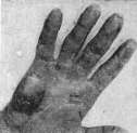
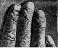
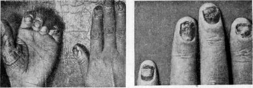
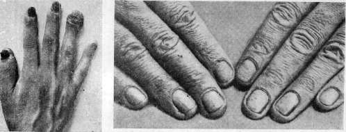
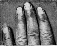
Figure 1. Callus on a violist from the fingerboard and scab. Figure 2. Callus on a cork-stopper worker in glass
production. FIG. 3. Koilonychia (with accompanying eczema)! Figure 4. Ulcerated syphilitic lesions of the nail fold: Figure 5. Onychomycosis trichophytina. Figure 6. Onychomycosis blastomycetica. Figure 7. Onychia syphilitica sicca. Fig. 8. Leuconychia totalis. To the article Onychia. According to Geller, spoon-shaped nails may be observed as a symptom of localized eczema of the nails. In one case, koilonychia developed during an attack of lichen planus. Koilonychia is frequently observed in rheumatism and acanthosis nigricans. Sometimes koilonychia is combined with leuconychia (Joseph). Apparently due to a changed ratio in pressure, ischemia of the vessels of the nail bed occurs, simulating leuconychia. Both sexes are affected to the same degree. In the pathogenesis, subungual hyperkeratosis often plays a certain role. Siemens was able to establish a familial predisposition. He found koilonychia once in 3, once in 2 generations, and once in 2 brothers. Wilson observed koilonychia as a familial disease in 7 members of a family in 3 generations. Geller observed congenital koilonychia in 2 brothers, and Riecke-in a mother and 4 children. Leuconychia (leucopathia, s. canities unguium), changes in the nails in the form of either single or multiple white spots of various sizes, or longitudinal or transverse stripes. The spots usually appear near the lunula and move toward the free edge as the nail grows. Three forms are distinguished: a) punctate leuconychia (leuconychia punctata), usually observed in children and adolescents, as well as in persons who traumatize the nails too assiduously by manicuring; b) diffuse whitening of the entire nail plate (leuconychia totalis) (see separate table, figure 8); c) striped (leuconychia striata), in which transverse white stripes alternate with normal areas. One or several nails are affected. White spots apparently appear due to the penetration of air bubbles between insufficiently keratinized nail cells (Heller). Leuconychia is often a consequence of severe general illness, nervous shock, neuritis, arsenic poisoning, etc. A slight injury to the nail matrix is the most frequent cause. The infiltration of certain parts of the nail plate with air is encountered in various nail diseases (psoriasis, onychogryphosis) as a secondary symptom. This, so-called false leuconychia (leuconychia spuria), is almost pathognomonic for trichophytosis (leuconychia trichophytica). Leuconychia has also been described as a professional sign from the effect of pickle brine. True leuconychia, affecting the entire nail, was observed by Brauer (K. Brauer) in 4 generations as a proper dominant hereditary trait. In this case, 18 persons were affected by leuconychia, and 10 were healthy. With the exception of two (father and son), all affected individuals simultaneously had multiple atheromas on the hairy scalp.-Prevention: avoid traumatic injuries to the nail matrices.-Helconyxis (A. Fournier), insufficient formation of horn substance on limited areas of the nail matrix in the form of oval, cup-shaped depressions, the bottom of which reaches the subungual dermis. A nail studded with such small pits resembles in appearance the surface of a thimble. These nail changes are often observed in eczema, psoriasis, and syphilis.-Dystrophic O., according to Thibierge and Dubreuilh, amounts to a complete cessation of nail growth, which acquire a dull, grayish-yellow coloration. It occurs rather rarely and is usually caused by syphilitic infection. O. syphilitica, a relatively rare disease, developing either in the early secondary period, or most often in the late stages (from 6 to 16 years, according to Geller and others). Several or all nails are affected symmetrically, more often on the hands than on the feet. Initial changes occur in the area of the matrix in the form of whitish, point-like depressions. In pronounced cases, this O. manifests itself as brittleness and fragility of the nail substance. The affected nails are dull, matte, rough, fibrous, and partially detached. Their thinned surface is covered with longitudinal grooves and cracks. Since syphilitic O. has no pathognomonic signs, it is necessary in differential diagnosis to exclude fungal disease, eczema, psoriasis, trophic disorders, etc. Milian described a form of syphilitic O. in the form of 'sand waves,' in which the nail plates of the fingers of the hands are strongly curved along their length and covered with thin, dark, transverse stripes and small grooves arranged in the form of sand waves; simultaneous chronic inflammation of the area of the nail matrices. Under the name onyxis atrophique, the same author described a congenital syphilitic lesion of the nails in the form of almost complete absence of the nail plates, the remains of which are replaced by skin pulp. The affected nails are of soft consistency and covered with transverse grooves. Under the influence of specific treatment, the nails may take on a natural appearance. Pigmented syphilis of the nails (onychia pigmentosa syphilitica). Due to specific damage to the nail matrix, a yellow-brown discoloration of the nail plates occurs. In one case, in an active syphilitic in the 3rd month of infection, persistent, almost black foci appeared in the area of the lunulae of the fingernails. The nail of the left little finger was diffusely colored black. The coloration of the other nails varied in intensity and form (formation of pigment stripes). The application of white mercury precipitate ointment eliminated the disease in three months.-Rag-onychia syphilitica arises from the deposition of a specific infiltrate around the nail plate. Depending on the period of the disease and the evolution of the infiltrate, dry (papular) and ulcerative (ectymatous or gummatous) paronychia are distinguished (see separate table, fig. 4 and 7). Insidious onset, absence of severe pain, and rapid success from specific treatment are the main characteristic features. Onychia scrofulosa, s. tuberculosa (scrofulous O.), a torpid form of ulcerative inflammation of the nail bed, also known under the name malignant O. (onychia maligna); it occurs mostly in atreptic children and young, exhausted subjects and is apparently sometimes of tuberculous origin. The possibility of the development of this form of O. on the basis of trauma, chronic irritations, and contamination is not excluded. The index finger is mainly affected. Chronic course, livid coloration of the surrounding tissues, almost ring-shaped form of the ulcerative process, fungoid growths of the ulcer base characterize this disease from a clinical side. In severe cases, a goblet-shaped thickening of the bones of the fingers (spina ventosa) was observed.-Diagnosis is possible only in connection with the entire sum of clinical, biological, laboratory, and experimental research and therapeutic results (specific therapy ex juvantibus for the purpose of excluding syphilis).-Treatment: scraping and surgical removal of the nail.-Onychia mycotica (onychomycosis) (see separate table, fig. 6). Changes in the nails caused by vegetable fungi are often observed simultaneously with disease of the skin and especially of the hairy part of the head. - Trichophytosis of the nails (trichophytia unguium) is caused by the penetration into the substance of the nail of fungi of the Trichophyton endothrix and ectothrix type (see separate table, fig. 5). The signs of affection are diverse and of a general nature of nutritional and growth disorder. The affected nails are uneven, bumpy, dull, brittle, lack luster, are thickened and longitudinally grooved. The anterior and especially lateral edges are irregularly notched, elevated, and partially detached from the underlying tissue. The nails of the right hand are more often affected, less often all nails on the hands or feet. Isolated trichophytous affection of the nail is observed rather rarely and mainly in adults of both sexes, especially in persons who intensively engage in manicuring and thereby traumatize the nail, which promotes the penetration of fungi. The presence of cases of familial onychomycosis without the participation of the skin and hair in the pathological process, for example in the case of E. K. Vasileva (5 sisters, 1 brother, daughters of one of the sick, as well as relatives on their mother's side), gives reason to think of a familial predisposition. Since analogous changes in the nails are partly characteristic of other skin diseases (eczema, psoriasis, syphilis), recognition of trichophytosis of the nails is hardly possible on clinical symptoms alone.-Diagnosis can be established in the presence of concomitant phenomena and mainly by microscopic examination of scrapings of the nail substance. Cultures of the scraped scales most often give growth of animal fungi (according to Bogrov and Chernogubov, Tr. acuminatum or Tr. crateriforme, according to Truffi-Tr. cerebriforme, according to Jessner-Tr. equinum, etc.). The disease lasts for years. Cases of spontaneous recovery are rare.-Treatment: prolonged local hot baths, soap-salicylic plaster, compresses of a 2-3% solution of caustic potash or depilatory paste for the purpose of softening the nail substance with subsequent removal by mechanical means (filing, etc.). Smearing with tincture of iodine in dilution 1:5-10, compresses of Lysol solution.
Excision and curettage of the affected parts of the nail followed by the application of antiparasitic agents. X-rays and radium have also been used with success. Tinea of the nails (favus unguium) (see separate table for the article on Tinea, fig. 5). Masses of favus fungus form in the thickness of the nail initially as small yellowish spots, scutulae, or the entire nail appears yellowish-colored, cloudy, dull, rough, and thickened. The substance of the nail becomes dry, brittle, and crumbles easily; the disease usually develops as a result of scratching the affected scalp. Isolated localization of favus on the nails is very rarely observed. A familial disease of the nails with favus was observed in 5 children of one family aged 8-16 years, in the mother and a 16-year-old daughter. Mycological examination of nail scrapings is decisive for the diagnosis. The therapy is the same as for trichophytia of the nails.-Blastomycosis of the nails (onychomyco-sis blastomycetica) occurs as a result of the introduction of budding, especially yeast-like fungi. The clinical picture is varied. The nails are thickened, in part insufficiently keratinized, uneven, bumpy, and brittle; colored yellow; often undergo destructive changes, especially in the area of the lunula; purulent inflammation of the nail folds and pustular dermatitis of the interdigital folds are not uncommon. Young subjects are mainly affected. Inoculation from person to person is apparently possible. Villaret described a professional blastomycosis of the nails in a brewer; according to American authors a/3, workers in fruit candy factories in certain localities of CACHI suffer from professional paronychia caused by yeast fungi. The question of the etiological role of these fungi is not fully clarified: Jessner and Kleiner found saprophytic yeast and mold fungi (Soor) in the nail plates and nail beds in 60% of healthy people. Alexander also confirmed saprophytic mold fungi in 20% of all healthy nails examined. Blastomycosis of the nails often heals spontaneously but frequently recurs. In one case, the lesion existed for about 20 years.-Diagnosis is possible only on the basis of biological, laboratory, and experimental studies.-Treatment-as in all onychomycoses.-Cases of onychia and paronychia of diverse clinical types have also been described, in which authors found various fungi of the endo-, oidio-, saccharomycetes type, isolated or in symbiosis, however the etiological role of the latter cannot be considered established. Pyogenic diseases of the nails-staphylococcal and streptococcal-occur in young subjects, especially in children, and may lead to confusion with acrodermatitis continua suppurativa (Hallopeau). One or more fingers of the hands or feet are affected, which is usually preceded by panaritium, impetigo, etc. The process begins with the formation of small abscesses under the nail, which may heal spontaneously. As the process spreads to the matrix, the nail plates become bumpy, loose, often detach and become deformed. Redness, swelling, and the formation of pustules in the area of the nail folds are often observed. Without treatment, the process can last for months, even years.-Treatment: local hot baths, wet dressings with Alibert's water, ointment of white mercury precipitate, etc.-Diphtheria of the nails (O. diph-theritica). Rolleston observed in a 5-year-old child suffering from severe diphtheria of the throat, along with temporary hemiplegia and aphasia, ulcerative lesions of the terminal phalanges, causing partial destruction and deformation of the nails. Cultures of diphtheria bacilli were obtained from the ulcer discharge. Injections of diphtheria antitoxin brought rapid recovery. Onychia caused by animal parasites is observed almost exclusively in so-called Norwegian scabies. All nails of the fingers of the hands and feet, especially the surrounding parts, may be affected. The nails are usually thickened, uneven, claw-like curved, yellow-brown. Thick hyperkeratotic masses around the nails, abundant in mites, can completely cover the nail plates and even lead to their atrophy.-Treatment: antiscabies and keratolytic agents. Lit ..'Vasileva E., On the doctrine of familial onychomycoses, Ven. and Dermatol., 1928, № 3; Mashkilleison L., Platonychia areata (psoriatic?) acuta, ibid., 1928, № 12; Feldman V. and Per M., On the question of Norwegian or crustous scabies, ibid., 1927, № 2; Heller J., Die Krankhelten der Nagel (Hndb. d. Haut-u. Geschlechts-krankneiten, hrsg. v. J. Jadassohn, B. XIII, B., 1927, lit.); Martinet J., L'etiologie des leucony-cnies partielles, P., 1920; Meyer E., Beziebungen der Tuberkulose zur Onychia maligna, B. M. lief.
Related articles
Mentioned in
Cite this page
“Onychia.” Soviet Medical Encyclopedia. English translation of Bolshaya Meditsinskaya Entsiklopediya, 1st ed. (Moscow, 1928–1936), ed. N. A. Semashko. https://sovietmedicalencyclopedia.pages.dev/article/onychia/