Typhus Fever
Historical document, translated for reference. It reflects medical knowledge of the 1920s–30s and is not medical advice.
Summary
Typhus fever is an acute, highly contagious, louse-borne infectious disease characterized by rapid onset, maculopapular rash, and severe neurological and circulatory system involvement. The article details its history, experimental transmission in animals, and laboratory techniques for studying the disease.
Encyclopedia article (1928–1936)
188 Specific prevention and serotherapy of typhus fever
192 Statistics and geographical distribution of typhus fever
226 Typhus fever (typhus exanthematicus) (from Greek typhos - smoke, fog, stupor), an acute, louse-borne infectious disease, highly contagious, cyclic, in which the characteristic symptoms are: rapid onset, maculopapular rash, deep lesions of the central nervous and peripheral circulatory systems, constant fever, resolving between the second and third week by shortened lysis or critically. Synonyms: famine, prison, war typhus. History. The first description of the characteristic signs of typhus fever belongs to Fracastorius (1483-1553). This is followed by the works of Moss, Mantuus, Jordanus, Ambroise Pare and others, who point out the distinctive clinical and epidemiological features of typhus fever. However, until the middle of the 19th century, many physicians, including Virchow, denied the existence of typhus fever as a separate disease. The question was finally resolved thanks to the thorough research of Jenner (1850), Grisinger (1856), Murchison, Wunderlich, Curschmann and others, who proved by analyzing the clinical and epidemiology of typhus fever that it represents a specific disease clearly distinct from other fevers: abdominal and relapsing.
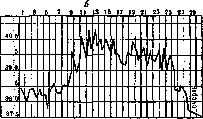
Experimental typhus fever. Mochutkovsky in 1900, by successfully infecting himself with the blood of a typhus patient, provided evidence that the infectious agent in typhus fever is present in the blood. Mochutkovsky's experiment was repeated and confirmed by Otero, Yersin, Vassal and others. In 1909, Charles Nicolle, Cornpte and Conscil in Tunisia reproduced experimental typhus fever in monkeys by infecting them with the blood of typhus patients. Similar results were obtained in 1909 by Anderson and Goldberger in Mexico. In 1910-11, Gavino and Girard established that guinea pigs, like monkeys, can be experimentally infected with typhus blood and give a characteristic picture of the disease. Infection in guinea pigs, as in monkeys, can be maintained indefinitely by passages on fresh animals. The observations of Gavino and Girard were soon confirmed by Charles Nicolle and his collaborators, developed and deepened by subsequent works of numerous authors. Thus, in the hands of researchers, typhus fever proved to be a valuable method for studying the nature of this infection on readily available material. The technique for reproducing experimental typhus fever in monkeys and guinea pigs 1 3 5 7 'J II 13 15 1? ]9 21 23 25 тшт щ т t- .1"-^ y-iv 1 3 5 1 9 11 13 15 17 19 21 23 25 i i "; " 40;Q \Ч \ ' !._Xl-1 -\ 39;° \ N \ "I rW ч IT jo Ww^~~vr^W 1 . - . If- JJ S7J5 1 1 1
typhus material, which is introduced due to its unreliable purity not into the abdomen, but under the skin of animals. Experimentally, typhus fever can be passaged indefinitely on new animals. The material for passages consists of blood, brain or spleen taken at the height of fever. Usually for passages, the animal is killed no later than the 3-4th day of fever, the sterile obtained brain (resp. spleen and other organs) is ground in 0.85% NaCl and injected into the abdomen of fresh animals. During passages, the virus adapts to a given animal species, the incubation period shortens, the fever becomes longer and higher, the percentage of animals susceptible to the passage virus sharply increases, reaching 90% and higher for monkeys and guinea pigs. Of the internal organs of passage animals, the large brain (gray matter) is especially rich in virus. In some typhus strains II 13 15 17 1(| 21 2325 УЧ; ЧК_: m already 7i fig 1 Various forms of typhus fever in guinea pigs: a-asymptomatic b-mild c-typical d-severe [thick line-primary infection, thin-secondary (after 1'/2 months)]. consists in introducing it subcutaneously or intraperitoneally 2-4 cm3 of blood from a typhus patient, taken sterilely from the cubital vein at the height of the disease. The incubation period on average is 8-12 days (ranging from 3 to 31 days), the fever period lasts about 1-I1/» weeks (ranging from 2 to 16 days). The fever is predominantly of the continuous type. It resolves by shortened lysis, and t° sometimes falls below normal and returns to normal only after 1-2 days. No other symptoms of the disease besides fever and slight weight loss are observed in either guinea pigs or monkeys. Experimental typhus fever in these animals can be severe, moderate or mild, as seen in the attached temperature curves (figure 1). Finally, in some cases, experimental infection can occur completely asymptomatically as an afebrile sepsis, as established in 1919 by Ch. Nicolle and Lebailly ("infection inapparente"). The blood of such animals, when inoculated fresh, gives them a typical febrile illness. Monkeys and guinea pigs infected with the blood of typhus patients do not all turn out to be susceptible to the human virus. Insusceptibility in monkeys is sometimes observed in 22% of cases, in guinea pigs - in 44% (Anderson and Goldberger). These figures are relative and depend on the origin and individuality of the typhus virus (Barykin and colleagues). Experimental infection of monkeys and guinea pigs is possible not only with typhus blood from sick people, but also with organs (especially brain and spleen) from cadavers of typhus patients. The brain of a passage guinea pig contains an infectious dose of virus for fresh guinea pigs. Other strains are less virulent, especially strains isolated from humans during sporadic typhus fever or obtained during non-epidemic seasons. Such strains, when serially passed through the guinea pig organism, not only do not increase in virulence, but gradually decrease in activity, becoming asymptomatic experimental infection by the 5th passage or completely losing the ability to infect guinea pigs. Depending on the individual typhus strain being passaged, mortality among passage animals varies, reaching for some strains 18.4% (Otto and Papamarku strain for guinea pigs). Recent research has established that ground squirrels (Spermophilus) are susceptible to the typhus virus, in which experimental infection proceeds the same as in guinea pigs (Lepine, Grünfeld, Serebryakaya and Neyman). The study of experimental typhus fever in monkeys and guinea pigs shows that they are much less susceptible to this infection than humans. Typhus infection in them is reproduced only artificially in laboratory conditions, has a benign course, is accompanied by almost the only symptom - fever, and gives negligible mortality. Experimental typhus fever proceeds even more easily in rabbits, rats and mice. In these animals, as shown by the observations of Ch. Nicolle, Conte and Conscil - for rabbits, Doer, Otto and Winkler - for rats, Ch. Nicolle - for mice, laboratory infection with typhus virus manifests itself in an asymptomatic form of infection. The blood of these animals, taken on the 7-15th day after their laboratory infection, when injected into the abdomen of guinea pigs, causes in the latter a typical febrile attack of experimental typhus fever. Rabbits when infected with typhus brain give a positive Weil-Felix reaction. Attempts to infect sheep, goats,
Fig. 1. Granulomas of typhus fever in the brain. Fig. 1. Low magnification. Granulomatous edema of the vessel wall and rupture of a vessel in the center). Fig. 2. Medium magnification. Granuloma, perivascular edema and scattered glial nodule. Fig. 3. Medium magnification. Hyaline granuloma at the site of vessel rupture. Fig. 4. Medium magnification. Edema around a small vessel. Fig. 5-6. Reticulum cells [reticulocytes]
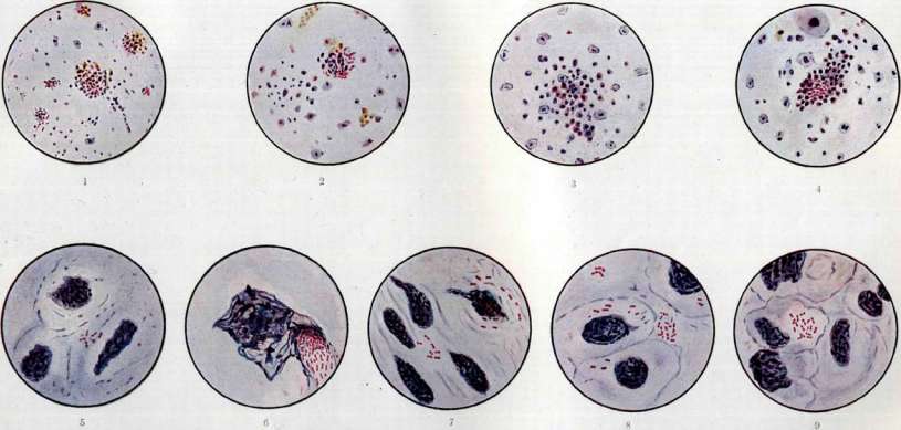
and of the organ* ЯКДвА, ПОГИ&1ПНХ from ci-imioro pyra. Rickettsia Prowazeki lie extracellularly, acr.....ened and in the blood plasma. Figure 1. Cell from the brain. Figure 2. Cell: p)csii* spogЮSHcia spread out, containing Rickettsia Prowazeki, located as if in a bunch. Fig. 3. Smear from the spleen. Rickettsia Prowazeki is scattered and proliferates in cells. Fig. 4. Smear from pftxua choroid. Intracellular and extracellular distribution of Rickettsia Prowazeki. Fig. 5. Smear on glass. Around the pericellular groups and Rickettsia Prowazeki-prosvetdzdni protavlaazhi. To st. Ordinary typhus. Attempts to reproduce typhus in cattle, dogs, and chickens ended in failure. In 1924-25, Selivanov succeeded in reproducing experimental typhus in chickens, ducks, pigeons, and guinea fowl. Confirmation of these data has not yet been forthcoming. As for the pathological anatomy of experimental typhus, it has been carefully studied by a number of authors: Levi, Otto, and Dietrich, Ritz Bauer, Barykin with colleagues, Ceelen, Davydovsky (Levy, Dietrich, Ritz, Bauer, Ceelen) and many others. It essentially does not differ from the pathological anatomy of human typhus. Macroscopic changes characteristic of typhus are not observed in experimental infection. Microscopically, it is a universal granulomatosis of small vessels and capillaries, especially of the central nervous system (brain, Ammon's horn) (see separate table, fig. 1-4). Granulomatosis is detected from the 4-5th day of fever. The involution of granulomas ends on the 2-3rd week of apirexia. The main distinguishing feature of experimental typhus granulomatosis from that of natural infection in humans should be considered the predominance of proliferative phenomena over destructive ones in the former. The severity and prevalence of experimental typhus granulomatosis depend: 1) on the individual typhus strain with which the infection is reproduced, 2) on the dose of infectious material, and 3) on the weight and individuality of the infected animal. According to Derry and Kirschner (Kirschner), in guinea pigs known to be suffering from experimental typhus, granulomatosis is not found in 15-20% of cases, according to Barykin, Kompanets, Zakharov, and Barykina- in 33%. In mild and atypical forms of experimental typhus, granulomas are extremely rare or completely absent. Typhus virus. The experimental reproduction of typhus allowed researchers to study the nature and properties of the typhus virus. Thus, it was established that this virus does not belong to the category of filterable viruses, although it stands on the border with the latter, passing in some rare cases through Chamberland L2 and Berkefeld V and N filters. It was further proven that the typhus virus is extremely unstable. It dies in typhus blood when heated to 55° for 15 minutes (Anderson and Goldberg, da Rocha-Lima), at room temperature t°- within 2-5 days (Olitzky), when frozen- within 2 days (Hamdi), while according to some authors within 15 days (Goldberger and Anderson). 30-80% glycerin, saponin, 5% carbolic acid, 1°/00 sublimate quickly kill the typhus virus. Attempts to microscopically detect this virus or to obtain its culture for decades proved fruitless. A number of researchers, from Haller (Haller, 1868) to Anigstein and Amzel (Anigstein, Amzel, 1927), described the most diverse microbes as the causative agents of typhus: Protozoa, cocci, bacilli, spirochetes, etc. Many of these microbes were isolated from typhus blood and gave immunity reactions with it, but none of them upon subsequent testing proved to be the true causative agent of typhus. Such diversity of findings in typhus is explained by the ease with which typhus blood, due to multiple granulomatosis destroying small vessels, becomes flooded with foreign microbes (Barykin). The search for the causative agent of typhus became more firmly established when it was proven that the transmitter of typhus is the body louse (Ш. Nicolle, 1909). As early as 1910, Ricketts and Wilder (Ricketts, Wilder) found in the intestines of body lice infected with Mexican typhus, oval and rod-shaped formations resembling bacteria of hemorrhagic septicemia. Identical formations were found in the typhus louse by Prowazek (Serbia, 1913), Sergent, Foley, and Vialatte (Tunis, 1914). At the end of 1914, da Rocha-Lima, systematically studying under the microscope body typhus lice collected from Russian prisoners of war in the Cottbus camp, established that these formations occur in enormous quantities in the intestinal epithelium of lice and are a constant characteristic sign of the latter's infection with the typhus virus. Rocha-Lima named them 'Rickettsia Prowazeki' in honor of the two aforementioned scientists who saw them and died of typhus in their research. According to Rocha-Lima's description, Rickettsia Prowazeki have a basic elliptical shape. Very short, almost round, they grow, elongate in length, and then divide into two new elements. A period of complete division is preceded by the formation of a constriction, which for some time still connects the young individuals and gives the entire microbe the appearance of a biscuit or a dumbbell. Along with such forms, rod-shaped forms staining at the poles and long, elongated specimens are found. There is nothing characteristic in their arrangement on the preparations. Individual formations are sometimes arranged in the form of short chains. It is characteristic of the huge number of formations resembling a pure culture of microbes on smears. Rickettsia Prowazeki are on the border of the resolving power of modern microscopes in size. Young forms are about 0.3-0.4 μ in length, dividing forms about 0.6-0.9 μ. Their morphology is visible on the microphotograph [see separate table (st. 199-200), fig. 1 and 2]. Rickettsia Prowazeki are difficult to stain and paler than ordinary microbes. The most reliable method for them is Giemsa's method, with which they are stained a pale pink with a raspberry tint (see separate table, fig. 5-9). They do not stain by Gram, do not have independent movement, when suspended in a 0.85% sodium chloride solution, they exhibit clear Brownian movement. A capsule has not been found in them, although on Giemsa-stained preparations they often appear surrounded by a transparent zone. The most important distinguishing feature is their localization in the epithelium of the midgut (stomach) of the louse. Penetrating this epithelium, they multiply vigorously, stretch it, and then rupture the epithelial cell, which undergoes profound degenerative changes. Together with fragments of cells, Rickettsia Prowazeki enter the intestinal lumen and are excreted outward with feces, as can be seen in the attached microphotograph. Sikora found Rickettsia Prowazeki in the salivary glands of infected lice, 187
TYPHUS FEVER
188 which is disputed by other researchers, Arkwright and Bacot - in the ovaries and eggs of female lice. The most convincing pictures of typhus rickettsiosis in lice are given by serial sections from their intestines, prepared by the Sikora or Weigl method. Rickettsia Prowazeki have been found in the blood and organs of typhus patients and guinea pigs. Thus, Ricketts and Wilder saw them in the blood of sick people, Bessau - in the blood plasma, Kuczynski, Jaffo, Barykin and Afanasyeva - in the endothelium of capillaries of internal organs, Wolbach, Todd and Palfrey - in the endothelium of skin granulomas, etc. Characteristic is the intracellular location of Rickettsia Prowazeki and their accumulation in the form of whole groups [see separate table (art. 183-184), fig. 5-9]. Woodcock considers Rickettsia Prowazeki as non-living formations of the granule, mitochondria or cell decay product type. Such an interpretation is contradicted by the entire body of facts proving the living nature of Rickettsia Prowazeki and their close relationship to the etiology of T. f. Rickettsia Prowazeki, unlike granules and cell decay products, are morphologically and tinctorially uniform. They are not destroyed even by strong acetic acid, boiling distilled water, 2% NaCl solution, ether, acetone, toluene, xylene, gasoline and alcohol (Epstein). They are digested by pancreatin. They are complete antigens. In favor of their etiological significance for T. f., a number of weighty and well-verified facts can be presented: 1) Rickettsia Prowazeki, localized in the intestinal epithelium, are found only in lice removed from typhus patients or fed typhus blood; 2) only lice containing Rickettsia Prowazeki cause experimental T. f. with immunity to subsequent infection with typhus blood; 3) Rickettsia Prowazeki in size, relation to temperature and other agents behave completely identically to the T. f. virus; 4) Rickettsia Prowazeki give all immunity reactions with typhus sera of humans and experimental animals; 5) optimum development of Rickettsia Prowazeki, as well as the T. f. virus, in the body of the body louse lies at about +32° (Rocha-Lima); at t°+23° in lice, development of both Rickettsia Prowazeki and the T. f. virus ceases. On the basis of these facts, the vast majority of researchers consider Rickettsia Prowazeki the causative agent of T. f. Unlike ordinary microbes, Rickettsia Prowazeki are not cultivated on ordinary laboratory media. In 1923, Kuczynski, as well as Krontovsky and Gakh succeeded in maintaining for one generation the T. f. virus in explants of the spleen of a typhus guinea pig by the Carrel method. Further improvement of this technique applied to T. f. in the hands of Sato Kiyoshi, Nigg and Landsteiner, as well as other authors, led to the final resolution of the problem of cultivating typhus virus in tissue explants. The technique of the authors ensures obtaining typhus culture in unlimited number of transfers. The culture completely retains all its virulent, antigenic and immunological properties. Morphologically it represents a culture of Rickettsia Prowazeki multiplying in the cells of the explant. In some cases, detection of these formations in tissue cultures requires special treatment (Sato Kiyoshi) or they cannot be detected at all. Tissue cultures, however, cannot give large quantities of typhus virus and therefore cannot serve as material for experiments on mass production of anti-typhus vaccine or serum. An original technique for mass cultivation of typhus virus was recently proposed by Zilber and Doser, who grow this virus in symbiosis with other microbes, in particular with kefir yeasts. The authors obtained 16 generations of typhus virus, but its biology apparently somewhat suffers, which is expressed by the loss of its antigenic properties (Barykin). It is interesting to note that Rickettsia Prowazeki are not found in yeast cultures. Since the establishment of the significance of these formations in the etiology of T. f., morphologically identical formations have been found in lice collected from both healthy people (Brumpt) and people suffering from trench fever (Jimgmann, Kuczynski) and a number of other infections. At present, it is proven that rickettsioses are widely distributed among blood-sucking and non-blood-sucking insects. They occur in both Insecta and Arachnoidea. However, all these types of rickettsioses ('Rickettsienarten') can be easily distinguished from Rickettsia Prowazeki by the totality of signs, especially by their relation to the intestinal epithelium of the insect (Rocha-Lima). The etiological significance for T. f. of Rickettsia Prowazeki was disputed by some authors. However, the attempt of Weil and Felix, Friedberger, Kuczynski, Fejgin and others to link the etiology of T. f. with the culture of B. proteus X19, specifically agglutinating typhus sera, proved untenable. Against the etiological significance of this culture for T. f. speak: 1) the extremely rare isolation of it from typhus blood, 2) the inability of B. proteus X19 and its variants to cause experimental T. f., 3) its absence in typhus lice, etc. The very ability of X-proteus to enter specific agglutination with typhus sera is considered by most authors as a property of a para-strain acquired as a result of its cohabitation with the virus, and is qualified as paraagglutination. Thus, summarizing the data on the etiology of T. f., it must be concluded that the causative agent of this infection is Rickettsia Prowazeki, although in some exceptional cases they cannot be detected, which may depend on their ability to give filterable invisible forms under certain conditions. Paths and methods of transmission of typhus fever The paths and methods of transmission of T. f. are determined by that basic and indisputable position that the transmitter of T. f. is the body louse (Pediculus vestimenti). This was first experimentally established on monkeys in 1909 by Ch. Nicolle, Comte and Conseil, and later found full confirmation both among experimenters and clinicians and epidemiologists of all countries. In favor of the main and decisive role of lice in the spread of T. f., numerous proofs can be presented. Of these, the most important are the following: 1) a deloused typhus patient ceases to be contagious to those around him, 2) destruction of lice among the population leads to the immediate disappearance of T. f., 3) outbreaks of T. f. are usually associated with cold times of the year, when the population lives more crowded, less frequently has the opportunity to bathe, wears woolen and fur clothing, difficult to delouse, and when due to all the indicated seasonal and domestic conditions, lousiness can reach its greatest dimensions. The main transmitter of T. f. is the body louse, living in underwear and moving to the person 3-4 times a day to feed on his blood. Along with the body louse, the head louse (Pediculus capitis) and possibly the pubic louse (Pediculus pubis) play a much less visible but still indisputable role in the spread of T. f. Transmission of typhus virus by lice can occur in three ways: 1) through the bites of an infected louse of a healthy person (Ch. Nicolle and colleagues); 2) through crushing this louse and rubbing it into the bites and scratches (Ricketts and Wilder and others); 3) through the same rubbing of the excrements of this louse (Ch. Nicolle and colleagues and others). The question of the ability of typhus lice to transmit their infection hereditarily is not fully clarified. Most authors, based on their experiments, deny the hereditary transmission of typhus virus by lice (Ch. Nicolle, Blanc and Conseil, da Rocha-Lima, Barykin and Kompaneyets and others). Rickettsia Prowazeki in young lice-offspring of typhus, according to the research of a number of authors, are not found. A louse that has sucked typhus blood does not become contagious to people immediately, but only after 3-5 days. During this period, active multiplication of the typhus virus occurs in the louse's body and the amount of this virus reaches a dose capable of causing infection in a person (da Rocha-Lima and others). The assumption of Ch. Nicolle and colleagues that the T. f. virus undergoes a special cycle of development in the louse's body, characteristic of protozoa, was not confirmed by further research. A louse infected with T. f. itself becomes ill and usually dies from it. The main changes produced by the typhus virus in the louse's body are concentrated in its intestine, the epithelium of which under the influence of multiplication of this virus (Rickettsia Prowazeki) in it is destroyed. The lifespan of a louse infected with T. f. is shortened due to this infection to 20-25 days, instead of the average lifespan of a healthy louse, which is 30-35 days. A louse infected with T. f. remains contagious until the end of its life.
It has not been determined whether typhus-carrying lice can transmit this infection to healthy insects, nor has the method by which such transmission could occur been identified. According to experiments by Barykin and colleagues, healthy lice placed together with typhus-carrying lice become infected from the latter in 2% of cases. In Weigl's experiments, such infection did not occur. Pavlovsky's observations establish that the sexual act between lice, lasting from 20 to 70 minutes, is accompanied by mixing of feces at the level of the anus of the male and female. It is possible that this act plays a role in the contact transmission of typhus infection among lice. Gakh claims that he was able to cause experimental typhus in guinea pigs by infecting them with lice collected 67-70 days after the last case of typhus in humans. As for other parasites that bite humans, it has already been established on the basis of epidemiological data that these parasites play no role in the development of typhus outbreaks. The season for typhus fever is the cold time of year (late autumn, winter, early spring), when mosquitoes are in a state of hibernation, and the number of fleas and ticks sharply decreases. According to observations of Ch. Nicolas in certain regions of Tunisia, endemic foci of typhus fever, abundant in lice, are almost completely free of human fleas and bedbugs. Measures aimed at the systematic destruction of lice, as world experience shows, are completely sufficient for the complete elimination of typhus fever epidemics and endemic foci. Nevertheless, bedbugs (Barykin and colleagues), fleas and ticks (Perfilev) are found to be susceptible to laboratory infection with the typhus epidemic virus and retain it in their bodies for some time (from 4 to 10-20 days). Their infection can be transmitted to a guinea pig either by bite (rarely) or by injection (rubbing) of crushed insects. Fleas play an important role in the spread of the so-called endemic virus of rat typhus fever (see below). Thus, from the analysis of the data presented, it is obvious that for the spread of epidemic classical typhus fever, the decisive importance remains with the human louse. This follows the basic requirement in the fight against this typhus—delousing the population. The louse becomes infected from an infected person. Therefore, success in the fight against typhus fever depends on the earliest possible recognition and complete identification of all cases of typhus infection in humans, careful delousing of both the patients themselves, who are immediately hospitalized, and those around them, their dwellings, and household items. Along with such clearly defined cases, during typhus fever outbreaks, mild, ambulatory, and atypical forms of the disease that occur without rash are observed, especially frequent among children and in endemic foci. They are sometimes completely inaccessible to clinical recognition. For epidemiologists, they represent a special danger, as they are a source of hidden spread of infection. Finally, cases of so-called typhus virus carriage, occurring without any symptoms, may be encountered. Such virus carriage by healthy people or those who have previously had typhus fever has been established by the work of Ramsin (Ramsin, 1929) in Serbia and independently of him in the USSR by the work of Barykin, Minervin, and Kompaneez, Bernhof, Kuteishchikov, and Dossor (1930). The epidemiological significance of typhus virus carriage (frequency, duration, influence on the biology of the virus), although not yet sufficiently clarified, must probably approach typhus virus carriage to mild forms of infection. All these forms can be recognized in time with the help of the Weil-Felix reaction (see). According to recent observations by a number of authors, repeated administration of the Weil-Felix reaction allows one to assess the degree of hidden and clinically undetectable infection (Durchseuchung) of a typhus focus and therefore gives important indications for its complete sanitation. A deloused typhus patient or carrier ceases to be dangerous to others. Some researchers suggested that besides lice, typhus infection can be spread by the excretions of the patient: sputum, urine, etc. (Zabolotny, Rabinovich, Fridberger and others). By direct experiments of Anderson and Holberger and others, it has been proven that the excretions of typhus patients are not infectious, except for those rare cases that have no epidemiological significance, when the blood of a typhus patient during an operation gets into abrasions and scratches on the skin and mucous membranes of healthy people (surgeons, nursing staff, etc.). Since typhus fever is sometimes complicated by pneumonia with the release of bloody, infectious sputum, it must be disinfected with disinfectant solutions. Caution is also necessary when caring for the oral cavity of a patient with bleeding gums, etc. The early recognition of typhus fever, leading to the hospitalization of a deloused patient, is the most important condition for the successful fight against typhus fever. This condition can be fulfilled through systematic, mass administration of the Weil-Felix reaction organized according to epidemiological indications (see). For epidemiological purposes, according to observations by Barykin and colleagues, this reaction should be administered repeatedly, which allows one to reveal its dynamics and to determine whether it is a matter of a positive non-specific and retrospective reaction or the presence of typhus infection, in whatever hidden form it may occur (virus carriage). With careful selection of cultures of Proteus X19, among which preference should be given to local, serologically adequate strains, and with proper technique, the Weil-Felix reaction provides invaluable service for identifying hidden paths of spread of typhus fever. According to experiments by Barykina and Ge, the Weil-Felix reaction after 1 month occurs only in 63%, after 1 year in 54.5% of those who had typhus fever, sometimes persisting for over 2 years. Thus, in some cases, it is suitable for the retrospective recognition of previously contracted typhus fever. In order to accelerate the Weil-Felix reaction, its modification proposed by Nobl (Nobl) for serodiagnosis of the typhoid group, meningococci, etc., and tested by Bergman and Rabinovich, as well as Mayofis for typhus fever, can be recommended. Bringing the Weil-Felix reaction to primary rural cells that have no laboratories is possible by supplying them with killed cultures of Weil-Felix in the form of typhus fever typhus diagnosticum, prepared by the method of Bien, Sontag, Sacks, Sodor and others. Serological recognition of typhus fever is also successfully carried out with the help of Rickettsia prowazeki as the causative agent of this infection, which gives specific agglutination, fixation of alexin, and tropinization (Epstein and others) with sera from both typhus fever patients and experimental animals. However, the difficulty of obtaining large quantities of Rickettsia prowazeki makes this test little accessible for broad practical purposes. Specific prevention and serotherapy of typhus fever. The questions of specific prevention and serotherapy of typhus fever are still in the study stage. They are closely connected with typhus immunity, the nature and mechanism of which have been studied far from sufficiently. It is known that a successfully contracted typhus fever as a rule leaves behind a firm and long-lasting, though not absolute, immunity to re-infection. In some rare cases, clinicians observe repeated cases of typhus fever (Pletnev, Flerov and others). According to systematic observations by Khinsky, repeated cases of typhus fever occur the more frequently the longer the time that has passed since the first attack of this infection, sometimes reaching 50% of primary cases. Usually, a repeated case of typhus fever runs milder than the primary one (Moroskin). In experimental animals (guinea pigs), the intensity of immunity is also closely related to the severity of the first contracted disease. Nevertheless, even an asymptomatic typhus fever can sometimes create immunity to subsequent infection. Some authors claim that such immunity appears as a result of any, even the mildest form of primary typhus fever in animals. Others prove that immunity to typhus fever in animals that have had it cannot be detected at all, since the brain of these animals contains the virus a long time after their clinical recovery. Sergan distinguishes two stages in the formation of immunity: 1) periode de premunition, occurring immediately after the temperature drop and characterized by incomplete elimination of the virus from the body (non-sterile immunity), and 2) the subsequent period of sterile immunity, when there is no virus in the body. It has been proven that the typhus virus can persist in the brain of a guinea pig after the end of the febrile typhus attack from 24 to 46 days, in rats—from 9 to 68 days. No less important a point for clarifying typhus immunity is the question of the nature of this condition itself. Apparently, the predominant cellular nature of the body's protection against the typhus virus is beyond dispute. The characteristic for typhus fever universal granulomatosis of small vessels speaks for this as a phagocytic reaction to the virus.
But alongside such cellular protection, one cannot disregard the humoral reactions that accompany recovery. Besides serum reactions such as agglutination, alexin fixation, and tropinization, it has been proven that sera of typhus convalescents, both human and animal, possess preventive and virucidal properties, although weakly expressed. The preventive and virucidal properties reach their maximum between the 16th-23rd days of convalescence, but they are so weak that they can only mitigate the infection, transforming it from a febrile form to an afebrile one. From what has been said, it is easy to understand what obstacles stand in the way of preparing effective typhus vaccines and sera. The main difficulty in obtaining a reliable typhus vaccine is that even live typhus virus, causing a mild illness, usually does not provide immunity. According to Schnabel, Landsteiner and Hausmann, and others, killed typhus virus loses its immunizing properties. Attempts to use typhus serum or blood as material for vaccines after their inactivation at 55-60°C (Nicole's serum vaccine, Hamdi's blood vaccine) ended in complete failure and now have only historical interest. The same fate befell vaccines prepared from inactivated typhus leukocytes (Neukirch) or internal organs of typhus guinea pigs (Blanc), as well as from intestines of typhus lice (Rocha-Lima). Nevertheless, researchers persistently continue to seek ways of preparing an effective typhus vaccine. Experiments are proceeding in 2 main directions. For some researchers, the starting material for vaccine preparation is the brain and other organs of typhus guinea pigs, subjected to preliminary treatment (avitaminous diet, injection of benzene, irradiation, etc.) with the aim of weakening their natural resistance and causing them a severe experimental infection with maximum virus accumulation. Other researchers prefer to accumulate typhus virus in lice and use their intestines as material for vaccine preparation. Both groups of researchers have had partial successes, however, these successes are not yet great enough to allow the authors' vaccines to be taken from laboratories into human practice. These vaccines only partially protect a certain percentage of guinea pigs from experimental typhus fever. The exception is Weigl's vaccine. As material for his vaccine, Weigl uses ground and suspended in a 0.85% NaCl solution intestines of typhus lice. Each intestine contains 10-100 million Rickettsia prowazekii. To inactivate the vaccine, Weigl adds 0.5% carbolic acid. The vaccine on ice retains its activity for up to 3 years. The vaccine is administered subcutaneously in doses of 20-40-60 typhus lice intestines with intervals of 3-5 days. In these doses, it protects guinea pigs and monkeys from several thousand infectious doses of passaged typhus virus. For human immunization, about 120 typhus lice intestines or about 5 billion Rickettsia prowazekii are required. According to observations by Varela, Parada and Ramos, Weigl's vaccine causes severe reactions in humans, resembling those from typhoid vaccine. Its effectiveness for humans is beyond doubt. It has been verified by Weigl himself, as well as by Nicole and Sparrow. During 1931-32, Weigl's vaccine was administered in Poland to 648 medical personnel working in typhus foci and 1,805 people who had direct contact with typhus patients (family members, etc.). Of the vaccinated, 28 people contracted typhus fever during the vaccination itself or in the first 2-5 days after its completion; 4 people contracted typhus fever 2-5 months after the vaccination ended. In all cases, the infection was mild and ended in recovery. The duration of immunity in vaccinated people is about 1 year. A significant drawback of Weigl's vaccine is the impossibility, due to the difficulty of preparation, of using it for mass vaccinations. No less difficulties are encountered in attempts to prepare a specific therapeutic anti-typhus serum than in the preparation of anti-typhus vaccine. The main obstacle here, as stated above, is the very nature of typhus immunity, predominantly cellular with weakly expressed serum protective properties. The first experiments with serum treatment of typhus were conducted with convalescent sera. These experiments showed that the action of such sera generally amounts to some mitigation of the severity of nervous symptoms and improvement in the patients' condition. Convalescent sera have no effect on either the duration or the outcome of the disease. Therefore, as early as 1916, Nicole and Blaizot undertook hyperimmunization of horses and donkeys with organs of typhus guinea pigs and rabbits. The immune serum obtained, according to the authors' observations, gave 'favorable results' in humans. When tested by other researchers, it proved ineffective. Pasevich and Triodin in 1919 made an experiment of immunizing a horse with serum from typhus patients. According to reports from these researchers, the serum they obtained showed good therapeutic effect. However, this was not subsequently confirmed. Finally, Barykin, Klimontova and Afanasieva in 1919 subjected 2 horses to hyperimmunization with organs of typhus guinea pigs. Testing the authors' serum on 30 typhus patients with a control group of 32 typhus patients gave the following results. In the serum-treated group, the duration of illness was reduced on average by 2 days compared to the untreated, mortality among the treated was 3.3%, among the untreated 12.5%, i.e., decreased almost 4 times. The serum-treated group was older in age (average 28.5 years) than the control group (average 24 years). The modest results of the therapeutic effect of the serum are far from those usually observed in specific serotherapy (diphtheria, dysentery, etc.). In recent times, Zinser and Castaneda subjected a horse to prolonged immunization with formalinized Rickettsia prowazekii, accumulated in the bodies of typhus guinea pigs, previously benzolized or irradiated with short X-ray rays. The authors' serum agglutinated Rickettsia prowazekii to a titer of 1:640, and Proteus X19 to a titer of 1:320. When tested, it proved capable of protecting guinea pigs from subsequent or previous (24-43 hours before) infection with typhus virus. The therapeutic effect of the serum has not been tested on humans. From what has been presented, it is clear that if in the field of specific prevention against typhus fever there have been certain achievements in recent years, in the field of serotherapy there are as yet no significant successes. V. Barykin. Statistics and geographical distribution. In the 19th century, typhus fever was endemic in most Western European countries. From time to time, under the influence of wars, famine, and other social disasters, typhus fever took the form of widespread epidemics. Such were the epidemics during the Napoleonic wars, covering a large part of Western Europe, during the Crimean campaign (1854-56), during the Russo-Turkish war (1877-78), in connection with famine in 1846-49 (a large part of Western Europe), in 1865-70 (Prussia, Sweden, Finland, etc.), and also repeatedly in tsarist Russia. By the beginning of World War I, typhus fever maintained at more or less high levels in Hungary, Balkan states, Spain and Portugal. The world war gave a significant rise to typhus fever in a number of countries. Of these, Germany should be mentioned first. The number of cases in the German army fronts rose from 0.03 per 1,000 in 1914 to 0.3 in 1918, i.e., increased 10 times; at the same time, on the Turkish front the morbidity reached 10.7, on the Balkan front - 0.9, on the eastern front - 0.8 per 1,000. In German prisoner-of-war camps during the 4 years of war, over 45,000 cases of typhus fever were registered. Among the civilian population, over 2,600 people died from typhus fever in 1914-19. But typhus fever developed particularly strongly in those countries where it was endemic before the war. At the same time, in a number of countries it reached its maximum development after the war in connection with the return of demobilized armies, prisoners, and refugees. On the territory of present-day Yugoslavia, during the first 2 years of the war, about 675,000 people contracted typhus fever and about 135,000 died. Of 534 Serbian doctors, 132 died from typhus fever, almost all others had the disease. The epidemic continued until 1921. In Bulgaria in 1917, about 7,000 people fell ill (20 per 10,000), the epidemic continued until 1920. In Austria-Hungary in 1915, 14,586 cases were registered, epidemics were observed in all states formed from it (Austria, Hungary, Czechoslovakia). In Romania (fig. 2), typhus fever took the form of a severe epidemic in 1919-20 (in 2 years, over 110,000 cases and about 15,000 deaths). The largest outbreak of typhus fever occurred in 1919-20 in Latvia, Lithuania, Poland and Estonia.
A significant increase was noted in some states that did not directly participate in the war, for example, in Holland, Greece, Spain, and Portugal. Western European states began to overcome the wartime and post-war epidemics, but typhus fever in Western Europe did not completely disappear. In a number of Western European countries, typhus fever in recent years not only does not show a tendency to decrease, but on the contrary, shows a noticeable increase (Bulgaria, Poland, Romania, Yugoslavia) (Table 1-3). Table 1. Morbidity from typhus fever in some countries in 1933 (absolute numbers). Countries Years 1923 | 1924 | 1925 | 1926 | 1927 | 1928 | 1931 | 1932 Europe ! Bulgaria .... 5,671 2,885 - - 5,302 3,004 3,408 210,988 168,097 49,547 42,724 11,185 7,706 4,186 3,568 3,049 2,369 1,921 1,633 1,929 2,283 2,942 Romania .... 66,242 46,216 8,189 3,902 5,173 3,312 2,012 2,439 3,122 1,456 1,790 Czechoslovakia . 2,457 3,019 138 Yugoslavia . . . - Asia - 1,230 116 Africa 1,078 6,860 16,970 13,279 4,476 2,484 1,683 1,141 2,268 7,901 Morocco..... .- - - - - 4,132 4,03 Tunisia...... - - - - 386 S. Africa Union . 4,828 9,157 8,531 2,923 1,114 1,778 134,7 1,387 3,083 America 1,942 Mexico* .... - - - - - 1,684 1,250 - 2,649 1,644 1,198 3,623 * Number of deaths. Table 2. Morbidity from typhus fever in Yugoslavia in 1927-1934 by quarters of the year (absolute numbers). Years . Number of diseases I quarter II quarter III quarter IV quarter 1927........... 1,016 74 46 47 44 60 79 229 961 Table 3. Morbidity from typhus fever in Poland in 1927-1931 by quarters of the year (absolute numbers). Years Number of diseases I quarter II quarter III quarter IV quarter 1927.......... . 1,232 1,043 722 762 593 784 1,016 2,146 1,365 1,036 1,164 1,389 194 228 169 130 94 168 242 258 337 198 163 260 295 520 In Asia, among those countries where typhus fever is subject to mandatory registration, the highest morbidity is observed in Korea (Table 1). In Japan, typhus fever gave a major outbreak in 1914 (7,309 cases). A major outbreak was observed in 1928 in Kwantung (1,709 cases, or 21.5 per 10,000). Foci of typhus fever are observed in China and Manchuria. In Iran in 1931 Table 5. so 25 20 15 10 Mortality from typhus fever in ,9 (per 10,000 population) \ \ / \ 5 ,4 0£. \ 2,4 зд 'i2 0,Л O» 0,8 0,0 2,« 1, 1915 1917 1919 1921 1928 1925 1921 1929 1981 1916 1918 J920 1922 1924 1928 1928 19SO 1932 FIG. 2. 1,167 cases were registered, in 1932 - 1,171 cases; in Palestine in recent years 30 to 100 cases are registered annually; in Turkey - from 100 to 300 cases annually. The largest foci of typhus fever are in Africa (Table 1). A sharp increase in typhus fever in recent years is observed in Algeria, the Union of South Africa, Egypt (Table 1 and 4, Fig. 3). In the USA in recent years, a noticeable increase in typhus fever is noted. Constant foci of typhus fever are observed in Mexico (Fig. 4), Chile. In the latter, typhus fever in 1933 gave an extremely high rise. Epidemics of typhus fever in recent years have been observed in Bolivia and Peru. In Australia, about 50 cases of typhus fever are registered annually. The increase in typhus fever in a number of foreign countries should be closely linked to the economic crisis in capitalist countries. Table 4. Morbidity from typhus fever in Egypt in 1927-1934 by quarters of the year (absolute numbers). Years Number of diseases I quarter II quarter III quarter IV quarter 1927........... 1,765 3,535 475 367 683 138 182 981 4,806 3,580 ' 6 1934........... Typhus fever in the USSR. In pre-revolutionary Russia, typhus fever was endemic throughout the country. The largest epidemics of it were during the Crimean campaign of 1854-56 (in the southern and western armies 79,533 contracted typhus fever and 15,571 lower ranks died) and the Russo-Turkish war of 1877-78 (in the Danube and Caucasian armies 48,011 contracted and 16,587 people died). Official data on the morbidity of typhus fever among the civilian population are available from 1881. Summarized by five-year periods, they give the following indicators: Years Morbidity per 10,000 Years Morbidity per 10,000 1881-85 1886-90 1891-95 181.6-1900 5.4 1901-05 5.5 1906-10 8,5 1911-13 3,5 4,0 6,8 When analyzing the curve of typhus fever by individual years (Fig. 5), several sharply expressed rises of it can be seen. The epidemics of 1881 and 1892-1893 are connected with the famine that covered most of the country in those years, the epidemic of 1908 and the following years - with the overcrowding of prisons in connection with mass repressions after the first revolution. At the beginning of the world war, Russia was severely affected by typhus fever (Fig. 6). It began to develop rapidly in the army, where the morbidity rate rose from 0.13 in 1913 to 2.33 per 1,000 as early as 1915. From the fronts, typhus fever, along with the sick and wounded, prisoners of war and refugees, began to seep into the interior of the country. As early as 1916, the morbidity rate rose throughout Russia to 8.3; in 1917, a significant increase in typhus fever was noted in a number of regions (Central Industrial, Ukraine, etc.). The return of troops from the front in 1918 gives the strongest impetus to the further development of typhus fever. In this year, the morbidity rate reaches an unprecedented height - up to 21.9. In the following 2 years, in connection with the civil war and intervention, typhus fever takes the form of a cruel pandemic. According to far from complete information, it is registered in 1919 and 1920 annually over 2 million, or about 340 per 10,000. All forces of the country are mobilized to fight it ('Comrades, all attention to this question. Lice will defeat socialism or socialism must defeat the lice!', from V. I. Lenin's report in December 1919). Typhus fever is reduced to 60 per 10,000 in 1921, but in connection with the famine and the flight of the starving, it gives a new outbreak in 1922 (158.9 per 10,000). For the period from 1918 to 1922, according to approximate calculations, about 20 million people contracted typhus fever in the USSR. Only gradually is it possible to eliminate the epidemic, and in 1925 typhus fever decreases to 5.2, in 1928-29 to 2.1, and in 1933 to 1.5 per 10,000, i.e. almost 5 times lower than the pre-war level. It has particularly decreased in the northern and central regions of the RSFSR, the Lower Volga, the North Caucasus, Crimea, Ukraine, and Transcaucasia (Fig. 7). In recent years, typhus fever still gives individual outbreaks of the disease throughout the country, and the fight for its elimination stands as one of the current tasks in the work of health authorities. Morbidity by age and sex. The highest morbidity from typhus fever is observed in the age group from 15 to 40-50 years. This characteristic age-specific morbidity has also been preserved during the typhus fever pandemic (Table 6). Table 6. Morbidity from typhus fever in Moscow by age groups (per 10,000 of corresponding age). Among men, mortality is higher than among women. During the world war, unequal mortality was observed among different nationalities: in German prisoner-of-war camps, it was 7.5% among Russians who contracted typhus fever, and 21.6% among Frenchmen. Seasonality of morbidity. The monthly distribution of typhus fever has clearly expressed seasonal fluctuations. In individual countries, depending on climatic and related living conditions (crowding, lice infestation), the maximum falls on different times of the year. In the USSR, the maximum morbidity falls on the winter and early spring months (February - March) (Fig. 8). It is characteristic that during the pandemic period, typhus fever preserved the type of its monthly curve. Fig. 3. people contract typhus fever significantly more often than is usually registered among them, because in this age it occurs atypically and is often not diagnosed by doctors.
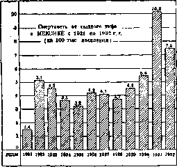
Figure 4.
The mortality rate for T. f. varies among different age groups: the rate increases with age, but in younger age groups (under 10 years) it is higher than in the immediately older groups. Pathological anatomy of typhus fever. The pathological anatomy of T. f. was developed mainly through the works of E. Frenkel, I. V. Davydovsky, and Sh. I. Krinitsky. It was established that from a morphological standpoint, T. f. is above all a disease of the circulatory system as a whole. Upon microscopic examination of this system, especially in the skin and nervous system, the following types of changes were discovered (Davydovsky). The first type - warty endovasculitis - is characterized by the appearance on the inner surface of blood vessels, including such as the aorta, of small warty deposits consisting of fibrin and often containing an admixture of nuclear detritus, i.e. disintegrated endothelial cells. Essentially, this is a mural thrombosis of blood vessels (see separate table, fig. 5) on the basis of destruction of individual endothelial cells or groups of these cells. In small vessels, the thrombi often have an obstructive character, accompanied by extensive destructive changes, sometimes transforming the entire vessel wall mass into amorphous detritus. This second form can be designated as destructive thrombovasculitis. Either in combination with the previous forms or independently develops the third form of vasculitis, consisting of focal proliferative reaction along the peripheral vascular system (capillaries, arterioles, venules) and leading to the development of typhus fever granulomas around these vessels. The proliferation proceeds either at the expense of endothelial and intimal cells or at the expense of adventitial cells, depending on which either pictures of proliferative endovasculitis or perivasculitis (periphlebitis, periarteritis) arise, or, when both factors are combined, pictures of endoperivasculitis. At the site of granuloma development, the vessel wall loses its contours, becomes fibrillated, and the lumen is often thrombosed. The tissue surrounding the vessel also participates in the proliferation; in particular, in the nervous system such
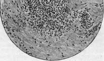
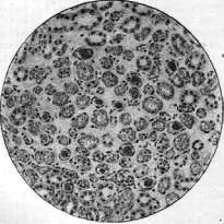
" fit ad i *<.. ;;:. &.. -/.; V: ' «r ] *. .-.*; -4; Sec; Figure 1. Rickettsia Prowazekl; intracellular location (according to Otto-Munter'y). Figure 2. Ricliettsia Prowazeki; extracellular location (according to Otto-Munter'y). Figure 3. Myocarditis in typhus fever. Figure 4. Granuloma in the skin in typhus fever. Рис. 5. Massive thrombosis of vessels of the renal medulla. Figure 6. Encephalitis-granulomas and perivascular sheaths in the medulla oblongata. 20-З
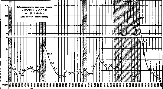
Figure 5.
participation is observed from the side of neuroglial elements. The above-mentioned vascular changes are observed mainly in the peripheral vascular system and as a rule without exception, although with significant qualitative and quantitative variations. Mild and moderate cases (this was verified by Davydovsky on biopsied exanthema) are characterized by the predominance of proliferative reaction, more severe cases - by the presence of significant, sometimes sharp destructive processes, massive thrombosis of vessels, hemorrhages, etc. As a fourth and moreover the latest type of vascular changes (usually not earlier than the 3-4th week) should be mentioned nodular periarteritis, morphologically indistinguishable from the pictures of periarteriitis nodosa Kuss-таиГя. The process either affects the entire circumference or part of the vessel wall and is located mainly along the course of relatively large muscular arteries (e.g. in the area of the adrenal gland capsule). The reverse development of vascular changes varies depending on the intensity of the process. Thrombotic (warty and obstructive) processes, since they are not accompanied by deep destruction of the wall, apparently do not leave special traces: autolysis of thrombi and regeneration of endothelium occur. With significant destruction, limited, or circular hyalinosis of the wall occurs, and in places apparently complete disappearance of the vessel with the development of fibrous connective tissue in its place. As the onset, quantitative increase, and reverse development of vascular changes in T. f. are often greatly stretched in time, that is why sometimes after P/г-2 months from the beginning of the b-ni along the course of vessels residual processes of incomplete regeneration are still found; thus, the morphological convalescence significantly lags behind the clinical convalescence. Upon detailed examination of typhus fever granulomas in the protoplasm of proliferating cells, in particular in the endothelium, the smallest coccal formations resembling rickettsiae were discovered. These findings, however, are not constant. Whether the rickettsia-like formations are parasites or a product of cell disintegration - this question also remains open. In organopathological terms, T. f. is characterized by the following features. The heart almost without exception shows pictures of myocarditis either in the form of diffuse infiltrates of large and small monopitoid, plasma cells or in the form of nodules-granulomas. In general, myocarditis has an interstitial character [see separate table (pp. 199-200), fig. 3]. Sometimes inflammatory changes, edema, thrombosis of vessels are selectively concentrated along the course of the atrioventricular system, which, like myocarditis in general, can be the cause of cardiac death sometimes developing in T. f. Endocarditis is not observed.-The most prominent place in the organopathology of T. f. is occupied by lesions of the nervous system. This lesion is universal, affecting the brain, cerebellum, medulla oblongata, spinal cord, sympathetic ganglia, ganglia in general, and peripheral nerves. The type of these changes is twofold. On the one hand, vulgar phenomena are noted, such as: hemorrhages (petechial and large, of apoplexy character), degenerative processes in ganglion cells, severe hyperemia of vessels, dilation of lymph spaces, sometimes rupture of the latter. On the other hand, and this is more characteristic, focal inflammatory processes are discovered along the course of the affected vascular system. Every T. f. is an acute encephalitis with predominant lesion of the gray matter of the brain. There are especially many foci of lesion in the medulla oblongata and precisely in its part lying below the level of striae acusticae, in the olives and in the area of the nuclei of the X and XII pairs of cranial nerves. The foci have the appearance of round granulomas [see separate table (pp. 199-200), fig. 6], in the central parts of k-ryr a diseased vessel passes in a state of b. or m. significant destruction; sometimes only
Related articles
Mentioned in
Cite this page
“Typhus Fever.” Soviet Medical Encyclopedia. English translation of Bolshaya Meditsinskaya Entsiklopediya, 1st ed. (Moscow, 1928–1936), ed. N. A. Semashko. https://sovietmedicalencyclopedia.pages.dev/article/typhus-fever/