SPLANCHNOMEGALY
Historical document, translated for reference. It reflects medical knowledge of the 1920s–30s and is not medical advice.
Summary
Splanchnomegaly refers to the enlargement of internal organs, particularly affecting the cardiovascular system, liver, spleen, kidneys, and gastrointestinal tract, while sparing the brain and often affecting reproductive organs. This condition is commonly associated with acromegaly and involves uniform increase in both stromal and parenchymal tissues of organs.
Encyclopedia article (1928–1936)
SPLENCHNOMEGALY (from Greek splanchnon - viscera and megas - large), gigantism of the internal organs. In a certain sense, cases of gigantism of individual internal organs, for example, megacolon in Hirschsprung's disease, can also be classified as S. In such cases, one can speak of partial or segmental S. But usually S. refers to the simultaneous increase in size of all internal organs or most of them, which occurs as a constant phenomenon in acromegaly. The increase in size in S. is observed especially in visceral organs: the cardiovascular system, liver, spleen, kidneys, gastrointestinal tract; exceptions are the brain, which remains unchanged, and the sexual organs, which often show degenerative-atrophic changes in acromegaly. The increase in organ size in S. is based on the simultaneous uniform increase in both the stroma and parenchyma of the organs, so that their histological structure remains normal. This condition of organs in true S. differs from the increase in organs due to their congestive induration or any specific processes. Various views exist regarding the origin of S. Some authors (Freund, Amsler, etc.) see the cause of S. in the changing conditions of blood circulation in acromegaly: chemical and mechanical irritation of the posterior lobe of the pituitary gland by its anterior lobe causes increased influence of the posterior lobe on the circulatory apparatus and as a result of increased blood flow to the organs - their hypertrophy and hyperplasia. In many cases of S., an increased amount of total blood mass and enhanced blood supply to organs are indeed noted. Other authors (W. Fischer et al.) explain the increase in organs in acromegaly not only by the increased blood flow but also by specific secretory influences from the pituitary gland changed in acromegaly. The concept of S. is opposed to the concept of splanchnomicrosis, the reduction of organs. This phenomenon is especially frequently observed in diseases of the pituitary gland, its anterior lobe, in particular in the so-called pituitary cachexia. SPLENCHNOPTOSIS, vsplanchnoptosis [syn. enteroptosis - Glenard's disease] (from Greek splanchnon - viscera and ptosis - falling), the prolapse of internal organs - a concept under which is implied a series of conditions, the common and characteristic feature of which is a lower position compared to the norm of one or many internal organs (partial and general S). Usually under S. is meant the prolapse of organs only of the abdominal cavity, although it can also affect the lungs, heart, testicles, uterus and other organs located outside the abdominal cavity. History. Already in Morgagni's work "De sedibus et causis morborum" one can find indications of changes in the position of internal organs in humans compared to the norm; Virchow also pointed to this, noting the close connection of S. with various dyspeptic and nervous phenomena. S. P. Botkin also attached great importance to the prolapse and excessive mobility of the kidneys in the development of various pain syndromes and all psychoneurotic conditions. However, Glenar was the first in the mid-90s of the 19th century ("Les ptoses viscerales") to develop a methodology for systematic palpation of the abdominal cavity, and then the clinical symptomatology of the prolapse of internal organs, calling this condition enteroptosis, resp. splanchnoptosis; Glenar described it as a separate more or less clearly defined disease, to a large extent explaining nervous dyspepsia. Glenar assumed that depending on the insufficient nutrition of a person, suffered severe diseases, and various congenital anomalies, as a result of relaxation of the suspending apparatus of the internal organs, the prolapse of the right flexure of the colon occurs, which also pulls the right kidney and duodenum, thereby contributing to the formation of bends in the intestinal loops and stagnation of their contents, on the basis of which dyspepsia and a neurotic condition develop. Glenar's doctrine quickly became known throughout the medical world and underwent comprehensive and thorough criticism (Ewald, Landau, Obraztsov, etc.), which forced a serious analysis of the causes and conditions on which the prolapse of internal organs depends, as well as to clarify the effect of the latter on the development of a number of pathological phenomena. Landau believed that S. develops secondarily in women who have given birth under the influence of excessive stretching of the abdomen during pregnancy and divergence of the rectus muscles, as a result of which the abdomen becomes pendulous and the internal organs prolapse. The condition of the abdominal muscles became the subject of further study, and by Curschmann, Lindner and many others (Curschmann, Lindner) it was clarified that the prolapse of internal organs, especially the kidneys, is observed not only in women who have given birth, but also in those who have never been pregnant, as well as in men. This observation required the study of the factors on which the normal position of human internal organs depends. The works of Russian researchers - Lesgaft and especially Volkov and Delitsin - deserve special attention. In their remarkable work devoted to the wandering kidney, Volkov and Delitsin presented the mechanics of the abdominal cavity as follows: the abdomen represents a closed cavity bounded on all sides by muscular walls - the abdominal wall, diaphragm and pelvic musculature, in which is placed a second balloon consisting of the peritoneum; in the latter in turn is enclosed a third balloon consisting of the hollow muscular organs. The muscular walls of the abdominal cavity and the internal (third) muscular balloon are in a changing interaction with each other. If the outer balloon (abdominal press) contracts, then the inner balloon (hollow organs of the abdominal cavity) automatically contracts, and conversely, if the inner balloon stretches, then the abdominal walls automatically relax and become softer. The correct function of the muscles in the walls of these balloons depends on the intra-abdominal pressure, which has the greatest significance in the statics and position of the abdominal organs. These authors for the first time clarified the significant dependence of the position of the diaphragm, and consequently of the internal organs, on the attraction of the lungs. Subsequently, Mathes also began to attach great importance to the elastic attraction of the lungs among other reasons for the position of internal organs. Parallel to these studies, which examined mechanical influences on the position of internal organs, another point of view developed, according to which hereditary and constitutional factors were given the greatest importance in the development of S. Weakness of tissues, easy stretchability of supporting ligaments, congenital biological inferiority of the organism began to be considered as those factors on the basis of which S. can develop (Glenar, Tuffier, Obraztsov, etc.). An extreme view of this order was expressed by Stiller, who believed that the statics of the abdominal cavity in the development of S. has a secondary significance, and the main cause of S. is the general congenital asthenia, which determines both the structure of the body and the position of internal organs, as well as various deviations in the functions of individual organs and entire systems. Stiller's doctrine, finding many adherents and at the same time being criticized, undoubtedly contributed to the clarification of some aspects of the pathogenesis of S, which in the modern light can be presented as follows: insufficient fixation of internal organs, especially the liver, right kidney, duodenum, with which a child may be born, gives rise to the prolapse of internal organs later, when the child begins to walk. The immediate cause of such insecure fixation is in most cases the insufficient fusion of the cecum and ascending colon on their posterior surface with the posterior abdominal wall, in view of which these parts of the colon receive a mesentery, which is a continuation of the mesentery of the small intestine, and become mobile. Subsequently, due to the accumulation in them of stagnant fecal masses and gases, as well as the ever-increasing stretching of the ligaments with enhanced intestinal peristalsis, parts of the colon descend under the influence of gravity, pulling the right kidney and liver with them. Such an anomaly of attachment of the ascending colon is observed in 20% of people, occurring mainly in individuals of asthenic constitution. The indicated developmental defect contributes to the gradual formation of S in adult life. Two types of S are distinguished: constitutional and acquired S. The first form is always associated with a special, abnormal body structure, weak development of muscles, with their perverted innervation and tone and usually insufficient nutrition and inferiority of the elastic elements of the organism, which in general are characteristic features of asthenic constitution. A narrow, long, often pear-shaped, downward-pointing chest (thorax piriformis) with a large inclination of the ribs and wider intercostal spaces, an acute epigastric angle, sometimes with a clearly expressed dorsal kyphosis, developing as a result of muscle weakness and insufficient innervation, characterize the asthenic constitution.
The described body structure is associated with a low diaphragm position, resulting from insufficient elastic tension of the lungs, excessive mobility of the lower opening of the chest cavity, and weakness of the abdominal press, which cannot, together with the poorly developed and insufficiently innervated muscles of the pelvic floor, resist the pressure of the abdominal viscera, causing the latter to descend, and the abdomen in profile takes the characteristic form of a pendulous abdomen in young women (fig. 1). In this regard, the descent of the small intestines, which in normal conditions fill the entire abdominal cavity, giving it a normal rounded shape, is of essential importance. The development of splanchnomegaly is promoted by wearing old-fashioned corsets or excessively tight lacing, as well as emaciation, associated with the disappearance of fat in the abdominal cavity and with the subsequent change in intra-abdominal pressure. The constitutional form of splanchnomegaly is most often observed among women, but sometimes it also occurs in tall men.
FIG. 1.
Acquired splanchnomegaly develops as a result of relaxation of the abdominal press, therefore it is primarily observed among women, in whom after repeated pregnancies and childbirth, an enlarged abdomen, divergence of the rectus muscles, and relaxation of the abdominal and pelvic musculature often remain. Such a condition is promoted by insufficient care for the abdominal wall in the last months of pregnancy and after childbirth, as well as heavy physical work soon after delivery, when the abdominal muscles have not yet been stretched and their normal tone has not been restored. In men, due to the absence of this factor, acquired splanchnoptosis occurs much less frequently or develops exclusively as a result of sharp emaciation in obese individuals, especially in the presence of pulmonary emphysema, as well as after the release of a long-existing ascites or after removal of a very large tumor. Of course, these same factors play a significant role in the development of enteroptosis in women as well, promoting the formation, due to the descent of the viscera in a weak abdominal press, of the characteristic in shape acquired pendulous abdomen (fig. 2). Symptomatology. Many cases of splanchnomegaly pass completely asymptomatic. This is most often observed in mild degrees of constitutional splanchnomegaly or in acquired splanchnomegaly. Often the only sign of splanchnomegaly is a tendency to constipation. The constitutional form of splanchnomegaly often gives a varied clinical picture in its manifestations, which is explained not so much by splanchnomegaly itself as by disorders of the functions of the autonomic nervous system. Occasionally, pronounced symptoms can also be observed in young nervous women with acquired splanchnoptosis. The main symptom of splanchnoptosis is a tendency to constipation, which depends not so much on the descent of the large intestine—coloptosis—and the formation of kinks in them, mechanically hindering the movement of fecal masses, as on the disorder of innervation of the intestinal musculature, the consequence of which are spasms or atony of the large intestine, and sometimes simultaneously spasms in some areas and atony in others (dyskinesia). Along with constipation, various dyspeptic disorders often occur. Constipation and dyspepsia are promoted by various abnormalities and deviations in the secretion of digestive juices, causing abnormal digestion and assimilation of food.
Figure 2. 84
Figure 3.
and dyspepsia various abnormalities and deviations in the secretion of digestive juices, causing abnormal digestion and assimilation of food. Long-term persistent constipations favor the development of enterocolitis, sometimes membranous colitis, accompanied by severe pains and promoting persistent nausea, loss of appetite, and all kinds of reflexes, sometimes accompanied by severe painful sensations, such as cardialgia, sometimes palpitations, spasms of the esophagus, etc. Due to these sensations, patients completely lose their appetite, lose weight, feel tired, exhausted, become hypochondriacs, begin to suffer from headaches and insomnia—various nervous disorders thus develop. In individual cases, the condition reaches a true hysterical state, when patients, especially women, completely refuse to eat, severely suffering from spasmodic pains, nausea and vomiting after almost every meal. To these general phenomena are often added symptoms from individual organs, which, due to certain local abnormalities in their attachment or after special circumstances, e.g., trauma, have descended more than neighboring organs. In severe gastropptosis of such patients, they are tormented by a feeling of fullness in the epigastric region, which remains for a very long time after food is introduced into the stomach, pains in the left hypochondrium, cardialgia and spasms of the esophagus, as well as constant belching (see Stomach, Belching). A relatively rare descent of the cecum is observed with its excessive mobility; according to Wilms, its descent depends either on excessive distensibility of the cellulose behind the intestine, or on its congenital elongation, or finally on the preservation of the mesentery of the ascending intestine; patients suffering from constipations are often bothered by a feeling of bloating and fullness in the right iliac region, dull pains here—from stretching of the cecum and its catarrhal inflammation, which often extends to the mucous membrane of the vermiform appendix. The works of Obraztsov, Gausman, Wilms, Strazhesko, Klose, etc., have drawn special attention to this condition of the cecum, as it often gives rise to the development of the described clinical symptom complex, which often leads to confusion of the 'mobile cecum' (coecum mobile) with appendicitis. Excessive mobility of it also predisposes to stagnation of fecal masses and sometimes to special painful attacks in the right iliac region, resembling attacks of acute appendicitis. Sometimes to them are added symptoms of intestinal obstruction, which
&Фт
Figure Figure 5. depends on kinks of the long intestine or partial twisting of it (Strazhesko) or even on its complete volvulus (Zegge von Manteuffel) (fig. 3). The hepatic flexure of the large intestine (flexura hepatica), due to the weak ligaments that strengthen it and in the presence above of such a heavy organ as the liver, often stands lower than normal. According to Gleaner, every splanchnomegaly begins with the descent of the right flexure of the large intestine, on the contrary, other authors believe that the first to descend is the right kidney and it involves in the process of descent the covering the intestine kidney, as a result of which a certain difficulty in the passage of intestinal contents through the right flexure may occur, which favors stasis of the ascending colon, and consequently promotes the development of catarrh in it. A much more common anomaly is in the location of the transverse colon, which, having a congenitally greater length and greater width of the mesentery, easily descends and acquires a U-shape, often fixing itself with adhesions in the pelvic region (fig. 4), to which Virchow (1890) first drew attention and which was then confirmed by Kuschmann and Simmonds. These adhesions are formed as a result of local peritonitis, the cause of which is transferred colitis or appendicitis, in women disease of the appendages, trauma of the abdominal cavity, etc. Such anomalies in the location of the transverse intestine, especially when it is excessively long and arranged in the form of parallel loops, favor stasis of intestinal contents and often the development of severe suffering in the form of attacks of intestinal colic, sometimes even with symptoms of obstruction, and in rare cases promote volvulus of the transverse colon (fig. 5). The splenic flexure of the large intestine, being firmly fixed by ligaments, rarely shifts and therefore with a sharp descent of the transverse intestine, severe disorders from kinking at this place easily occur, the typical clinical symptomatology of which was described by Paire (Paug). Stagnation of intestinal contents, already physiologically delayed in the region of the splenic flexure, significantly increases and then conditions are created for the development of an inflammatory local process here—pericolitis, as a result of which the descended left knee of the transverse intestine and the descending colon lying nearby often adhere. As a result of adhesion, an even greater difficulty in the passage of contents through the flexure occurs, fecal masses accumulate behind the narrowing in the descended transverse intestine, burden it and promote the intensification of coprostasis, which sometimes extends to 34» itself the Bauhinian valve. The developing stasis causes not only constipations, periodically alternating with diarrhea, due to the accompanying enterocolitis, but also constant pains in the left hypochondrium in the form of stabbing or dull pain, with a feeling of constant heaviness and bloating in the umbilical region, which is sometimes accompanied by reflex angina-like cardiac attacks, headaches and general lethargy. The condition of patients improves only after the passage of gas and defecation. The easily occurring under these conditions 'transversitis' makes the existence of such patients miserable (see Colitis). Sometimes to these symptoms
Figure 6. Descent of the kidney.
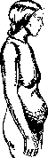
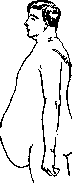
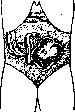
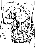
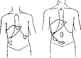
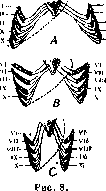
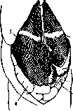
Figure 7. Wandering kidney. True attacks of obstruction-intestinal crises often occur, frequently at night, beginning with painful colic throughout the abdomen, which depends on the intensified peristalsis of the intestines filled with gas and contents, and is sometimes accompanied by nausea, urges to vomit, elevated temperature, and pain in the right iliac region. After several hours of suffering, all these phenomena can quickly disappear if gases pass and there is an abundant stool, then the patient seems to recover, only to repeat the above-described attack after some time.--Op u she niye S-Romani occurs when it is congenitally elongated and provided with a broad mesentery, which easily leads to retention of solid fecal masses in it-constipation develops, which eventually leads to inflammation of the mucous membrane-sigmoiditis (see). In this case, the previously observed feeling of heaviness in the left iliac region is complicated by pains and periodic diarrhea with the excretion of large amounts of mucus, sometimes with an admixture of blood. A long S-Romanum often gives rise to partial twisting of it, accompanied by abdominal bloating, intestinal colic with elevated temperature, and sometimes to complete volvulus and the development of an attack of complete intestinal obstruction, requiring surgical intervention. On the prolapse of the kidney (figs. 6 and 7)-see Kidneys, changes in the position of the kidney. The liver is fixed in its position, on the one hand, by pressure on it from the viscera and fat pads, which in turn are supported by the tonic tension of the abdominal muscles, and on the other hand, by its own ligaments. Depending on the body build, the liver occupies different positions (fig. 8), because the spatial relationships in the right hypochondrium vary with different structures of the chest cavity, which with more or less the same volume of the liver causes different positions of it and variations in the size of the area of the upper anterior surface of the liver not covered by the bony skeleton. Under physiological conditions, the liver descends in the vertical position of the human body and during deep inspiration. In pathological cases, it can change its position secondarily-in various diseases of the thoracic and abdominal organs. An increase in the volume of the right lung in emphysema, accumulation of fluid or gas in the right pleural sac contribute to the descent of the liver, while accumulation of fluid or the presence of a large tumor (pregnant uterus, cyst, etc.) in the abdominal cavity causes it to rise. In other cases, the descent of the liver may be primary. This hepatoptosis can be complete or partial. The descent of the entire liver (hepatoptosis totalis), when it becomes so mobile that it even deserves the name wandering, is relatively rare (fig. 9). Hepatoptosis is most often observed in women with general S. A flabby body build, sedentary lifestyle, lack of physical exercise, repeated pregnancies, wearing unhygienic old-fashioned corsets that constrict the waist, rapid and significant weight loss-are the main factors contributing to the development of total hepatoptosis.
But in practice, partial descent of the liver (hepatoptosis par-tialis) is much more common, being a manifestation of general S, in which either only the lower edge of the liver descends and forward, while its posterior edge is elevated, or the left lobe of the liver descends in isolation, reaching the level of the navel, while the right remains almost in place, or finally the right lobe descends predominantly. In all these cases Figure 9. Descent of the li- simultaneously with the descent of the liver, its deformation and a significant increase in its mobility in the lateral direction, its mobility during inspiration occur, why the liver begins to ballot. A slight hepatoptosis may not give any symptoms and is discovered only upon physical examination of the abdomen. With more significant hepatoptosis, patients often complain either of dull sensations in the form of heaviness or of sharper pains in the area of the liver with their irradiation into the shoulder, under the right scapula or into the spine. These unpleasant sensations usually decrease when the liver is lifted by hand and when wearing appropriate belts.
Significant descent of the liver, causing difficulty in portal circulation and free entry of bile into the intestine, contributes to the development of various dyspeptic conditions, disturbances of intestinal function, and, mainly, the occurrence of various nervous phenomena, such as headaches, dizziness, noise in the ears, and even various mental disorders. Glenar attributed all these symptoms to disturbances of various liver functions and various reflexes coming from the liver, calling this condition hepatism. However, in most cases it is difficult to determine to what extent all these phenomena depend on the descent of the liver and to what extent on general S, accompanying hepatoptosis. Other organs of the abdominal cavity may descend, such as the spleen (see).
liver: 1-right lobe; 2-gall bladder; 3-stomach; 4-cecum; 5-sigmoid colon; 6-transverse colon
its mobility in the lateral direction, its mobility during inspiration occurs, why the liver begins to ballot. A slight hepatoptosis may not give any symptoms and is discovered only upon physical examination of the abdomen. With more significant hepatoptosis, patients often complain either of dull sensations in the form of heaviness or of sharper pains in the area of the liver with their irradiation into the shoulder, under the right scapula or into the spine. These unpleasant sensations usually decrease when the liver is lifted by hand and when wearing appropriate belts. Significant descent of the liver, causing difficulty in portal circulation and free entry of bile into the intestine, contributes to the development of various dyspeptic conditions, disturbances of intestinal function, and, mainly, the occurrence of various nervous phenomena, such as headaches, dizziness, noise in the ears, and even various mental disorders. Glenar attributed all these symptoms to disturbances of various liver functions and various reflexes coming from the liver, calling this condition hepatism. However, in most cases it is difficult to determine to what extent all these phenomena depend on the descent of the liver and to what extent on general S, accompanying hepatoptosis. Other organs of the abdominal cavity may descend, such as the spleen (see).
/-3'
its mobility in the lateral direction, its mobility during inspiration occurs, why the liver begins to ballot. A slight hepatoptosis may not give any symptoms and is discovered only upon physical examination of the abdomen. With more significant hepatoptosis, patients often complain either of dull sensations in the form of heaviness or of sharper pains in the area of the liver with their irradiation into the shoulder, under the right scapula or into the spine. These unpleasant sensations usually decrease when the liver is lifted by hand and when wearing appropriate belts. Significant descent of the liver, causing difficulty in portal circulation and free entry of bile into the intestine, contributes to the development of various dyspeptic conditions, disturbances of intestinal function, and, mainly, the occurrence of various nervous phenomena, such as headaches, dizziness, noise in the ears, and even various mental disorders. Glenar attributed all these symptoms to disturbances of various liver functions and various reflexes coming from the liver, calling this condition hepatism. However, in most cases it is difficult to determine to what extent all these phenomena depend on the descent of the liver and to what extent on general S, accompanying hepatoptosis. Other organs of the abdominal cavity may descend, such as the spleen (see).
Related articles
Mentioned in
Cite this page
“SPLANCHNOMEGALY.” Soviet Medical Encyclopedia. English translation of Bolshaya Meditsinskaya Entsiklopediya, 1st ed. (Moscow, 1928–1936), ed. N. A. Semashko. https://sovietmedicalencyclopedia.pages.dev/article/splanchnomegaly/