Femoral Nerve
Historical document, translated for reference. It reflects medical knowledge of the 1920s–30s and is not medical advice.
Summary
This article from the 1928–1936 Soviet Great Medical Encyclopedia provides a detailed anatomical description of the femoral nerve, its origin, course, branches, and functions, as well as its clinical significance and pathology.
Encyclopedia article (1928–1936)
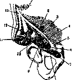
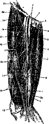
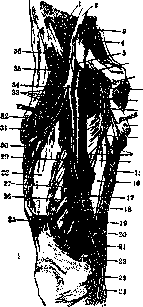
FEMORALIS NERVUS, s. cruralis, femoral nerve, begins with three roots from L4, the thickest nerve of the lumbar plexus, about 5--6 mm in diameter (Fig. 1). It descends down and outward between the m. psoas major and the m. iliacus under the fascia iliaca, enters behind the lig. inguinale into the lacuna musculorum, where it lies medially to the thigh muscles and laterally to its blood vessels (Fig. 2). Here or below it divides into its terminal branches (Fig. 3), which can be reduced to two bundles: the anterior, predominantly sensory, and the posterior, predominantly motor. Even before exiting the femoral opening, the F. n. gives off short muscular branches in the region of the fossa iliacae, running over the lig. inguinale to the m. iliacus and to its lower part, common with the m. psoas major, as well as a branch to the m. pectineus and the n. arteriae femoralis proprius, accompanying the artery along its entire length and giving twigs to the femur and the periosteum. Sensory branches (rami cutanei ant.) can be divided into two groups, medial and lateral. The medial twigs run along the inner edge of the m. sartorii and, piercing the fascia at various heights, branch in the skin of the anterior-inner surface of the thigh to the knee, anastomosing with the n. cutaneus n. obturatorii and the n. cutaneus femoris medialis. The lateral twigs partly pierce the m. sartorius, partly run over it and branch in the skin of the anterior surface of the thigh to the patella, anastomosing with the n. lumbo-inguinalis and the n. cutaneus femoris lateralis. Motor branches (rami musculares) go to the mm. rectus femoris, vastus medialis, interme-dius and lateralis and the m. sartorius (Fig. 4). Several twigs go to the m. articularis genu, as well as to the periosteum and the capsule of the knee joint. The longest branch of the n. femoralis is the n. saphenus, forming its immediate
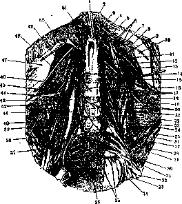
Fig. 1. Branches of the lumbosacral plexus: 1-pars lumbalis diaphragmatis; 2-hiatus oesopha-geus; 3-truncus sympathicus; 4-costa XII; 5-ramus anterior n. lumbalis I; e-rami muscujares; 7-n. intercostalis XII; 8-n. ilio-hypogastricus; 9-m. quadratus lumborum; 10-m. transrersus abdominis; 11-n. lumbalis II; 12-n. genito-femo-ralis; 13- n. cutaneus femoris lateralis; 14-.plexus lumbalis; IS-n. lumbalis III; 16-n. ilio-inguina-lis; 17-rami musculares; 18-n. cutaneus femoris lateralis; 19- д. lumbalis IV; 20-n. genito-femora-lis; 21- rami communicantes; 22 и 37-n. femoralis; 23-ветвь к plexus sacralis; 2i-mm. psoas major et minor; 25-n. obturatorius; 26-m. iliacus; 27-n. femoralis; 28-ramus anterior n. lumbalis V; 29-n. obturatorius accessorius; SO- n. glutaeus superior; SI-plexus sacralis; 32-n. ischiadicus; 33- n. cutaneus femoris posterior; 34-n. pudendus; 35-rami anteriores nn. sacralium I-V; 36-ganglion coccygeum; 38- n. obturatorius; 39- rami musculares n. femoralis; 40- m. iliacus; 41-n. spermaticus externus; 42-n. lumbo-inguinalis; 43-m. psoas minor; 44-m. psoas major; 45- n. cutaneus femoris lateralis; 46-m. quadratus lumborum; 47-m. transversus abdominis; 48-n. ilio-inguinalis; 49-n. ilio-hypogastricus; 50-n. intercostalis XII; 51-hiatus aorticus.
continuation (Fig. 5). It descends initially laterally to the a. femoralis, covered above by the m. sartorius. A little above the middle of the thigh it pierces the fasc. ilio-pectinea and lies in front of the a. femoralis, enters together with it into the canalis adductorius (Hunteri), then pierces the anterior wall of the latter and descends along the posterior edge of the m. sartorii, behind the epicondylus medialis. While passing along the thigh it gives a thin branch to the skin of the inner surface of the thigh and the popliteal fossa. At the level of the tuberositas tibiae it divides into its terminal branches-ramus infrapatellaris and rami cutanei cruris mediales. The first spreads in the skin in front of the patella and the tuberositas tibiae, the last, constituting the continuation of the n. saphe-ni, distribute in the skin of the anterior medial and posterior surfaces of the shin to the medial edge of the foot, anastomosing with the n. peronaeus "18
superfic.-Function of F. n. F. n. participates in the flexion of the pelvis together with the trunk with the thigh fixed; flexes and adducts the thigh,
rotating it outward, extends the knee, and also participates in its flexion, turning the shin inward (t. sartorius). Skin innervation is considered above. The symptomatology of lesions of F. n. follows from the distribution of its motor and sensory branches described above. In mild forms, in which the lesion does not spread to the entire thickness of the nerve, phenomena of irritation predominate, pains along the course of the F. n. and the n. sapheni. In far-advanced forms, hypesthesia and Fig. S. Cutaneous branches of the femoral nerve: 1-a. et v. femoralis; 2-lym-phoglandula Rosen-miilleri; 3-tubercu-lum pubicum; 4- m. peetineus et -a. circumflexa femoris medialis; 5-m. adductor longus; 6-m. gracilis; 7-ramus n. obturator!! to m. gracilis; 8 - anterior wall of the adductor canal; 9-a. ge-nu suprema et n. sa-phenus; JO- projection of the lower opening of the adductor canal; 11-a. po-plitea; 12~ patella; 13-m. vastus medians; 14-п. saphenus; IS-m. sartorius; IS-a. profun-da femoris; 17-a. circumflexa femor. lat.; IS- cutaneous and muscular branches of the femoral nerve; 19-т. ilio-psoas; 20-п. cutaneus femor. lat.; 21- spina iliaca ant. sup. anesthesia in the area of distribution of the cutaneous branches, motor disturbances, expressing themselves mainly in a greater or lesser limitation of flexion of the thigh and extension of the knee up to their complete paralysis. Severe forms are accompanied by noticeable wasting of the m. quadrici-pitis and changes in its electrical excitability.
Wasserman described a symptom in lesions of the F. n., analogous to that observed in neuritis
Fig. 4. Muscular branches of the femoral nerve: 1-m. iliacus; 2- n. femoralis; 3-т. psoas major; 4- n. obturatorius; б-ramus muscularis for m. peetineus; в-m.pecti--? neus (cut off); 7- n. obturatorius (ramusan-' terior); 8- m. obturator ~8 externus; 9- n. obtu--i&torius (ramus posterior); 10- m. pectine-_,)us (cut off); 11- m. -12 adductor longus; 12- m. adductor brevis; u 13- branches of rami po--n sterioris n. obturatorii; 14- m. adductor longus (cut off); 15- ramus cutaneus n. obturatorii; 16- m. adductor magnus; 17- entrance into canalis adduc-torius (Hunteri); 18- ramus cutaneus anterior n. femoralis; 19- m. gracilis; 20- n. saphenus; 21- ramus infrapatellaris; 22- m. sartorius; 23- v. sa-phena magna; 2^- ramus infrapatellaris a. sapheni; 26- m. rec-tus femoris; 28- m. vastus medialis; 27- n. saphenus; 28- m. yastus intermedius; 29- ramus muscularis; 30- m. vastus lateralis; 31- a. circumflexa femoris lateralis (ramus descendens); 32- m. rectus femoris; 3 3- rami musculares for m. quadriceps femoris; 34- a. femoralis; 35- ramus muscularis; 36- m. sartorius. n. iscliiadici and consisting in the appearance of pains in the groin area with dorsal flexion of the thigh in a patient lying on the abdomen. To intensify the effect, one can simultaneously press on the nerve in the groin area. Especially significant for the diagnosis of lesions of the F. n. are the lowering or extinction of the knee reflex. The cause is most often tumors of the spinal column, retroperitoneal lymph. glands, the pelvis and the internal organs located in it, the femur, then abscesses of the m. psoas, arising on the basis of appendicitis, femoral hernias. Among toxic moments, alcoholic polyneuritis must be noted, in which sensory twigs are most often affected; however, a complete bilateral paralysis of the F. n. was also described in this case. Cases of lesion of the F. n. were also observed in diabetes mellitus, rheumatism, and some other diseases. Traumatic injuries of the F. n., due to its protected position, are observed significantly less often than those of other nerves. In addition to bullet and other wounds, their causes can be fractures of the femur and pelvis bones. More often there is a lesion of individual branches. In differential diagnosis, one must keep in mind poliomyelitis and myopathy (see.). Therapy and prevention in lesions of the F. n. do not differ from those in lesions of other nerves and nerve plexuses (see. Neuritis, Polyneuritis and individual nerves).
Related articles
Mentioned in
Cite this page
“Femoral Nerve.” Soviet Medical Encyclopedia. English translation of Bolshaya Meditsinskaya Entsiklopediya, 1st ed. (Moscow, 1928–1936), ed. N. A. Semashko. https://sovietmedicalencyclopedia.pages.dev/article/femoral-nerve/