Hunter's Canal
Historical document, translated for reference. It reflects medical knowledge of the 1920s–30s and is not medical advice.
Summary
This article from the 1928-1936 Soviet Medical Encyclopedia describes the anatomy, contents, and surgical access to Hunter's canal (adductor canal). It details the boundaries, the vessels and nerves passing through it, and the operative approach for ligating the femoral artery in cases of trauma or aneurysm.
Encyclopedia article (1928–1936)
HUNTER'S CANAL, canalis Hunteri, also known as canalis femoro-popliteus or adductorius, is the continuation of the anterior femoral sulcus. The inner boundary of the latter is formed by the adductor brevis, longus, and magnus muscles, and the outer boundary by the vastus internus muscle. From above downwards and from without inwards, the sulcus and Hunter's canal are crossed by the sartorius muscle. At the level of the attachment of the adductor longus muscle, the common vascular sheath of the thigh thickens due to fusion with fibrous fibers running from the tendons of the muscles forming the anterior femoral sulcus. The bridge of these fibrous fibers—the vasto-adductor membrane (fascia vasto-adductoria)—forms the anterior wall of Hunter's canal (see Figure 1). The lower opening of the canal is located where the vessels pass into the popliteal fossa, piercing the adductor magnus muscle. In addition to the opening for the vascular bundle, the canal has another anterior opening, variously located, through which the greater saphenous nerve and the highest genicular artery emerge. In Hunter's canal, the vein lies behind the artery, and the saphenous nerve lies lateral and anterior to the artery. The usual length of Hunter's canal is 6-7 cm, and its midpoint is located at a distance of about 13 cm above the medial epicondyle of the femur (see Figure 2). Wounds of the femoral artery in the middle third are quite common (mainly wartime gunshot wounds). Wounds of the femoral artery are generally the most frequent: according to Punin's statistics (1,443 cases), in 20%.
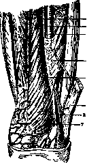
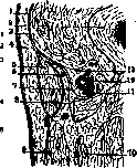
Figure 1. Hunter's canal: 1 - femoral vein; 2 - saphenous nerve; 3 - femoral artery; 4 - anterior wall of Hunter's canal (vasto-adductor fascia); 5 - vastus medialis muscle; 6 - sartorius muscle; 7 - highest genicular artery. Figure 2. Cross-section of the thigh in the lower third in the region of Hunter's canal (inner section of the peripheral segment of the right thigh): 1 - fascia lata of the vastus medialis muscle; 2 - femur; 3 - vastus medialis muscle; 4 - skin with subcutaneous tissue; 5 - sartorius muscle; 6 - saphenous nerve; 7 - vasto-adductor fascia; 8 - tendinous part of the adductor magnus muscle; 9 - semitendinosus muscle; 10 - semimembranosus muscle; 11 - femoral vein; 12 - femoral artery; 13 - Hunter's canal (After Testut-Jacob). When the artery is wounded in Hunter's canal, if ligation is necessary at this level in the presence of an aneurysm of the peripheral section of the artery, the following operative approach is used, which leads to the goal more reliably than others usually described. The patient lies on the back, the limb is flexed at the knee, the thigh is abducted and externally rotated. The operator stands on the side of the wounded limb, the assistant opposite. An 8 cm incision through the skin, subcutaneous tissue, and superficial fascia is made along the edge of the tendon of the adductor magnus muscle. The midpoint of the incision is 7 fingerbreadths upwards from the medial epicondyle. After incising the fascia, the sheath of the sartorius muscle is opened, and the muscle is retracted medially. Then, having spread the edges of the wound, the fibers of the vasto-adductor membrane are found. Having found an opening in it from which the artery, nerve, or both emerge, a grooved probe is introduced into it. The anterior wall is incised along the probe for the entire length of the canal. The tissue surrounding the vessels is torn with the probe, the artery is isolated, and a ligature is applied. It is important to note that ligation is not performed according to Quain's line.
Related articles
Mentioned in
Cite this page
“Hunter's Canal.” Soviet Medical Encyclopedia. English translation of Bolshaya Meditsinskaya Entsiklopediya, 1st ed. (Moscow, 1928–1936), ed. N. A. Semashko. https://sovietmedicalencyclopedia.pages.dev/article/hunters-canal/