Femoral Artery
Historical document, translated for reference. It reflects medical knowledge of the 1920s–30s and is not medical advice.
Summary
The femoral artery, a continuation of the external iliac artery, runs along the anteromedial aspect of the thigh from the inguinal ligament to the popliteal fossa. This article describes its anatomical structure, embryological development, branching patterns, and clinical significance in surgical procedures.
Encyclopedia article (1928–1936)
Femoral Artery, femoral artery, being a continuation of the external iliac artery, goes along the anteromedial side of the thigh in the area from the inguinal ligament to the popliteal fossa (Fig. 1). The wall of the F. a. histologically differs by the more powerful development of the muscular layer. The development of the femoral artery is closely connected with the fusion of individual metameres forming the outgrowth of the lower extremity. As the base of the outgrowth narrows and the extremity takes on an elongated form, the arteries merge with each other. The posterior displacement of initially neutrally positioned extremities led to the loss of connection with the anteriorly lying arteries (a. saphena) and the establishment of connection with the posteriorly lying arteries. Therefore, the F. a. can be considered as having developed from the fusion of individual metamers, where the participation of anterior and posterior branches is determined by the dorsoventral position of the extremity. These comparative anatomical data well explain the variability of the main branches and the types of their origin: main, scattered, and transitional. One of the features of the scattered type for the F. a. is the so-called high division of the vessel: the anterior and posterior tibial arteries arise not from the popliteal, but directly from the F. a., dividing immediately under the inguinal ligament.
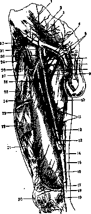
More often another form of the scattered type is encountered: the main trunk of the F. a. reaches the popliteal fossa, however all branches up to the deep femoral artery depart at a distance of 1-2 cm from the inguinal ligament. In the main type, a sequential pattern in the departure of branches is observed.
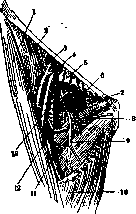
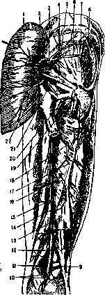
The deep femoral artery (a. profunda femoris) originates from the F. a. at various levels within the iliopectineal fossa. Departing from the posterior periphery of the artery, it lies in front of the iliopsoas, pectineus, and adductor muscles, covered by the adductor muscle (Fig. 2). The branches of the deep femoral artery: the circumflex femoris medialis, with its deep branch, widely anastomoses with the obturator artery system (Fig. 3). The external circumflex femoris artery surrounding the femur anastomoses with the gluteal arteries through its ascending branch, while the descending branch, going under the rectus muscle, sometimes reaches the knee joint plexus (rete genu) (Fig. 2). The terminal branches of the deep femoral artery: the first perforating artery (perforans prima) goes to the posterior surface of the thigh, at the lower edge of the pectineus muscle it anastomoses with the inferior gluteal and internal circumflex arteries (Fig. 4). The second and third perforating arteries, passing to the posterior surface of the adductor musculature (Fig. 3), anastomose with the artery accompanying the sciatic nerve (a. comitans n. ischiadici)—a vessel requiring isolated ligation during thigh amputation.
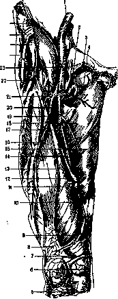
All these anastomoses have great practical significance in ligation of the F. a., contributing to collateral circulation. G13 FEMORALIS ARTERIA
The branches of the deep femoral artery form numerous anastomoses connecting the F. a. with other main arteries: the internal circumflex femoris lateralis; 22-ramus ascendens; 23-a. epigastrica superficialis; 24-m. glutaeus medius; 25-m. glutaeus minimus; 26-m. tensor fasciae latae; 27-a. circumflexa ilium profunda.
Figure 3. Anastomosis of the circumflex artery with the obturator artery: 1-m. sartorius; 2-a. circumflexa ilium prof.; 3-a. femoralis; 4-v. femoralis et a. epigastrica inf.; 5-m. pectineus; 6-a. obturatoria et n. obturatorius; 7 - membrana obturatoria; 8-m. adductor magnus et n. obturatorius; 9 - m. adductor brevis et n. obturatorius; 10-mm. adductor longus et gracilis; 11-m. pectineus; 12-a. circumflexa femoris medialis; 13-m. obturator ext. especially if the F. a. is ligated above the origin of the deep artery of the thigh. Anastomoses of the femoral artery with the system of the popliteal artery and the anterior tibial (a. tibialis anterior) are carried out by the first artery of the knee joint (a. genu prima, s. suprema), entering into the composition of the network of the knee joint (fig. 1). Anastomoses of the F. a. with the system of the internal iliac are formed by the external iliac arteries. The greatest value is acquired by intramuscular anastomoses, well developed in the thigh region and absent in the knee joint region, which is clearly reflected in the results of ligations of the corresponding arteries. The projection of the F. a. is determined by Quain's line, connecting the middle of the distance between the anterior superior iliac spine and the symphysis with the adductor tubercle (see Quain's line). The entire course of the F. a. is topographically divided into three sections: the first - 8-10 cm from the Poupart's ligament to the apex of Scarpa's triangle. In this section, the deep artery of the thigh originates from the F. a. The middle section - from the apex of Scarpa's triangle to the upper opening of Hunter's canal. Here, behind the F. a. lies a vein, in front - the internal cutaneous nerve of the thigh. The last section - in Hunter's canal, from which the F. a. passes into the popliteal. The nutrition of the walls of the F. a. is constructed according to the general type of vascularization of the vessel walls. (See also Thigh, Blood vessels.) Vasomotor fibers of the femoral artery approach from the side of the mixed nerves of the limbs and are distributed along the vessel according to the principle of segmentation. For the F. a., vasoconstrictors arise in the Dxi-hiи Li-sh segments of the spinal cord, pass as myelin-containing fibers in the composition of the white connecting branches (rami communicantes albi) to the ganglia of the border column and end in the cells located there. Vasoconstrictors exit from the cells of the border trunk in the form of gray connecting branches (rami communicantes grisei) and, joining the spinal nerves, go with them to the periphery. To the F. a., vasoconstrictors approach in the composition of vascular branches (rami vasorum) under the Poupart's ligament from the femoral nerve and in the popliteal region - from the sciatic. Most of the nerves approaching the F. a. then go to its branches and to the vein, forming the femoral plexus (plexus femoralis, fig. 6), connecting with the plexus on the common iliac artery, but not originating from it: long sympathetic conductors going from the aorta to the F. a. are not proven either anat.-histologically or physiologically. Branches of nerves in the adventitia of the F. a. form networks of Remak's fibers; in the deep layers on the muscular membrane there is a deep network, part of the fibers of which is of local origin - from ganglion cells in the perivascular cellular tissue. The static-dynamic position of the lower limbs affects the condition of the lumen of the F. a.: in a state of rest (in a lying position) the lumen of the artery is narrowed, in a standing position the pressure on the walls of the F. a. is significantly increased. The F. a., supplying branches to all sides of the thigh (see above), is a rich
Figure 4. Arteries of the right thigh: 1-a. circumflexa femoris lateralis, superior branch; 2-a. glutaea superior; 3-a. piriformis; 4-branch of F. a. __ ramus superior; 5-ramus inferior; 6-m. gluteus minimus; 7-a. glutaea medius; 8-a. genu superior medialis; 9-aa. surales; 10-a. genu superior lateralis; 11-m. biceps femoris (caput breve); 12-m. semitendinosus; 13-m. semimembranosus; 14-a. nutricia femoris inferior; 15-a. perforans tertia; 16-a. perforans secunda; 17-a. nutricia femoris superior; 18-a. perforans prima; 19-a. circumflexa femoris medialis, ramus profundus; 20-m. quadratus femoris; 21-a. comitans n. ischiadici; 22-a. glutaea inferior. The ligation of the F. a. above the origin of the deep artery gives about 20% of gangrene, ligation below the place of origin - 10%. In ligations of the F. a., it is necessary to remember about possible anomalies: the area of branching of the F. a. may be both enlarged and reduced. Branches belonging to the area of the external iliac artery and even the internal (iliac) may depart from the F. a. On the other hand, a replacement of the branching of the F. a. by a strongly developed inferior gluteal or sciatic (a. glutaea inf., s. a. ischiadica) is observed. The matter can be reduced to the complete absence of the trunk called
20 «15 F. a.,-atavistic type, characteristic of the group of reptiles and birds (Sauropsidia). In these cases, the anterior and posterior tibial and fibular arteries depart from the sciatic, passing to the popliteal fossa and having the usual diameter for the popliteal artery. The next group of anomalies - the absence of the deep artery and the exit of branches belonging to it from the F. a.; this type extends to the platyrrhine monkeys. In humans, it is rarely encountered, more often a partial reduction of the branches of the deep artery of the thigh is observed, i.e. a number of branches, usually belonging to it, depart from the F. a.: the circumflexa femoris medialis - in 20% (Erb)-23% (Quain), the circumflexa femoris lateralis - in 13% (Erb)-17% (Quain), the descending branch of the circumflexa femoris lateralis - 5% (Erb), the third perforating. With insufficient development of the deep artery, the F. a. gives supernumerary branches: the accessory lateral circumflex femoral artery (a. circumflexa femoris lateralis accessoria), the accessory inferior perforating artery (a. perforans inferior accessoria), the accessory deep femoral artery (a. profunda femoris accessoria) and the gluteal branch (ramus glutaeus). The third variant - the preservation of an atavistic trunk, the a. saphenae (great saphenous artery of the thigh), - is characteristic of many mammals. The trunk, departing from the beginning of the external iliac artery, again flows into the femoral artery under the place of departure of the deep femoral artery; such a division of the F. a. (formation of rings) can also be lower, giving a characteristic form - a. femoralis bifida. Finally, the trunk a. saphenae can almost throughout its course accompany the internal cutaneous nerve of the thigh (n. saphenus); with simultaneous underdevelopment of the femoral artery, it can be replaced by a. saphena, giving corresponding branches to the leg.
Related articles
Mentioned in
Cite this page
“Femoral Artery.” Soviet Medical Encyclopedia. English translation of Bolshaya Meditsinskaya Entsiklopediya, 1st ed. (Moscow, 1928–1936), ed. N. A. Semashko. https://sovietmedicalencyclopedia.pages.dev/article/femoral-artery/