Knee Joint
Historical document, translated for reference. It reflects medical knowledge of the 1920s–30s and is not medical advice.
Summary
The knee joint is formed by the condyles of the femur, the upper articular end of the tibia, and the patella. It includes menisci, cruciate ligaments, and collateral ligaments that provide stability and limit movement.
Encyclopedia article (1928–1936)
KNEE JOINT (articulatio genu) is formed by the condyles of the femur, the upper articular end of the tibia, and the patella. The condyles of the femur - lateral and medial - are separated by a fairly deep notch (fossa intercondyloidea), to which the cruciate ligaments are attached. The medial one is longer, narrower, and comparatively lower than the lateral in the sagittal direction; however, in the joint both condyles are at the same level, as the long axis of the femur and tibia form an obtuse angle, open outward, more pronounced in women. Cartilage covers the condyles on all sides, and on the anterior surface it passes from one condyle to another along the patellar surface (facies patellaris), to which the patella with its cartilaginous posterior surface adheres during extension; posteriorly the cartilaginous surfaces do not merge, as they are separated by a notch. On the side, above the border of the cartilaginous surface on each condyle, there are bony protrusions - epicondylus medialis et lateralis, of which the first is more developed and can be palpated through the skin coverings. The articular surfaces of the tibia are separated by the intercondylar eminence (eminentia intercondyloidea), which is a rough ridge, in front of and behind which there are small depressions - fossa intercondyloidea ant. et post. Both articular surfaces are covered with cartilage, are flat and concave, and correspondingly the medial one is longer and narrower than the lateral. The patella is embedded in the tendon of the m. quadriceps and with its smooth, cartilage-lined posterior articular surface adheres to the cartilaginous anterior surface of the femur. A vertically running ridge divides it posteriorly into an outer - larger and inner - smaller section. When the limb is extended in the knee joint, the patella adheres to the facies patellaris; during flexion it moves downward and somewhat into the space between the condyles of the femur. Figure 1. Tibia from above: 1 - meniscus med.; 2 - lig. cruciat. post.; 3 - lig. cruciat. ant.; 4 - meniscus lat.; 5 - lig. transversum genu. Between the articular surfaces of the femur and tibia are cartilaginous formations - menisci, or semilunar cartilages (see; Articular Menisci). Between the anterior convex edges of the menisci is stretched a bundle of fibrous fibers - lig. transversum genu (fig. 1). Additionally, within the joint cavity there are two ligaments called cruciate (lig. cruciata), connecting the femur and tibia and located in the sagittal plane in such a way that they mutually cross each other. Lig. cruciatum ant. is located in front of the posterior one, originates from the fossa intercondyloidea tibiae ant., goes obliquely upward, backward, and outward, and attaches to the inner surface of the lateral condyle of the femur (fig. 2); lig. cruciatum post. originates from the fossa intercondyloidea tibiae post., goes obliquely upward, forward, and medially to the outer surface of the medial condyle of the femur (fig. 3 and 4). Both ligaments limit the inward rotation of the tibia and are the main ligaments of the K. j. The auxiliary apparatus consists of lig. collaterale tibiale and lig. collaterale fibulare. The first is stretched between epicondylus medialis femoris and the posteromedial edge of the tibia. Its fibers in the posterior part are usually so well expressed that they form a separate ligament - lig. mediale accessorium, which weaves into the posterior edge of the medial meniscus. Lig. collaterale fibulare is usually a well-expressed, strong, round bundle stretched between epicond. lat. femoris and the head of the fibula. Sometimes here too more or less expressed fibers are found, going to the outer edge of the lateral meniscus in the form of lig. accessorium lat. (Rauber); generally it is not embedded, like the previous one, in the thickness of the joint capsule itself, but is separated from the latter and from the outer semilunar cartilage by a layer of fatty tissue and partly by the tendon of the m. poplitei. All these ligaments tense when the limb is extended in the joint, limiting hyperextension and rotation of the tibia. In front is located the powerful ligament of the patella (lig. patellae), which is in fact a continuation of the tendon of the m. quadriceps femoris, in which the patella is included as a large sesamoid bone. This flat and rather broad ligament, starting from the lower edge of the patella, gradually narrows and attaches to the tuberositas tibiae. With it merge the adjacent ligaments, s. retinaculum patellae mediale et laterale, which are a direct continuation of the tendon of the quadriceps muscle and are located on the sides of the patella. As a result, in fractures of the patella, if the integrity of these latter ligaments is not violated, the possibility of extension in the K. joint is preserved. On the posterior surface, the joint capsule is strengthened by ligaments: lig. popliteum obliquum - a rather strong ligament going from the lateral condyle of the femur obliquely downward and medially; its fibers are partly woven into the capsule, partly into the fasc. m. poplitei, and partly into the tendon of the m. semimembranosi; lig. popliteum arcuatum - from the lateral condyle curves upward to the middle part of the capsule under the previous ligament, giving off from its convex edge a supporting ligament - retinaculum lig. arcuati (Spalteholz), which attaches to the head of the fibula. The joint capsule consists of fibrous and synovial membranes. The former is expressed only on the posterior surface, while on the anterior it is formed partly by the tendon of the m. quadriceps, partly by the broad fascia of the thigh. The synovial membrane lines the fibrous membrane from within and forms the cavity of the K. j. Covering the posterior surface of the tendon of the quadriceps muscle of the thigh, it
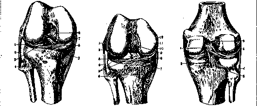
Figure 2.
Figure 3.
Figure 4.
Figure 2. Knee joint from the front: 1 - meniscus lateralis; 2 - lig. collat. fibulare; 3 - lig. cruciatum anterior; 4 - lig. capit. fibulae; 5 - lig. transvers. genu; 6' - meniscus medialis; 7 - lig. collat. tibiale; 8 - lig. cruciatum posterior. Figure 3. Knee joint from the front: 1 - epicond. lat. femor.; 2 - condyl. lateralis femor.; 3 - meniscus lateralis; 4 - lig. collat. fibul.; 5 - section of lig. cruc. anterior; 6 - lig. capit. fibulae; 7 - lig. transvers. genu; 8 - meniscus med.; 9 - lig. cruciat. post.; 10 - lig. collat. tibiale; 11 - condyl. med. femor.; 12 - epicond. medialis femor. Figure 4. Ligaments of the knee joint from behind: 1 - lig. cruciat. post.; 2 - lig. collat. tibiale; 3 - lig. capit. fibulae; 4 - art. tibio-fibul.; 5 - lig. collat. fibul.; 6 - lig. cruciat. ant.
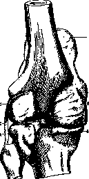
"adheres to the edges of the patella, as a result of which the latter appears inserted into the anterior part of the joint capsule, like in a frame" (Lysenkov). The fusion with the tendon of the m. quadriceps, especially near the patella, is so firm that it is difficult to separate the capsule from the tendon, and often impossible without opening the joint cavity (Shatsky). The attachment of the capsule to the femur bone passes 1-1.5 cm above the edge of the cartilaginous articular surface on both sides to the condyles of the femur in the form of an arched line, convex downward to the level of the collateral ligaments, located medially from them. When the capsule passes from the posterior surface Figure 5. Pouches of the tendon capsule onto the femur of the joint (posteriorly): 1 and 5 - posterior superior pouches (lateral and medial); 2 - lig. collat. fibul.; 3 and 4 - posterior inferior pouches (lateral and medial); 6 - lateral superior pouch; 7 - suprapatellar pouch (recessus suprapatellaris) (fig. 5 and 6). These formations are independent of each other, but often (in 85%, Shatsky) their cavities communicate through an opening of varying size, sometimes to the point that there are almost no boundaries between them (Poirier, Schwarz, Sappey, Rauber, Malinovsky), and this circumstance is more often observed in women. Between the synovial fold of the pouch and the anterior surface of the femur there is usually a rather thick layer of fatty tissue. Descending from the condyles downward, the capsule is fixed along the upper edge of the lateral (outer) surfaces of the menisci, thereby forming protrusions - superior lateral and medial pouches (recessus super. lateralis et medialis, Shatsky). From the lateral and anterior surfaces of the semilunar cartilages, the capsule descends and attaches along the articular edge of the tibia, forming pouches Testut: anterior inferior lateral and medial (recessus anter. infer, lateralis et medialis - Malinovsky, Shatsky). Both pouches communicate with the joint cavity from the side of the sharp edges of the menisci. Posteriorly, the joint capsule attaches along the edge of the articular surfaces of the condyles of the femur, then passes onto the menisci, with the edge of which it fuses, and from here - onto the tibia, where it attaches immediately under the articular edge. The synovial membrane at the same time lines the anterior surface of the cruciate ligaments, leaving them thus behind itself, i.e. outside the joint cavity, and penetrating between them in the form of a blind pouch, sometimes completely detached.
As the membrane transitions from the femoral condyles to the menisci and from these latter to the tibia, recesses are formed: posterior-superior-medial and lateral (recessus poster, sup. medial, et lateralis) and posterior-inferior-medial and lateral (recessus post, infer, medialis et lateralis). Thus, the synovial membrane has a total of nine recesses: 1) anterior superior, 2 and 3) lateral medial and lateral, 4 and 5) anterior inferior medial and lateral, 6 and 7) posterior-superior medial and lateral, and 8 and 9) posterior-inferior medial and lateral. From the point of attachment of the synovial membrane along the inner and outer edges of the articular surface of the patella, well-defined folds extend, between the layers of which are located fatty masses - plicae alares; these folds, converging downward, give rise to a longer synovial fold - plica synovialis patellaris, which, located in the sagittal plane, extends backward toward the lower edge of the intercondyloid fossa of the femur and, together with the cruciate ligaments, divides the joint cavity into two compartments - external and internal. The level of attachment of the joint capsule on the femur is such that the capsule reaches the epiphyseal line anteriorly and posteriorly, while laterally (on the condyles) the capsule only partially covers it. This circumstance explains the possibility of rupture of femoral epiphyseal foci forward and backward into the joint capsule, and the extreme rarity of their rupture sideways. The epiphyseal line of the tibia is located below the joint line (laterally): i-upper attachment of the capsule; 2-anterior recess; 3-lateral recess; 4-plica alaris; 5-lig. patellae. 6-posterior inferior recess; 7-bursa infrapat.; 8-menis-cus; 9-posterior superior recess; 10-lateral superior recess. These latter (numbering 3) are located 1) subcutaneously - bursa praepatellaris subcutanea; 2) between the fascia and the tendon of the quadriceps muscle - bursa praepatellaris subfascialis; and 3) between the tendon of the quadriceps muscle and the periosteum of the patella - bursa praepatellaris subtendinea. The first two usually communicate and form a two-chamber cavity. Between the lig. patellae and the tibia, there is usually a well-developed bursa - bursa infrapatellaris profunda. At the attachment site of the homonymous muscle, there is bursa m. poplitei, s. bursa mucosa poplitea. Its peculiarity compared to others (except b. mucosa suprapatellaris, see above) is that it often communicates with the cavity of the knee joint on one side and with the cavity of the joint between the tibia and fibula on the other. The bursa located between the attachment of the tendon of m. semimembranosi and the medial head of m. gastrocnemii, bursa mucosa semimembranosa Gruberi, communicates with the cavity of the K. s. in 50% of cases. The remaining bursae do not communicate with the joint cavity. The blood supply to the K. s. is provided by a. femoralis, a. poplitea, and a. tibialis. From the first, in the area of the Hunter canal (see), a. anastomotica genu suprema branches off, supplying the upper-medial area of the joint; from a. poplitea, aa. genu sup. med. et lat., aa. genu inf. med. et lat., and a. genu media branch off; the first of these supply the upper and lower lateral areas and extend to the anterior surface, while the middle unpaired one (a. genu media), piercing the capsule, penetrates and branches into the cruciate ligaments. The branches of a. tibialis post. - aa. recurrentes tibiales post. et ant. - supply: the first - the lower-lateral area of the K. s. and the area of the joint between the tibia and fibula; the second - the area of the lateral part of the patella. The branches of all the aforementioned arteries, anastomosing abundantly, form a well-developed network - rete genu, especially developed on the patella and playing a significant role in the collateral circulation of this area. The veins vary extremely but generally follow the arteries. Lymphatic vessels of the anterior part of the K. s. follow the course of the femoral artery to the inguinal glands (Malinovsky); lymph from the posterior and part of the lateral surfaces collects in 4 lymph nodes, one of which is under the fascia at the site where v. saphena parva flows into v. poplitea, and three are in the adipose tissue of the popliteal fossa, along the course of a. poplitea (Sappey). From these nodes, lymphatic pathways go to the inguinal glands. The area of the K. s. is innervated by branches of n. tibialis, n. peronaei, and n. femoralis. The first two supply the corresponding areas of the posterior and lateral surfaces, while the anterior surface receives branches from n. femoralis, from which branches also extend to the skin; the skin of the lower-medial and posterior areas is supplied by branches from n. saphenus (ram. infrapatellaris) and cutan. surae med.; the lower-lateral area is supplied by branches of n. cutanei surae lat. The distribution of innervation fields is very variable, and often there is the so-called 'overlap of innervation fields' (Forster, A. S. Vishnevsky). The anterior surface of the K. s. area is free of muscles. The patella lies against the corresponding articular surface of the lateral femoral condyle and, covered externally by fibers of the quadriceps tendon, is separated from the subcutaneous tissue and skin only by the fascia extending here from the thigh. In the posterior part, there is a series of muscles, their attachment in the K. s. area limiting the diamond-shaped popliteal fossa - fossa poplitea. Its upper-outer edge is formed by the biceps femoris, attaching to the head of the fibula. The upper-inner edge is composed of the semimembranosus (m. semimembranosus) and the more superficially located semitendinosus (m. semitendinosus) muscles, which together with m. gracilis and m. sartorius attach to the tuberositas tibiae in the form of the 'goose's foot' (pes anserinus); in this case, the tendon of m. semimembranosi weaves its fibers into the lig. popliteum obliquum. The lower-outer and lower-inner edges of the popliteal fossa are formed by the heads of m. gastrocnemii, attaching to the posterior-superior parts of the femoral condyles. The floor of the fossa is formed by the area on the posterior surface of the femur (planum popliteum) and the posterior wall of the K. s. capsule. In the K. s., 1) flexion and extension, 2) rotation of the leg outward at the end of flexion and inward at the beginning of extension, and 3) rotation of the leg around its longitudinal axis with the knee bent are possible. During flexion and extension, movements occur between the articular end of the femur and the menisci along an axis drawn through the femoral condyles perpendicular to the structural (stem) axis of the lower limb. (The structural, or stem, axis passes through the center of the head of the femur, through the fossa intercondyloidea femoris, and then through the eminentia intermedia tibiae and malleolus lateralis.) During rotational movements, the lateral meniscus is fixed relative to the femoral condyle, and sliding occurs between the meniscus and the articular surface of the tibia. Flexion in the K. s. is achieved by contraction of the gastrocnemius muscle and the combined action of the biceps, semimembranosus, semitendinosus, gracilis, and sartorius muscles. Of these latter, the biceps, when contracting separately with the K. s. bent, rotates the leg outward, while the others rotate it inward. Extension is performed by the powerful quadriceps femoris muscle.
The joint capsule of the knee is a fibrous sac that surrounds the knee joint, providing stability and containing the synovial fluid that lubricates the joint. It is strengthened by ligaments both inside and outside the joint. The capsule is attached to the femur, tibia, and patella, and it forms pouches or recesses that allow for the movement of the joint. The synovial membrane lines the inner surface of the capsule and covers all the internal structures except the articular cartilages. It secretes synovial fluid, which nourishes the cartilage and reduces friction. The knee joint has several bursae, which are small fluid-filled sacs that reduce friction between moving parts. The most important of these are the prepatellar bursa, infrapatellar bursa, and popliteal bursa. The knee joint is innervated by branches of the tibial, peroneal, and femoral nerves, which provide sensation to the skin and muscles around the joint. The blood supply comes from the femoral, popliteal, and tibial arteries, which form a rich network of vessels around the joint. The lymphatic drainage of the knee joint is primarily to the inguinal lymph nodes. The muscles that act on the knee joint include the quadriceps femoris, which extends the knee, and the hamstrings, which flex the knee. The gastrocnemius muscle also assists in flexion. The knee joint is a complex joint that allows for a wide range of movements, including flexion, extension, and rotation. It is one of the most commonly injured joints in the body, particularly in athletes.
P. Kupriyanov. Method of examination of the K. s. 1. Changes in the configuration of the diseased K. s. compared to the healthy one are determined by comparing both limbs. -2. The presence of swelling and fluctuation in the joint is determined by palpation; the latter can be true in the presence of fluid and false in the filling of the joint cavity with fungous masses. Fluctuation in the K. s. is determined by encircling with the fingers of one hand the fold of the joint capsule from above and inside, and with the other-from below and to the side. Pressing with one hand on the capsule, a lifting sensation is felt with the other hand. False fluctuation in a fungous joint differs from true fluctuation in that it is detected on a small area of the knee, does not transmit from one side of the joint to the other, and with it there is no clear ballottement of the patella. Ballottement sometimes gives a tactile sensation, sometimes an auditory sensation from the impact of the patella (choc rotulien of French authors) against the femoral condyles, obtained only in the presence of fluid between the patella and the condyles, which, when pushed, moves to the lateral areas of the joint capsule. -3. The condition of the synovial membrane, which in normal state is not palpable, but when thickened is palpated in the form of a ridge at the borders of the folds of the capsule. -4. The ligamentous apparatus of the knee is examined for the presence of a) painful points at the attachment sites of the collateral ligaments; b) lateral mobility in the extended position of the limb (rupture or stretching of the collateral ligaments, themselves or together with the cruciate ligaments); c) the presence of rotational movements of the tibia relative to the femur in the extended state (damage to the collateral and cruciate ligaments); d) the amplitude of flexion and extension in the knee joint; e) the condition of the transverse mobility of the patella (its disappearance indicates ankylosis of the K. s, in which the patella participates); f) the presence of anterior-posterior movements (mouvements de tiroir-drawer movements) when the cruciate ligaments are damaged: the subject lies on the table, flexing the thigh to the pelvis and the leg to the thigh until the foot stands flat on the table; at this moment the examiner, grasping the upper part of the leg with both hands, pulls it toward himself (resembles pulling out a drawer). With damaged cruciate ligaments, a distinct forward movement of the tibia occurs-sliding of the planum tibiae over the condyles. The presence of the opposite movement (pushing in the drawer) occurs when the cruciate ligaments are damaged plus the joint capsule, especially the collateral ligaments, lateral or medial. -5. Condition of the m. quadriceps. -6. Condition of the popliteal fossa. Pathology of the K. s. Developmental defects of the K. s. are divided into congenital anomalies a) of the patellar apparatus and b) of various other components of the K. s. - A. Congenital anomalies of the patellar apparatus according to the classification of Kirmisson (Kirraison) are divided into absence of the patellar apparatus and dislocation of the patella. Absence of the patella manifests in that with bent knees their anterior surface flattens. Palpation reveals both condyles with a groove between them. With active extension, absence of the patella is found with almost normal function. Functional disturbance is observed when there is simultaneous weakness of the knee apparatus. -Therapy-prolonged electrization and massage; such treatment sometimes gave impetus to the subsequent appearance of bone foci where even X-rays established complete absence of any bone substance (Kirmisson, Nove-Josserand). This is probably explained by the fact that in a certain percentage of cases this anomaly is only a delayed development. Where absence of the patella causes extreme relaxation of the joint and impairs its function, the wearing of knee pads should be resorted to. Congenital dislocations of the patella almost always occur outward. According to Bessel-Hagen, they are divided into luxatio incomplete, intermittens et completa permanens. In incomplete dislocation the patella is placed in front of the lateral condyle. Intermittent dislocations occur only with special movements, usually with strong flexions of the knee. In complete dislocation the patella remains constantly dislocated and is placed on the lateral surface of the condyle. Dislocation of the patella usually manifests after the child begins to walk, sometimes at the age of 10-12 years. -Symptomatology: flattening of the knee, depression of the intercondylar space, displacement of the patella above or to the lateral side of the lateral condyle; functional disturbances, if there is no pain and effusion, are often mild. Luxatio intermittens is usually accompanied by sudden pains and functional disturbances-the leg is instantly lifted, buckles. Hence in such children fearfulness and uncertainty in gait. -Treatment: wearing of Haudek knee pads, massage, electrization. In persistent cases-operation on bones (due to the young age of the patient and unfinished growth is irrational), on the joint capsule; operation for fixation of the patella with the help of fascial formations, free or on a pedicle; such are: 1) operation of Krogius (Krogius) (fig. 7 and 8), 2) operation of Gobell (Gobell) (fig. 9 and 10), 3) operation of Klapp (Klapp) (fig. 11).
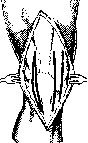
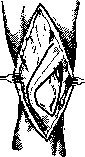
Figure 7.
Figure 8.
Formation of a double flap according to Krogius. The meaning of the operations is clear from the accompanying figures. -Patella bipartita was first described by Gruber (Leningrad; 1883); this anomaly-division of the patella lengthwise, crosswise or obliquely into 2 or more parts-may present clinical interest when discovered after trauma, when it can be confused with a fracture (for differential diagnosis X-ray is needed, a film of the other limb) or when accompanied by painful apophysis with subsequent hydrarthrosis, sometimes simulating tbc. Treatment of patella bipartita is not required. -B. Congenital deformities of the knee, a) Genu recurvatum-unilateral or bilateral-is characterized by hyperextension of the K. s, with the tibia forming with the femur an angle, open forward, obtuse, straight or even acute-up to the possibility of contact of the anterior surfaces of the tibia and femur. Genu recurvatum is usually accompanied by other congenital defects of the knee and is considered as the first degree of congenital anterior dislocation of the tibia. -Treatment: in reducible-after reduction immobilization in the position of flexion. In difficult cases reduction is performed under general anesthesia. In case of impossibility of reduction-osteotomy of the tibia with lengthening of the tendon of the m. quadriceps and others. b) Congenital recurrent dislocation of the knee on the basis of weakness of the ligamentous apparatus. Cases of healing by means of sutures and plication of the joint capsule have been described.
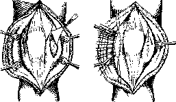
Figure 9.
Figure 10.
Free aponeurotic plastic surgery according to Gobell. Acquired deformities of the K. s: genu valgum, varum et recurvatum. a) Genu valgum, X-leg. In the normally obliquely running femur connects with the vertically standing tibia at an angle, open outward, of 170-177°. Reduction of this angle, which usually does not exceed 145°, creates conditions for genu valgum. -The degree of genu valgum is determined by the distance between the medial malleoli with the knees together; these indicators are usually used in mass examinations. Genu valgum can be unilateral or bilateral. As a rule with the knee bent this deformation disappears. -Subjective symptoms: pains, fatigue when standing and walking, especially when carrying weights, disturbance of gait, which becomes straddling to avoid knocking the knees together. At the same time pes valgus exists, rarely-varus, oblique position of the pelvis and scoliosis. According to the modes of origin genu valgum is divided into: 1) congenital (rare); 2) traumatic-in ruptures of the medial collateral ligaments, improperly united fractures of the femur and tibia (rarely); 3) inflammatory (sometimes in tbc, osteomyelitis, arthritis deformans and neuropathica); 4) paralytic-in spinal infantile paralysis; 5) rachitic-very often in children 1-5 years old with florid rickets in the presence of other rachitic curvatures; 6) static, as deformation from overloading (Belastungsdeformitaten)-very often at the age of 13-18 years, especially in boys of delicate build in standing professions (in bakers, blacksmiths, carpenters, porters, waiters, etc.). Only the last two groups have great practical importance, which are still often denoted by age
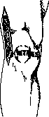
Figure 11. Fascial
plastic surgery according to Klapp's principle-genu valgum infantum et adolescentium.-Treatment should, if possible, be causal. In rickets-anti-rachitic treatment; early walking should be prevented.-In genu valgum in adults, the main treatment is a change of profession. Since many authors also associate this form with late rickets, general strengthening of the body by constant exposure to fresh air and sun is recommended here, as well as avoiding prolonged standing with legs spread apart; walking with toes turned inward, orthopedic inserts, and heel pads on the inner side are recommended. For severe rachitic deformities-restraining splints and apparatus with gymnastics and massage due to the possibility of relaxation of the ligamentous-capsular apparatus. For severe deformities and florid rachitis-reduction with a plaster cast. For completed rickets and failure of splint treatment-surgical intervention: usually osteotomies-wedge-shaped or linear.-b) Genu recurvatum-a rare deformation-divided into: 1) congenital; 2) traumatic-from rupture of the ligamentous apparatus, especially the cruciate ligament; 3) inflammatory, especially in arthropathia tabetica; 4) paralytic-in frequent or complete paralysis of the knee extensors; 5) static, especially after fractures; 6) rachitic.-Absence of tibia-partial, usually of the lower part, or complete.-Symptomatology-severe constant flexion of the knee. Palpation and X-ray reveal the presence of only the fibula. Cause (according to Riese)-arrest of development on the basis of a tight amniotic sac. Due to the severe impairment of function, a number of osteoplastic operations have been performed. Injuries to the K. s. are divided into closed and open.-1. Closed injuries to the K. s. are distinguished according to the mechanism of origin and the nature of the injury sustained, a) According to the mechanism of origin, injuries to the K. s. are divided into contusions and sprains, which differ in the following signs: in contusion there is direct trauma, in sprain-indirect-tin contusion there is usually some injury to the skin, in sprains there is none; tenderness on pressure is found in contusion to the greatest extent at the site of contusion, in sprains-at the sites of attachment of the collateral ligaments.-Determining the mechanism of origin is important for establishing the diagnosis of so-called intra-articular injuries (Binnenverletzungen), especially since X-rays can only be useful in injuries to the bones of the joint.-Injuries from contusions and sprains of the K. s. 1. Paraarticular: simple ecchymoses, hematomas in the subcutaneous tissue or in the numerous bursae, ruptures of tendons or the patellar ligament.-Treatment according to the usual principle-first cold and rest, then heat and movement. For extensive hemorrhages to accelerate absorption-punctures; for ruptures of tendons and ligaments-suture. 2. Injuries to the various components of the K. s., namely: the synovial membrane, ligaments, menisci, bones. Injury to the synovial membrane leads to hemorrhage into the joint-hemarthrosis. In the treatment of hemarthrosis of the K. s., two methods are used: 1) the old classical'-rest, cold, immobilization for 15 days (Lecene) with or without puncture, etc. Subsequent intensive physical therapy-massage, baths, air baths, mud baths. 2) Willems' method-early repeated punctures and active movements without splints and cold.--Injuries to the ligaments-collateral, posterior, and cruciate. The mechanism of injury and diagnosis are clear from the anatomy and physiology of the ligamentous apparatus. In particular, tender points, mobility are determined. A. Symptoms of injury to the medial collateral ligament (ligamentum collaterale tibiale). 1) Severe tenderness on palpation of its lower attachment on the upper part of the medial surface of the tibia, slightly above the level of the tuberositas ant. tibiae, the area of its upper attachment, and along the entire ligament. 2) Impairment of mobility, which is determined as follows: the limb is extended, the left hand firmly fixes the lower end of the thigh, the right hand deviates the leg outward. Absence of outward inversion indicates the integrity of the ligament, tenderness during this act indicates stretching, clear inversion to a right angle-indicates simultaneous rupture of the cruciate ligaments (Lejars).-B. Symptomatology of rupture of the lateral ligament: 1) tender points at the attachment sites; 2) mobility, tested as in injury to the medial ligament, but in the opposite direction.-C. Injury to the posterior ligament is determined by the presence of a small and painful hyperextension compared to the healthy leg, firmly fixing the thigh to the bed and simultaneously lifting the leg. A large amplitude of hyperextension indicates injury to the cruciate ligaments.-D. Injury to the cruciate ligaments. Symptomatology in rupture of the anterior cruciate ligament is hyperextension of the knee, and the thigh is displaced backward on the tibia or the tibia slides forward. In rupture of the posterior ligament, the thigh may be pulled forward or the tibia backward on the thigh. Rupture of both of these ligaments occurs less frequently. Diagnosis is determined by the presence of the drawer sign (see above), increased rotation with flexion of the knee, lateral displacement with the knee bent (rupture of both cruciate ligaments and one or both collateral), avulsion of bony parts on X-ray, and pain and effusion in the joint.-Rupture of the cruciate ligaments is rare, only in severe injuries (dislocations), and is often associated with avulsion of bony fragments at their attachment sites and rupture of the collateral ligaments, especially the medial.-Treatment. Initially, a plaster cast in a semi-flexed position can give excellent results; then usual physical therapy procedures. If unsuccessful-arthrotomy with removal of bone fragments and subsequent suture or plastic repair of the cruciate ligaments.-E. Injury to the menisci-see Joint menisci. Injuries to the patella are divided into dislocations and fractures.-Dislocations of the patella most often occur outward under the influence of direct trauma (a blow to the medial edge of the patella with the knee bent) or, more often, indirect: contraction of the quadriceps muscle at the moment of abduction of the leg with the knee bent, an awkward jump, a fall. Predisposing factors: small size of the patella, X-shaped legs, flattening of the edge of the lateral femoral condyle, relaxation and stretching of the joint capsule on the basis of chronic effusion, etc. Dislocations can be complete and incomplete.-Symptoms of complete dislocation: the patella protrudes outward from the K. s.; upward goes the tendon of the quadriceps muscle, downward-the patellar ligament; the knee is slightly bent, in an X-shaped position, the leg is rotated outward, there is effusion in the joint. Reduction is performed by direct pressure with the quadriceps relaxed; a pressure splint is applied in the extended position for 2-4 weeks.-If the treatment course is unsatisfactory and the capsule is relaxed, habitual dislocation should be expected, for which surgical treatment is indicated (see above-congenital dislocation of the patella); dislocations inward are very rare, as well as dislocation with twisting around the vertical axis.-Fractures of the patella-see Patella. Free bodies in the K. s., or joint mice, can be single and multiple. By origin they are divided into traumatic and pathological. Traumatic ones arise from sprains, contusions, intra-articular injuries, etc., and the formation of these bodies sometimes occurs very gradually as a special process of osteochondritis dissecans, usually in young individuals between 15 and 35 years of age. Pathological ones form in various chronic arthritis accompanied by proliferation of synovial membrane papillae and deposition of fibrin in the joint.-Symptoms: 1) presence of a more or less movable foreign body in the joint; 2) often chronic serous effusion; 3) attacks of incarceration: sudden cutting pains, sometimes with a click, sudden cessation of movement in the joint with more or less prolonged painful flexion contracture. Such attacks often persistently repeat. Complications: chronic synovitis, joint instability, deforming arthritis.-Diagnosis is sometimes confirmed by X-ray, which reveals a foreign body in the joint if it has a bony nucleus.-Treatment consists in removing the free bodies by arthrotomy. For single bodies-a small incision over the previously fixed body, for multiple bodies-a more or wide arthrotomy. Among open injuries to the K. s., it is advisable to distinguish gunshot wounds. They prevail over gunshot wounds of other large joints, constituting, according to statistical data from the last three major wars (Franco-Prussian, Russo-Japanese, and World War 1914-18), from 28.6% to 54% of all joint wounds. The frequency of knee wounds is explained by the large space that the K. s. occupies on the lower limb: one recessus superior extends along the entire lower third of the thigh.-According to anatomical features, gunshot wounds of the knee are divided into the following groups: 1) penetrating wound of the synovial capsule, 2) blind wound of the synovial capsule (with a bullet lodged), 3) wound of the synovial capsule and bone, 4) wound with fracture of the articular ends, 5) wound with comminution of the articular ends (according to Leriche-Lecene).
The first four groups in mass casualty care for the wounded (field warfare) require conservative treatment with immobilization. For wounds of the fifth group, primary debridement with immobilization is necessary. The positional war, with relatively few gunshot wounds and the proximity of medical organizations, prompted the French during the 1914-18 war, under suitable conditions, to apply more active treatment methods in the first hours after injury to prevent infection and to restore joint function more quickly. When one synovial sac was injured, successful use was made of excision of the entrance and exit gunshot wounds with inspection of the joint cavity through the enlarged openings and the most meticulous hemostasis to prevent hemarthrosis, after which both wounds were sutured tightly. When a bullet was present in the cavity of the K. s., it was removed either through the excised gunshot channel or through a separate incision. Types of inflammation of the K. s. in wounds complicated by infection. -1. Synovitis sero-fibrinosa. The joint is filled with fluid with large flakes of fibrin. The synovial membrane is red and covered with a sticky fibrous layer consisting of a mixture of synovial fluid with fibrin. In the first 24 hours, bacteria are usually not found, and the process can even end favorably with a significant rise in temperature. -2. Empyema of the K. s. usually develops from the previous type and is characterized by the presence of free pus in the joint with the melting of the inner layers of the joint capsule. The joint is tense and sharply painful. -3. Phlegmon of the joint sac most often arises from the previous form, sometimes primarily, and represents progressive purulent inflammation of the entire connective tissue joint apparatus. Phlegmon of the sac ends in total suppuration of the joint with frequent involvement of the bony articular ends. Treatment of severe purulent lesions of the K. s. comes down to wide opening of the joint, drainage and immobilization. -According to various German authors, mortality from injury to the K. s. ranges from 4.3% to 20.4%. The functional results are shown by the following data of Erlacher (Ph. Erlacher), who studied 260 convalescents after injury to the K. s. and found among them: 139 ankyloses, 8 loose joints, 74 with severely limited mobility, 36 with moderately limited mobility, 3 with normal mobility.
_
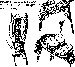
Stab and incised wounds, to which most so-called accidental injuries belong. Differential diagnosis between empyema and phlegmon of the sac (according to Landois-Payr). Empyema Phlegmon of the sac Consistency of the joint Elastic fluctuation. The joint is swollen. Fluctuation is often unclear. External appearance of the joint Initially absence of inflammatory changes in the area around the joint. Strong inflammatory edema of the soft parts and skin, perforation of the sac, para-articular swellings. Para-articular abscesses. Puncture Much pus Little pus or none at all. Active mobility In the initial stage good and relatively painless; active muscular fixation is lost with progression of the process. Worse. The same with muscular fixation. Pain Initially insignificant. Very severe pain with passive movements. Position of the joint The patient assumes an active position, in which he has the least pain in the joint (mid-position) and passively fixes the joint reflexively with muscles. Palpation Painful only over the joint capsule; no pain on traction and jolting. The entire joint is painful on palpation. Severe pain on traction and jolting. The popliteal fossa is often swollen and painful. General condition Not particularly disturbed; temperature up to 39°. Heavily disturbed; septic condition, dry tongue, high temperature and chills. Embolisms.
Stab and incised wounds of the K. s. can give the same pathological picture and require the same treatment as the previous ones. Contractures, ankyloses and paralytic deformities of the knee. - Contractures can be dermatogenic, myogenic, neurogenic and arthrogenic. Dermatogenic arise on the basis of injuries, burns, ulcers, etc. Myogenic are based on a) ischemia due to tight and long dressings, ligation of blood vessels; b) muscle scars after injuries and inflammations; c) nutritional shortening with prolonged rest due to fractures, joint diseases, etc. - Arthrogenic contractures are caused by shrinkage of the soft parts (joint capsule, ligaments) in joint diseases. Due to the predominance of flexors over extensors, contracture of the knee occurs predominantly in the form of flexion, which is very unfavorable for the supporting function of the lower limb in contrast to contracture in extension. Flexion is often combined with abduction. - Therapy distinguishes between recent and old cases. The former are treated with active and passive movements, heat (baths, hot air, diathermy, etc.), massage and electrization, injection of substances that dissolve scars (fibrinolysin, etc.). In more stubborn cases - continuous traction, splint apparatus with redirecting devices, careful forcible redressement under anesthesia. In old cases, one has to resort to surgical treatment: excision of scars and plastic closure of defects (Italian plastic surgery, tubular flap according to Filatov) and to lengthening, plastic surgery and sectioning of tendons and muscles in myogenic contractures. In arthrogenic contracture, osteotomy in all its modifications and resection are used. Ankyloses. The only cause of ankylosis is arthritis in various forms. Immobilization itself without arthritis cannot cause ankylosis. According to the nature of the adhesions, ankyloses are divided into incomplete fibrous and complete bony. Indications for operation in ankylosis of the K. s. are 1) fixation of the joint in a faulty position, unfavorable for the function of the limb, 2) the patient's persistent desire to restore lost mobility of the joint. - There are many ways to mobilize an ankylosed K. s. surgically (Figs. 12 and 13). Long-term results
Figure 12. Figure 13. Figure 12. Fascial pad according to Raug. Figure 13. Arthroplasty according to Raug. Inflammations of the bursae of the K. s. area are divided into acute and chronic. Acute ones arise either through direct injuries or per continuitatem from neighboring infected parts. Most often one has to deal with inflammations of the prepatellar bursae and the bursae of the popliteal fossa. - Acute inflammations of the prepatellar bursae are characterized by general and local symptoms of inflammation, the latter corresponding to the anatomical localization of the bursa. Besides localization in front of the patella, bursitis praepatellaris differs from inflammation of the K. s. by the absence of swelling of the entire joint, ballottement of the patella and sharp limitation of mobility. - Chronic inflammation arises either as an outcome of acute ones or independently on the basis of chronic trauma (in parquet floorers, roofers, foundry workers, etc.). The diagnosis of bursitis praepatellaris is determined by the presence of a cystic tumor with typical localization, firm-elastic or fluctuating, little or not painful, covered with normal movable skin. Bursae of the popliteal fossa have the appearance of cystic op
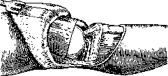
Figure 14. Joint transplantation according to Lexer.
chole and are accompanied by tension, pain, fatigue, difficulty bending, walking, climbing stairs, and often by paresthesias and neuralgias. Such cysts are better seen with the knee extended, but better palpated when bent. It is necessary to differentiate them from aneurysms, cold abscesses, tumors, etc. In differential diagnosis, one must keep in mind the possibility of specific diseases of the bursae (tuberculosis, syphilis, gonorrhea, gout, etc.), which in stubborn cases may also require surgical intervention. - Treatment: suppurated cysts must be incised; chronic cysts as a rule do not yield to therapeutic treatment. Surgical intervention is indicated when there is impairment of function, large size of the cyst, and pain. Puncture is useless and sometimes dangerous; the only rational intervention is extirpation. Tumors in the area of the K. s. From the skin, subcutaneous tissue, and aponeurosis originate lipomas, fibromas, myxomas, sarcomas, and carcinomas. In the mucous bursae, fibromas, chondromas, osteomas, myxomas, and sarcomas have been observed. From the joint capsule and in particular from the synovial membrane originate fibromas, chondromas, sarcomas, and those having a special clinical interest—lipomas. Lipomas are observed either as limited tumors the size of a walnut or cherry, or in the form of the so-called lipoma arborescens—a tree-like lipoma occupying larger or smaller areas of the synovial membrane and originating from fat in the duplicatures of the synovial membrane.-Diagnosis of intra-articular tumors is difficult due to indefinite symptoms resembling those of joint bodies, but without pain. From cartilage and bone originate cartilaginous exostoses, chondromas, and sarcomas. The latter, by breaking into the joint, may give rise to confusion with tuberculous arthritis. Diagnosis is established by X-ray; the same tumors may originate from the patella and in that case are easily diagnosed and operated on. Among the operations performed on the K. s., mention should be made of transplantation not only of individual bones but of the entire K. s., taken from a freshly amputated limb or from a cadaver, proposed and successfully performed by Lexer (fig. 14). v. nedokhlebov.
Related articles
Mentioned in
Cite this page
“Knee Joint.” Soviet Medical Encyclopedia. English translation of Bolshaya Meditsinskaya Entsiklopediya, 1st ed. (Moscow, 1928–1936), ed. N. A. Semashko. https://sovietmedicalencyclopedia.pages.dev/article/knee-joint/