Gonitis
Historical document, translated for reference. It reflects medical knowledge of the 1920s–30s and is not medical advice.
Summary
Gonitis is an acute or chronic inflammatory process of the knee joint, which may affect the synovial membrane or the bony structures. This 1930s encyclopedia entry details the etiology, clinical forms, and treatment methods for various types of gonitis, including rheumatic, gonorrheal, and syphilitic.
Encyclopedia article (1928–1936)
GONITIS, gonitis (from Greek gonu—knee), an acute or chronic inflammatory process in the knee joint, affecting either its capsule (synovial membrane) or the bones forming the joint; it occurs either in a capsular form (synovial) or in a bony form, depending on whether it begins with the capsule or with the osteo-cartilaginous ends of the joint. This classification is maintained even when, at the height of the process involving adjacent tissues, one form transitions into another. The extensive volume, magnitude of load, and complexity of the knee joint's structure predetermine both the frequency of its involvement compared to other joints and the diversity of clinical and pathological-anatomical forms. According to large statistics, gonitis accounts for 35% to 52% of all joint inflammations. Most often, the etiological factors causing gonitis are infections (tuberculosis, syphilis, gonorrhea, diplococcus, staphylococcus, streptococcus). In view of the impossibility of establishing the etiology bacteriologically in every individual case, diagnosis often has to be based exclusively on the clinical form with one or another pathological-anatomical manifestation. Although different infections often cause similar pictures, the latter still present certain features characteristic of each infection. The diversity of clinical, and consequently pathological-anatomical, forms of any given etiology is a function of two variables: the polymorphic virulence of the microbe or toxin, on the one hand, and the constitution, age, and reaction of the organism and tissues, on the other. In this regard, acute gonitis manifests more often in the form of a capsular type, and chronic in a bony type. Among acutely developing synovial gonitis, the following are pathologically distinguished: 1) a purely hyperemic form (synovitis hyperaemica genu), which is for the most part the initial stage of almost every arthritis, often hidden under the diagnosis of arthralgia, and gives no other symptoms besides pain in the joint. The subsequently joining 2) serous synovitis (synovitis serosa genu) manifests with swelling of the synovial membrane, proliferation of villi, and the formation of an effusion, and is clinically determined by swelling of the joint with contouring, fluctuation, and more acute tenderness of the three synovial recesses of the knee and a balloting patella. Active and passive movements of the knee are difficult and painful, temperature is elevated. It is observed more often in common rheumatism and gonorrhea. Gonitis rheumatica does not present difficulties in recognition, as it manifests most often in the presence of polyarthritis with a high intermittent temperature following some staphylococcal or streptococcal infection, most often tonsillitis. In the initial stage, rheumatic catarrhal synovitis is easily amenable to treatment (especially with the use of salicylate preparations) and then does not cause persistent changes in the joint, but it can drag on and transition into the stage of serofibrinous, fibrinous, and adhesive arthritis. These fibrinous forms with a more or less sharp onset, temperature, and pain are characteristic not only of rheumatism but also of gonorrhea, whereby the multiplicity of joint involvement with the catarrhal form of synovitis and the good effect of salicylate preparations speaks more in favor of rheumatism. Gonitis gonorrhoica is observed predominantly in adults in the form of monarthritis, usually with fresh urethritis, often in parallel with the retention of discharge (see Arthritis). The knee is affected more often than other joints—approximately in 40% of cases. No other process causes such rapid and complete destruction of cartilage and formation of ankylosis as the gonorrheal one. Finding gonococci in urethral or vaginal secretion or in the punctate of a trial knee puncture is not always successful even when cultured on ascitic agar, but this is not so important given the characteristic picture of gonorrheal arthritis. Treatment in the acute period—rest (plaster cast or traction); salicylate preparations do not give an effect. Gonococcal vaccine with urotropin is used with success, intramuscularly or intravenously (2 series: from 10 to 100 million—6 injections, and from 100 to 1,000 million—also 6 injections; 3 times a week with a weak reaction, 2 times a week with a more sharp reaction).
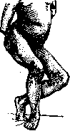
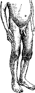
Figure 1. Bilateral gonitis with ankylosis (septic, after scarlet fever). Figure 2. Syphilitic gonitis—capsular form, with lengthening of the limb and with almost normal function. In the subacute period, diathermy and Bier's active hyperemia for 20–22 hours with small intervals. In case of ankylosis—attempts at plastic resection with fascial interposition. A transitional stage from a purely synovial to a purely purulent form is Volkmann's purulent catarrh, pathologically resembling diffuse conjunctivitis with ridges and folds of the synovial membrane. In purely purulent gonitis, with purulent infiltration spreading to the surrounding tissues, necrosis of the cartilage begins early, and after it, the ends of the epiphyses are involved in the destruction, causing osteomyelitis. On the other hand, primary acute osteomyelitis of the articular ends can cause empyema of the joint. Both of these latter forms of acute purulent gonitis (gonitis acuta purulenta) are most often caused by pyogenic microbes (staphylococci, streptococci, or diplococci), as well as by the dysentery bacillus and toxins of eruptive diseases (typhus and scarlet fever). Clinically, gonitis acuta purulenta is characterized by a sharply elevated temperature, with severe, often septic phenomena, significant swelling of the joint, and diffuse pain, which intensifies upon attempts at movement. The joint assumes a shape characteristic of fluid accumulation in the knee joint (the so-called Bonnet position)—flexion at a right angle—and is subsequently held in this position due to muscle tension. These gonitides leave behind severe contractures, ankyloses (see Figure 1), and scars from fistulas that suppurate for a long time or from arthrotomy incisions, which are indicated mainly for empyemas caused by pyogenic microbes, especially in the presence of general sepsis. In the latter case, the use of electrargol is recommended—intravenously or intramuscularly, 5–10 cubic cm daily or every other day, until the temperature drops. Finally, according to the character of the effusion, hemorrhagic synovitis is distinguished. Being an admixture to ordinary exudate in acute infectious arthritis, blood can indicate the intensity of the inflammatory process. Furthermore, the knee is a favorite site for synovitis haemorrhagica in hemophilia and scurvy: clinically, in these cases, besides the bloody effusion, there is involvement of the gums and ecchymoses on the skin of the knee.
All the listed capsular forms of acute gonitis of the most varied infections can cause secondary changes in the bone. Primary changes in the skeleton of the knee, however, most often represent the product of chronic gonitis. Chronic gonitis is also subdivided into capsular forms (effusive), most often caused by syphilis, and bony forms—fungous or dry, caused more often by tuberculosis. Chronic dropsy of the knee, like that of any other joint, in the form of either synovitis serosa or sero-fibrinosa, develops from acute gonitis or proceeds from the very beginning in the form of chronic synovitis, more often on the basis of syphilitic, less often tuberculous, infection. Gonitis syphilitica (see Figure 2) manifests most often with signs of benign chronic synovial arthritis. In a growing individual, one finds a lengthening of the diseased limb by 2–3 cm, and with the help of X-rays, also a widening of the bony ends of the diseased knee by 0.5 to 1 cm. The fluid as a rule gives a positive Wassermann and Meinicke reaction, which are sometimes obtained in the blood of these patients as well. If the chronic process drags on, then the irritant acting on the synovial membrane for a long time creates changes in the villi with swelling, infiltration, and fibrous degeneration—synovitis chronica sero-fibrinosa; with the further development of this process, synovitis chronica villosa hy...
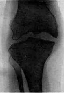
perplastica. Fibrin deposits or overgrown villi at the site of necrosis of the detached synovial membrane may separate from it and form bodies freely floating in the joint. Due to the movement of the joint, they are polished and form so-called free rice bodies, or joint mice (corpora oryzoidea), which are subject to incarceration with pain during movement. A process in which these villous formations come to the fore in the form of voluminous growths (lipoma arborescens), starting from under the synovial membrane or from under the synovial villi (fibroma arbor, genu), is distinguished as a special pathological-anatomical type. Clinically, instead of a rounded, well-mobile, and slightly painful joint, there is a bumpiness and a kind of lobulation of the protruding capsule on the sides of the kneecap and in the area of the recesses; being elastic, they are palpable in the form of a separate tumor or a series of them, of various sizes, of cartilaginous consistency. Their determination is important: if simple, slightly painful capsular forms of chronic gonitis, with good joint function, give the right to suspect a syphilitic infection, then fibroma s. lipoma arb. gives this right even more so. On an X-ray, with insignificant changes in the bone and joint space, there are focal erosions at the sites of ligament attachments, mainly the collateral and cruciate ligaments. In terms of treatment, the administration of large doses of potassium iodide, according to Velyaminov, is indicated here in adults and Sirupus Ferri jodati in children, as well as opening the joint and removing the pathological growths. In cases with the formation of fibrous and bony ankylosis with an incorrect position of the articular ends, resection is indicated. Gonitis tuberculosa constitutes half of all knee diseases, predominantly in childhood, equally often in girls and boys; in the vast majority, the process is unilateral; bilaterality of the disease in doubtful cases may speak against tuberculosis. A bony, or dry form (tbc sicca) is distinguished, in which the capsule is little affected and only fibrotically degenerated, and a fungous form, in which the growth of granulations in the capsule is sharply pronounced. Clinically—at a normal or slightly subfebrile temperature in an asthenic child aged 2 to 14 years, often several weeks after a minor injury (it should be noted that a more significant injury with hemorrhage or bone fracture usually does not cause tuberculosis), pain, atrophy of the thigh and lower leg muscles appear with a gradually developing tumor of the joint, giving the leg a spindle-shaped appearance. At the beginning of tuberculosis, the toxin irritates the epiphyseal line and, like syphilis, causes an increase in bone growth and lengthening of the entire limb; later, at the height of the process, the presence of sharp atrophy is accompanied (according to Novotelnov) by the suppression of the epiphyseal line: a retardation of the growth of the entire limb and even the foot occurs. Palpation determines tenderness of both epiphyses, especially at the sites of capsule attachment. The X-ray picture is characteristic: osteoporosis of the epiphyses, more often total, less often focal, without a hint of hyperplasia, with scalloping of the bone-articular ends, with blurring of the joint line (see separate table, fig. 1). The peculiarities of tuberculous gonitis also include changes in the joint produced by effusion, stretching of the capsule and ligaments, as well as connective tissue degeneration of the bursa, with filling of the joint with fibrous and scar tissue, which, together with painful, reflex, muscular contracture, creates persistent static changes. Inflammatory granulations spread to the cartilage; destroying it and replacing it with connective tissue, they fuse the articular surfaces, forming ankylosis genu fibrosa s. ossea. Most often with
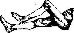
Figure 3. Tuberculous gonitis with posterior subluxation.
this, a flexion contracture of the knee with posterior subluxation of the lower leg (see figure 3), genu valgum or varum, depending on which condyle is more affected, is obtained. The more frequent formation of genu valgum in this case (see figure 4) is due to the greater load on the condyli lateralis in the presence of a normal genu valgum and participation in the reflex shrinking of the fasciae latae. Rare synovial forms of chronic tuberculous gonitis can produce clinical and pathological-anatomical pictures similar to syphilis. It is easy to differentiate them from syphilis: early-onset stiffness, more severe atrophy and pain, a negligible amount of effusion, negative Wassermann reaction and Meinicke reaction not only in the blood but also in the knee aspirate, and osteoporosis of the affected epiphyses characterize the granulation or fungous form of tuberculous synovitis. Due to the transition of the tuberculous process from opened bone or capsular foci into the soft tissues and the rupture of the skin, a long-suppurating fistula is formed. A secondary infection penetrating from the outside leads to osteomyelitis, often spreading to the diaphysis. Tuberculosis complicated by a syphilitic infection creates a very severely progressing hybrid form. - Treatment. The earlier the tuberculous process is detected, the sooner it yields to subsidence. Fresh gonitis in the presence of contracture requires, along with enhanced nutrition, constant traction or light, single-stage redressment followed by a plaster cast. Neglected cases with flexion and subluxation yield to straightening with the Yermolayev
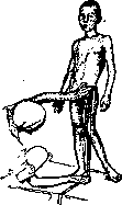
Figure 4. Tuberculous genu valgum.
apparatus (see figure 5) or are corrected by supracondylar osteotomy. With more severe contracture, a double osteotomy is performed
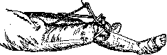
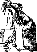
Figure 5. Straightening of flexion contracture of the knee with the Yermolayev apparatus.
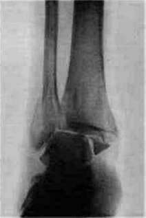
on the thigh and on the lower leg [see separate table (art. 675-676), fig. 2]. Osteotomy, by creating irritation near the epiphysis, is to a certain extent a replacement for the Lavalle operation, which has not yet received widespread acceptance. In cases of bony ankylosis of the knee at an angle [see separate table (vol. I, art. 756-757), fig. 1-3], a wedge-shaped resection must be performed before straightening the leg. If one tries to create a neoarthrosis with fascial interposition, then, to avoid flail joint in such a knee, it is necessary to spare the collateral ligaments and not create a large diastasis between the cut surfaces. To avoid severe shortening of the leg in a growing child, it is necessary to spare the epiphyseal lines, therefore, do not perform resections and neoarthrosis before the age of 17. Cold abscesses should be punctured, preventing the formation of a fistula. The latter, in the absence of sequestra, yield to iodoform filling according to Mosetig-Moorhof. Such fistulas close significantly faster in a southern climate, in the sun, or during treatment with a quartz lamp. In addition to the forms listed above, there are forms of gonitis of a neurogenic or trophoneurotic nature: osteochondritis genu juvenilis, observed in adolescence (see separate table, fig. 3), and arthropathia tabetica (see figure 6) in adults—proceeding benignly and being transitional forms to arthritis deformans. In general, this is a degenerative and hyperplastic process in the articular ends of the cartilage, bone, and soft tissues. But in some cases, as with osteochondritis, the process of degeneration predominates, in others, as with arthropathia tabetica, the hyperplastic process predominates (see Arthritis).
S. Novotelnov. GONOSAN, Gonosan, kava-santal, a preparation containing 20% alpha- and beta-resin of kava-kava (analgesic and hemostatic) and 80% East Indian sandalwood oil (diuretic, disinfecting the urinary tract). Used for acute and chronic gonorrhea, 0.3 in capsules, 3-5 times a day. Side effects: belching, heaviness in the stomach, diarrhea (very rarely).
Related articles
Mentioned in
Cite this page
“Gonitis.” Soviet Medical Encyclopedia. English translation of Bolshaya Meditsinskaya Entsiklopediya, 1st ed. (Moscow, 1928–1936), ed. N. A. Semashko. https://sovietmedicalencyclopedia.pages.dev/article/gonitis/