Arthritis
Historical document, translated for reference. It reflects medical knowledge of the 1920s–30s and is not medical advice.
Summary
A comprehensive overview of arthritis from the 1928-1936 Soviet medical encyclopedia, covering various types including infectious, rheumatoid, and non-inflammatory forms, with emphasis on etiology, clinical manifestations, and treatment approaches of the period.
Encyclopedia article (1928–1936)
Arthritis (from Greek arthron - joint), an extensive group of diseases of the joints of inflammatory and non-inflammatory origin (the latter would more correctly be called arthroses). The phenomena of arthritis can be caused by very diverse irritants of bacterial, toxic, and chemical types; these irritants lead to certain changes in the individual elements of the joint, and it has been established that different irritants can cause the same patho-anatomical changes in the joint, which further complicates the precise classification of A. by groups. At present, it is still not possible to consider and classify A. exclusively according to the causal factor. Similarly, the patho-anatomical nomenclature does not satisfy in all cases. Therefore, it is more convenient to adhere to the classification of A. according to clinical characteristics, combining them, as far as possible, with patho-anatomical and bacteriological data. Among the numerous types of A., there are those where it is possible not only to establish with certainty the inflammatory nature of the disease, but also to determine the etiological factor. Such are, for example, gonorrheal, pneumococcal, typhoid, and paratyphoid A. The role of the gonococcus as an etiological factor in many A. of the 'rheumatoid' type was established long ago (1896 - Thayer, Blumer and later Hordes).-P nevmokokkovye A. are caused by the diplococcus of Fränkel, although cases of mixed infection with diplococcus and other pyogenic cocci have been noted. This form of A. is most often observed in childhood, proceeds relatively peacefully in the form of 'rheumatoid' (arthralgia) serous synovitis and more rarely in the form of purulent A. L e-ch e-n i e-repeated aspirations and vaccine therapy (Tasso). For severe purulent forms-arthrotomy with subsequent washing of the joint. In recent years, especially Russian authors (Kozlovsky, Novotel'nov), note the importance of pneumococcal infection in the development of tuberculosis of the joints.-A. after typhoid fever and paratyphoids are caused by the corresponding bacteria, which was established by a number of authors (Klemm, Hübener, Schneider) and confirmed by Russian scientists in recent years. Typhoid A. manifest in the form of indefinite rheumatoid-type pains in the joints (pseudorhumatisme typhique of the French), in the form of synovitis with non-purulent effusion and in the form of purulent A. with destructive changes in the articular surfaces, with subsequent dislocation or subluxation, especially frequent in the hip joint. Joint disease in connection with relapsing and typhus fevers was also observed by Russian authors during the epidemic of 1919-23. Non-purulent A. proceed favorably, without giving serious complications. The main thing in such forms is the prevention of contractures and subluxations. For purulent forms, interventions applicable in general for purulent A. (aspirations followed by washing with various solutions, arthrotomies, in severe cases-resections) can be used. During the disease, it is necessary to prevent contractures and path. dislocations by traction, k-rye also have their effect in fresh path. dislocations, especially in the hip joint. In chronic cases, surgical treatment is indicated. A whole series of other acute infectious diseases also give complications in the form of A.: such are scarlatinal A., in which streptococcus is almost always found. They proceed in the form of polyarthritis, often with subsequent formation of contractures and ankylosis of many joints. Serous synovitis take a more favorable course. Relatively rare are joint diseases in measles, smallpox, diphtheria, cholera. Malleus also causes joint diseases.- Arthritis in dysentery deserve special mention. Kofoid and Swi-zi (Kofoid, Swezy) in a careful study of the resected head of the femur found amoeba cells in necrotic foci directly at the eburnated articular surface. They tend to consider this amoeba as Entamoeba dysenteriae, the combination of k-roy with chronic A. is a frequent phenomenon. It should be assumed that A., arising in connection with the disease of bacillary dysentery and proceeding more often in the form of polyarthritis, also have a specific etiology. In treatment, all types of physiotherapy in combination with orthopedic manipulations are indicated. It should be mentioned about A. in acute and chronic osteomyelitis, when the focus is localized in the metaphysis or epiphysis, and metastatic A. in septicopyemia (see). To acute lesions of the joints can be attributed their pains and swellings, which are observed as an anaphylactic phenomenon during serum treatment. A large group of chronic A. with specific etiology deserves more detailed description and is given in the corresponding sections. Such are tuberculous A. (see Tuberculosis) and syphilitic A. (see below). Mixed forms of joint damage with gonorrheal infection and syphilis and syphilitic-tuberculous A. (hybrid forms) also occur, which are carefully analyzed in the classical works of Veliaminov. Rarer A. in sporotrichosis, Madura disease deserve only mention.-Joint diseases in hemophilia do not yet have a sufficiently precise scientific explanation, just like hemophilia itself (see). Such A. manifest more often in children and adolescents in the form of hemarthrosis with a slightly thickened capsule or in the form of chronic A. with phenomena of capsule wrinkling, with a tendency to contractures and even fibrous ankylosis. The latter form can easily be confused with other types of chronic A., in particular with tuberculosis. A careful history in doubtful cases facilitates therapy, k-roya comes down exclusively to conservative measures and prevention of deformities.-A somewhat similar picture is given by A. in scurvy, which also represents one of the manifestations of hemorrhagic diathesis (see Scurvy). Among arthritis with unclear etiology, the so-called 'rheumatic' A. occupy a prominent place, k-rye can proceed in the form of polyarthritis rheumatica infectiosa acuta or in the form of polyarthritis rheumatica infectiosa chronica. For most researchers, the infectious nature of such A. is the most explainable, and many English and American authors attach great importance to the so-called focal infection ('focal infection'). However, Lindsay (Lindsay) in a very careful study of 172 cases found the supposed foci of infection only in 50%. Bassler (Bassler) in 35 cases of chronic polyarthritis out of 45 found the supposed source, in the other 10 cases no traces of infection could be established. Removal of foci in 35 patients could not stop the development of the disease. Individual researchers tend to recognize the role of non-hemolytic streptococcus in most polyarthritis. The source of focal infection is most often the tonsils, nasopharynx, root abscesses of teeth, sinusitis. Smith (Smith), studying 100 cases of polyarthritis, performed 60 abdominal operations and was convinced of the role of intestinal infection, which is also confirmed by Taylor (Taylor) on the basis of radiological studies of the intestine. Attempts by individual authors (Nichols and Richardson) to explain the clinical and pathological manifestations of such A. by incorrect metabolism (see) do not agree with the data of Ely and the very careful patho-anatomical studies of T. Fischer (T. Fischer). The small-cell infiltration, usually noted in such A., is evidence of the inflammatory process in chronic forms. Here, as a result of proliferation of the joint capsule, a layer of granulation tissue is formed, which sooner or later grows over the articular cartilage and causes its destruction. Proliferation of the perichondrium leads to the formation of new cartilage or bone at the edges. These true inflammatory changes, characterizing proliferative A., lead to partial or complete destruction of the joint with subsequent fibrous or bony ankylosis. One can share the opinion of Pemberton (Pemberton) about the influence of several factors in the development of the patho-substrate of such arthritis: various types of infection, the influence of humidity and cold, intestinal intoxication, violation of endocrine balance. One cannot also deny the importance of some constitutional predisposition to the so-called 'rheumatoid' A., which occur more often in subjects of 'visceroptotic' or hyper-onomorphic type of Bean (Bean). Acute 'rheumatic' A. usually respond to large doses of sodium salicylate (salicylic preparations in the vein, trypaflavin, collargol, etc.) and pass without leaving serious destructive changes in the joints. However, if they pass into a chronic form, the outcome is less favorable. Removal of the supposed sources of infection (tonsils, carious teeth, etc.) does not always give the desired effect and cannot be expected in advanced cases. Similarly, treatment with vaccine and non-specific protein therapy have not fully justified expectations. True, the latest data of Stockman and Campbell (Stockman, Campbell) are more encouraging. They used for intravenous injections standard typhoid vaccines in doses from 125 to 200 million, achieving a significant general reaction. In 80% of cases, there was undoubtedly improvement, in 57% the course of the disease stopped, and in some chronic cases there was a return. In k. cases, all types of physiotherapy are indicated: dry-air baths, non-irritating massage, active movements, diathermy, etc. Prevention of deformations by traction, splints, orthopedic appliances is a very important moment.
Prolonged fixation reduces blood supply to the area of the diseased joint and increases atrophy. Surgical intervention in chronic forms of rheumatic A. is also indicated. Synovectomy on the knee joint is used by a number of English and American surgeons (Swett and Jones, 1923, present a large amount of material). This operation is better applied early in order to preserve other elements of the joint. Key experimentally proved that the synovial membrane is replaced by a fibroblastic pathway without impairment of function. In the presence of significant destructive changes in the joint, resection is indicated. Arthroplasty on such joints has less success. The next large group of chronic joint diseases constitutes the so-called osteo-A. of English authors or degenerative (hypertrophic) A. of American authors. Although the etiology of this group of A. is just as unclear as the preceding group, the patho-anatomical changes and the distinctive clinical course justify considering 'osteo-A.' separately. These A. occur in patients of middle and above middle age. Climate, conditions, moments of cooling, 'colds' rarely play a role in them. The first changes in the joints, accessible to clinical and X-ray examination, are slight localized swelling of the joint, pointedness of the joint ends, bone-cartilage proliferations at the border of the cartilages. With further development of the process, the joint cartilage disappears, the eburnated bone is exposed, often of irregular shape. Changes of this kind on the fingers are called Heberden's nodes, changes in the hip joint-malum coxae senile. Similar changes are noted in the joints of the spine (see Spondylitis and Spondyloses). To this group should probably also be included the primary, chronic, deforming polyarthritis, in which the process affects the limbs more or less symmetrically, starting with the fingers of the hands (Golubov). Whether to consider such a polyarthritis as a trophoneurosis or whether it should be attributed to disorders of internal secretion-this question should for the present be considered not finally resolved. From such deforming polyarthrites one should strictly distinguish the disfigurement of joints after inflammatory processes in the joints-gonorrheal, typhoid, tuberculous, syphilitic, and others. From Fischer's point of view, osteo-A. is not a disease, but a series of pathologic and physiologic changes in the joint subjected to prolonged and repeated mechanical or toxic injuries of moderate degree. The main feature distinguishing osteo-A. from the more acute and presumably infectious 'rheumatic' A. is the almost constant absence of small-cell infiltration in osteo-A.-To the group of chronic non-inflammatory degenerative A. should also be included those forms of osteo-A. in women that coincide with the onset of the climacteric (A. of the menopause according to Cecil and Archer). The disease occurs more frequently in the knee joint, interphalangeal articulations, lumbar region of the spine in obese women of middle age. The course is slow. There is no tendency to involve other joints in the process. Treatment-regulation of nutrition, diathermy, iodine and thyroidin. Ovarian preparations usually have no effect. To the number of chronic arthritides standing in unquestionable dependence on anomalies of internal secretion should be included polyarthritis progressiva chronica thyreotoxica (Velyaminov), running in the form of polyarthritis on the basis of dysthyreosis; further-A. in hypothyroidism (Hagen-Thorn) and osteo-arthritis deformans endemica of Beck (joint diseases in metabolic diseases, see Gout).-Attention should also be drawn to a group of neuropathic A., among which arthropathia tabidorum and arthropathies in syringomyelia deserve the greatest attention (see Arthropathy).-As for traumatic A., in connection with more precise diagnosis, their circle has recently been constantly narrowing. To the number of a. traumatic should be included proper distortions and contusions with subsequent hemarthrosis (see). The so-called 'internal injuries of the knee joint' (derangement interne) at the present time are divided into 1) injury to the menisci, 2) injury to the cruciate ligaments, 3) rupture of the lateral ligament, 4) injury to plicae alares, 5) fracture of the patella, 6) habitual dislocation of the patella, 7) intra-articular fracture. In accordance with the established diagnosis, the corresponding therapy should be applied, mainly operative. The general position in traumatic A. is, as far as possible, to avoid prolonged fixation.-Summarizing the data presented regarding chronic A., it should be noted that in certain forms the etiological factors are not yet precisely established, and in connection with this the therapy of such A. is not yet on the absolutely correct path. However, the recent tendency toward the union of the internist and orthopedist in the study of A., more profound biochemical, bacteriological, and patho-histological investigations in individual forms of A. are bringing more clarity and definiteness to the pathology and clinic of chronic joint diseases.
G. Meshchersky. Gonorrheal arthritis was described in 1507 (long before the discovery of the gonococcus) by the Dutch physician Foreest. The connection between joint lesions and gonorrheal processes in the genital organs was subsequently proven by finding gonococci in the joint effusion of patients. Gonorrheal arthritis appears within the first two to three weeks from the onset of gonorrhea, but cases of gonorrheal arthritis occurring after several days have also been described. Cases of the appearance of arthritis during the incubation period are also known. A characteristic feature of gonorrheal arthritis is the involvement of the ligamentous apparatus and tendon sheaths; the cavity of the joint itself is involved in the process comparatively rarely. Lesions of the mucous bursae behind the olecranon, at the lower angle of the scapula, at the acromial process are often observed, but most commonly the calcaneal and subcalcaneal mucous bursae are affected, causing the typical lesion of gonorrhea—achillodynia. Pain on pressure at the points of attachment of the Achilles tendon to the calcaneus, on the sole in the location of the subcalcaneal bursa, at the points of attachment of tendons to bones, and lesions of tendon sheaths in other places are characteristic symptoms of gonorrheal arthritis. Statistical data on the frequency of involvement of various joints by the gonorrheal process are not the same among different authors. The knee joint is affected more often than others, followed by the ankle, wrist, shoulder, etc. Gonorrheal polyarthritis occurs significantly more often than gonorrheal monoarthritis. The multiplicity of joint involvement in gonorrhea is fully confirmed by modern views on the pathogenesis of this disease. Most believe that gonorrheal arthritis represents metastases from gonococcus-infected genital organs, but a positive blood reaction to the presence of gonorrheal toxin, in the absence of positive results from blood and joint effusion cultures, suggests that gonorrheal arthritis can also result from the toxemia of the body. Three main forms of gonorrheal arthritis can be distinguished. Toxic—due to the intoxication of the body in acute inflammation of the seminal vesicles; it is characterized by an acute course, multiple involvement of joints and tendon sheaths, and proceeds without effusion, without pus and gonococci in the affected joints. This form gives the highest degree of Bordet-Gengou reaction, and its course is directly related to the course of vesiculitis. The second form is metastatic gonorrheal arthritis, in which usually one joint is affected; pus with gonococci is present in its cavity. The course of this form of arthritis is independent of the course of the disease in the primary focus of infection and gives a weak degree of Bordet-Gengou reaction. The third form is septic gonorrheal arthritis, characterized by a sluggish course of the disease and multiple involvement of joints; the Bordet-Gengou reaction is usually negative. Many authors have pointed to the connection between gonorrheal arthritis and vesiculitis. Fuller indicated that after vesiculotomy, arthritis also disappears. Toxic gonorrheal arthritis proceeds very acutely, with high temperature, severe pain, and general severe condition. The acute and subacute course of gonorrheal arthritis is quite quickly replaced by a chronic state. A tendency of muscles near the affected joints to atrophy and limitation of joint mobility quickly appear, and a characteristic feature of gonorrheal arthritis is the greater limitation of active movements compared to passive ones. Metastatic gonorrheal arthritis is usually not accompanied by a general severe condition, and the patient's complaints are limited only to the affected joint. The prognosis in mild forms of toxic gonorrheal arthritis is quite favorable. In severe cases, it is directly related to the treatment method. Metastatic gonorrheal arthritis often ends in ankylosis. The most unfavorable prognosis is for septic gonorrheal arthritis, when the patient gradually develops muscle atrophy. The treatment of gonorrheal arthritis should be based on increasing the general protective forces of the body, the use of immunotherapy in all its forms, and appropriate intervention on the source of infection generalization. Clinical observations on the treatment of gonorrheal arthritis with vaccine (Fronshtein, Dembskaya and others) showed a very large difference in the success of treatment compared to the pre-vaccination period. Local treatment of gonorrheal arthritis consists in the use of all kinds of heat. Immobilizing bandages are contraindicated, as they promote the development of ankylosis in the affected joints. As soon as the acute phenomena diminish, passive movements should be resorted to first, and then active movements in the joints and massage should be applied. Many authors note the success of local treatment of gonorrheal arthritis with hyperemic congestion by Bier, iontophoresis, diathermy. The treatment of chronic gonorrheal arthritis with mud has always been very popular.
P. Timofeyev. Deforming arthritis (arthritis defor-mans), a disease of the joints accompanied by their deformation and disfigurement. In former times, all chronic diseases of the joints accompanied by their curvature and disfigurement were designated as deforming arthritis; however, later tuberculous, chronic rheumatic and gouty arthritis, as well as neuropathic arthropathies, were excluded from this concept, and the name deforming arthritis remained only for a special, primarily occurring disease of the joints, in which the basis of the process is a combination of atrophic-degenerative and proliferative
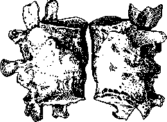
Figure 1. Bony ankylosis of the third lumbar vertebra in deforming arthritis (according to Schwalbe).
changes in the cartilage and bone. The disease can affect only one joint, usually the hip or knee (monoarticular form of deforming arthritis), or it can spread to many (sometimes all) joints (polyarticular form of deforming arthritis); in the latter cases, the disease often affects mainly the small joints of the upper or lower limbs. A corresponding disease of the spine (see Figure 1) is also observed, which is designated as deforming spondylitis (spondylitis deformans). The affected joints are curved and deprived of their normal configuration due to uneven thickening and displacement of the bone ends; joint mobility is limited. Upon opening an affected joint, changes in the articular cartilage are found in the form of fibrillation, softening, P 32B
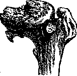
Figure 2. Destructive and productive process in the area of the femoral head in arthritis deformans (according to Küster).
disintegration and disappearance of the cartilaginous plates in places of greatest pressure from the opposing articular surface; in such places the bone is exposed, which is sclerosed and, due to friction during joint movement, as if polished and shiny. In addition, in the bone, in areas exposed from cartilage and under still preserved cartilage, focal resorption is observed, as a result of which small superficial defects and areas devoid of bony trabeculae arise in the bone of the articular heads. This reduction of bone substance results in the flattening of the articular heads and is the basis for the appearance of cracks and fractures in them. Along with these atrophic-type changes, proliferative processes also develop from the bone (see Figure 2); of these, the most characteristic for deforming arthritis is the overgrowth of cartilage-covered bone along the edges of the articular surfaces, which gives the formation of a typical for the patho-anatomical manifestations of this disease smooth "marginal ridge", as it were surrounding the altered articular surface like a crown. In addition, near the articular surfaces, irregular bony outgrowths often originate from the periosteum, sometimes reaching enormous sizes (see Figure 3). Thickening of the joint capsule is observed, either uniform or in the form of the formation of branching villi directed into the joint cavity; these changes in the capsule are usually most pronounced in its parts closest to the articular surfaces. Finally, rounded cartilaginous and bony formations (cap- Figure 3. Arthritis deformans of the right hip joint—cartilaginous chondromas and osteomata) can develop in the capsule; in those cases, when they, being connected to the joint capsule by a thin stalk, separate into the joint cavity, they acquire the significance of free joint bodies ("joint mouse"—see). Regarding the pathogenesis of deforming arthritis, there are many theories, from which some the beginning of the process
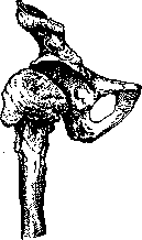
deformed head
of the thigh and the exostoses projecting along its edges (according to Aschoff). Some assume the process begins in the bone, others in the cartilage; at present, the most probable view is that the beginning and basis of the disease are atrophic-degenerative changes in the articular cartilage, while all other changes should be considered as secondary. The etiology of deforming arthritis is not yet fully elucidated. The functional theory (Pommer) has the most supporters, according to which the etiological factor is the functional mechanical overload of the articular cartilages, leading as it were to their wear, which manifests itself in atrophic-degenerative changes of the cartilaginous tissue; in particular, this theory is partly confirmed by the connection of deforming arthritis with certain professions (locksmiths, bakers, dishwashers). Views that emphasize the importance of intestinal autointoxication, constitution, or the influence of endocrine organs (for example, the thyroid gland) cannot be considered convincing. The physicochemical approach of Seeliger deserves attention, indicating an increase in the acidity of the synovial fluid in deforming arthritis, which can serve as the basis for degenerative changes in the cartilage. It should be added that some researchers also include in deforming arthritis cases of similar patho-anatomical manifestations affecting a single joint, arising after trauma (traumatic deforming arthritis), and, in addition, the so-called chronic ulcerative dry arthritis (arthritis chronica ulcerosa sicca), observed in old age usually in one joint, for example, in the hip joint (malum coxae senile), more rarely in several joints; in this form of the disease, the same atrophic-degenerative changes are present in the cartilage and bone as in typical deforming arthritis, but there is no proliferation of bone tissue.
Related articles
Mentioned in
Cite this page
“Arthritis.” Soviet Medical Encyclopedia. English translation of Bolshaya Meditsinskaya Entsiklopediya, 1st ed. (Moscow, 1928–1936), ed. N. A. Semashko. https://sovietmedicalencyclopedia.pages.dev/article/arthritis/