Paratyphoid
Historical document, translated for reference. It reflects medical knowledge of the 1920s–30s and is not medical advice.
Summary
This 1930s encyclopedia article covers the definition, history, and bacteriology of paratyphoid fevers, differentiating between paratyphoid A and B, their clinical manifestations, and characteristics of the causative organisms.
Encyclopedia article (1928–1936)
Paratyphoid (paratyphus) is the name of a general acute infectious disease that proceeds similarly to typhoid fever and is caused by microbes related to the typhoid fever causative agent, Eberth's bacillus. This term was first proposed by Achard and Bensaude in 1896. Observing two cases of a disease resembling typhoid fever, these authors isolated a bacillus—in one case from a sternoclavicular abscess and in the other from the purulent urine of patients—that was morphologically and culturally similar to Eberth's bacillus, agglutinated by the serum of the patients from whom it was isolated, but not agglutinated by typhoid patient serum. Similar observations were made by Gwyn in 1898 and Cushing in 1900. In the same year of 1900, Schottmüller in Hamburg isolated bacilli from 5 patients among those suffering from typhoid fever, which presented certain differences from Eberth's bacillus both in regard to their biological properties and in regard to agglutination. Following Achard, Schottmüller named these bacilli paratyphoid bacilli, and the diseases caused by them paratyphoids. Kayser, having established the distinction between the cultures isolated by Schottmüller, divided them into 2 types: paratyphoid A and paratyphoid B. The name Bact. paratyphi B Schottmuelleri became established for bacillus B, and Bact. parat. A Brion-Kayser for bacillus A. Achard's cultures proved identical to paratyphoid B, and Gwyn's cultures to paratyphoid A. Over time, it became clear that a number of microbes share great similarity with paratyphoid A and B bacilli, and therefore they were united into one large paratyphoid group (see bacteriology below); many of them proved pathogenic to humans. Failure to account for the fact that clinical terminology must reflect the clinical expression of the disease led to the term "paratyphoid" being applied to various diseases caused by paratyphoid bacilli. Meanwhile, not only different types of paratyphoid bacilli, but also one and the same type can cause forms that are very diverse in their pathogenesis and clinical expression. Therefore, the name paratyphoid should be retained for only one of the forms of paratyphoid diseases, namely, for the form analogous to typhoid fever. To emphasize the nature of this form, it should be called "abdominal paratyphoid" A or B (paratyphoid abdominalis A, resp. B). Along with this form, paratyphoid infection can lead to the development of acute and hyperacute gastroenteritis and enterocolitis (gastroenteritis, enterocolitis paratyphosa). The third form is septic (sepsis paratyphosa). Besides these forms, which are as a rule accompanied by pronounced phenomena of general intoxication of the organism, paratyphoid infection is very prone to cause various local inflammatory and suppurative processes: cholecystitis, cystitis, pyelitis, periostitis, perichondritis, etc.
G. Ivashentsov. Bacteriology. The group of microbes that occupied an intermediate position between the coli group on the one hand and the typhoid group on the other was named paratyphoid. The main limiting features separating the paratyphoid group from the coli group include the inability of paratyphoid microbes to ferment milk sugar and break down protein compounds with the formation of indole, as well as the presence of serological properties uniting the entire group that are absent in microbes from the coli group. From typhoid microbes, paratyphoid microbes differ first of all in greater enzymatic activity regarding carbohydrates and more modest requirements regarding nitrogen nutrition. Paratyphoid microbes are found not only as causative agents of various diseases or in the form of carriage in humans and animals, but to a significant extent also in the environment (water, milk, meat, and other products), which is favored by their relative stability. The question of the pathogenicity of the majority of microbes of the paratyphoid group for humans and animals is very complex. While some tend to see the essence of species distinction in the specific adaptation of the microbe to a given host (humans and various animal species) with the acquisition of corresponding specific pathogenic properties, others consider this kind of adaptation only conditional and allow for the bi- or polypathogenicity of the corresponding microbes. A more or less definite position, both from the point of view of bacteriological systematics and from the point of view of etiology, is occupied by the paratyphoid A microbe. It is fairly clearly demarcated from paratyphoid B microbes by both cultural and serological properties and, being the causative agent of paratyphoid A in humans, is detected only in corresponding patients and carriers, and in the human environment only insofar as it is directly contaminated by them. In animal pathology, the paratyphoid A bacillus plays no role and, with rarest exceptions, is not found as a saprophyte in them. The causative agent of paratyphoid A is a well-motile (peritrichous), easily staining, Gram-negative bacillus; a facultative anaerobe. It grows in broth and peptone water with the formation of turbidity. It does not liquefy gelatin, makes milk slightly acidic but does not curdle it; it grows on potatoes as a delicate coating. It does not form indole. Of carbohydrates and alcohols, it ferments with gas formation grape sugar, maltose, mannitol, arabinose. It does not ferment milk sugar, cane sugar, or xylose. In Petruschky's serum, it causes the formation of weak turbidity and slight reddening. It reduces Rotberger's medium. Colonies on agar and gelatin bring the paratyphoid A microbe close to the typhoid bacillus. A special form of the paratyphoid A microbe was described by Japanese authors as Bact. paratyphi A Sendai. Culturally, this bacillus differs from the previous one by the late formation of gas on media with grape sugar, maltose, and mannitol, and the transition of the acid reaction of milk to alkaline. Serologically, Bact. paratyphi A Sendai is characterized by diphasicity (see below) in contrast to the monophasic character of the paratyphoid A bacillus of the Brion-Kayser type. In its specific phase, Bact. Sendai is agglutinated to the titer of ordinary paratyphoid A serum, whereas in the nonspecific phase it is agglutinated predominantly by paratyphoid B sera and to a significantly lesser extent by the paratyphoid serum of the A type of Brion-Kayser. Encompassing a significant number of varieties, the paratyphoid B group is extremely extensive. The multitude of schemes proposed for subdividing the group are based either on the preferential consideration of the serological properties of the microbes or on a combination of cultural, serological, and biological properties with additional consideration of their etiological significance. This reflects the entire history of the development of the question of paratyphoid. An example of the first kind of subdivision can serve the English scheme, encompassing 12 serological types, or Aoki's Japanese scheme with subdivision into three types. An example of the second kind of subdivision can serve the scheme of Bitter and Holtz. A more detailed study of metabolism in paratyphoid bacilli of the paratyphoid B group was carried out by Kisch, Pesch, and in particular Braun. Of the salts, a mixture of chlorides and phosphates ensures the plastic needs of the microbes; of nitrogen compounds, ammonium salts, but not nitrites and nitrates; further—oxalic, succinic, citric acids, etc. Carbonic, oxalic, and acetic salts are not utilized by paratyphoid B bacilli. Typhoid-like diseases caused by representatives of the paratyphoid group are in the vast majority of cases caused by a bacillus called by some simply Bacillus paratyphi B, and by others Bacillus paratyphi B hominis (Schottmulleri). This bacillus is characterized by the same features as the paratyphoid A bacillus, but differs from the latter in its property of changing the color of Petruschky's milk serum after its preliminary reddening to blue due to the ability of this bacillus to break down the citric acid salts of the serum with the formation of alkali. A similar property is manifested by it on milk, which upon prolonged growth is peptonized. Of the pentoses currently used for the differential diagnosis of the entire typhoid-paratyphoid group, it ferments both xylose and arabinose with gas formation. In addition, serological reactions (agglutination and complement fixation) clearly demarcate it from the paratyphoid A bacillus. Extremely close to it stands a second microbe of the same group, which in the vast majority of cases is isolated from diseases in humans proceeding under the clinical picture not of typhoid, but of acute gastroenteritis and associated frequently (though not always) with the use of substandard products in food. In such cases of food poisoning, it is sometimes possible to isolate the same microbe from the products that served as the cause of the disease. This microbe is identified by some authors with Bact. paratyphi B, while other authors define it as a microbe sui generis and designate it
Bact. paratyphi Flügge-Kansche, Bact. Aertrycke, Bact. breslaviense or Bact. enteritidis Breslau, and others. This distinction, which at the present time has more of a principled than practical significance, is disputed by unitarians led by Uhlenhuth and defended by dualists led by followers of the Kiel school (Fischer, Bitter, Knorr, and others). The distinguishing features between Bact. paratyphi B Schottmüller and Bact. enteritidis Breslau are presented in Table 1. Table 1. Differential diagnostic features of Bact. paratyphi B and Bact. enteritidis Breslau. Features: Bact. paratyphi B, Bact. enteritidis Breslau. Formation of mucous colonies: +, +. Color change on medium: red, yellow. Phenomenon of overgrowth (Ver...): coral-like structure, etc. Pathogenicity for white mice upon feeding.... Extremely indicative in a differential-diagnostic regard are data concerning pathological-anatomical and histological changes observed in enteric infection of white mice and partly rabbits with cultures of the Breslau bacillus type (Waldmann). Serologically, the first bacillus (paratyphoid B Schottmüller) differs from the second (Breslau bacillus) in that in a simple and cross-agglutination test, the corresponding immune sera with a sufficiently high titer agglutinate only homologous cultures up to the limit. Even more clearly, the serological difference manifests itself in the Castellani absorption test, in which the corresponding sera are completely exhausted only upon absorption with the homologous strain. When analyzing the receptor apparatus, a heat-stable receptor common to both bacilli and two heat-labile receptors are revealed, of which one also serologically unites them, and the second delimits them. The serological scheme of Schiff is presented in Table 2. Table 2. Name of cultures, Thermostable receptor, Heat-labile receptors... Slightly more complex is the structure of the receptor apparatus depicted by Olitzki (Table 3). Table 3. Name of cultures, Thermostable receptor, Heat-labile receptors... The presence of cultural-biochemical and serological differences between both bacilli according to the data of dualists (Bitter, Knorr, and others) is aggravated by the presence in corresponding cases of clinical-bacteriological, anatomical-bacteriological, and epidemiological-bacteriological parallelism. Thereby, the question of the identity or independence of both microbes transcends the framework of a dispute between systematists, acquiring an actual-practical significance in the field of bacteriological diagnosis, therapy, prophylaxis, and epidemiology. The empirical and experimental material accumulated up to the present time in most cases confirms the correctness and practical significance of the position of dualists who delimit Bact. paratyphi B Schottmüller from microbes of the Breslau type. The correctness of such a delimitation is not denied by unitarians either, who, despite this, still consider it necessary to focus attention not on the "majority of cases" that fit well into this scheme, but precisely on the "minority" that fall outside these frameworks. In such cases, encountered more often in pre-epidemic or inter-epidemic periods, microbes are isolated that deviate in one way or another from typed microbes and represent, as it were, intermediate links between them. These observations and the generally recognized provision on the relatively low stability of differential features, which also bear partly (rhamnose, raffinose feature) only the character of a quantitative functional lag, force many even at the present time to regard the dualistic position as a utilitarian, but not a principled stance. Extremely close, if not identical with Bact. enteritidis Breslau, are a whole series of microbe-pathogens of diseases in humans and animals. This includes Bact. enteritidis Freiburg, encountered, just like Bact. enteritidis Breslau, in food poisoning in humans and differing from the latter by a richer heat-labile receptor apparatus, as well as microbes described by de Nobele during an epidemic in Mairébecke, van Ermengem during an outbreak in Ellezelles (Siro), Uhlenhuth in Greifswald, and others. To this same microbial group belong further: the mouse typhoid bacillus (Bact. typhi murium Löffler), the causative agent of paratyphoid epizootics in mice; Bact. psittacosis Nocard, the causative agent of epizootics among parrots (see Psittacosis), and a whole series of other microbes described as causative agents in sporadic and epizootic diseases in animals. Under favorable conditions, especially with mass infection through food products, these microbes can serve as a cause of diseases among humans as well. In addition to serological and cultural proximity, all these microbes, just like the paratyphoid B bacillus and the Breslau bacillus, possess the property of forming a heat-stable toxin in vitro. In vivo (on experimental animals) the toxin acts only upon parenteral administration. Per os even 15-fold lethal doses are ineffective (Bahr and Dyssegaard). Apparently, the serological types described by English bacteriologists, such as type Newport, Stanley, and others, have a wider distribution than was believed until recently. Lately, type Newport has repeatedly been found in Germany (Kauffmann). To the so-called "meat poisoners" (see Meat poisoning) belong further a whole series of microbes united in the Gaertner group, such as: Gaertner's bacillus (Bact. enteritidis Gärtner), Bact. Moorselie, Bact. enteritidis Gent, Brugge, Bact. Danysz, Bact. Dunbar, Bact. Issatschenko, Ratinbacillus, and others. The question of the identity or independence of individual representatives included in this group cannot be considered sufficiently clarified. The work of recent years outlines the possibility of subdividing the Gaertner group into 4 subgroups. The most fully studied is the Gaertner microbe, isolated in 1888. Morphologically and biologically, it stands close in all main features to the paratyphoid B bacillus and Bact. enteritidis breslaviense. Slime formation, characteristic of Schottmüller's bacillus, is also noted in Gaertner's bacillus, especially in freshly isolated strains. Old laboratory cultures often lose this feature. Colonies on gelatin are somewhat closer in structure to the colonies of the typhoid bacillus than to the colonies of the paratyphoid microbe. Attempts to find stable differential features for Gaertner's bacilli cannot be considered successful. The absence of gas formation on arabinose media, noted by Ruge in Gaertner's bacilli, is disputed by a number of authors. Sugar fermentation tests in the Gaertner group apparently can yield extremely variegated results up to the absence of gas formation on media with grape sugar. To this kind of culture belongs the strain "G" described by Viktorov and classified by the author as an intermediate form between the typhoid bacillus and Gaertner's bacillus. A more successful approach to delimiting bacilli of the Gaertner type from other representatives of the paratyphoid group consists in a detailed study of the assimilation properties of the microbe on synthetic media. In this way, it was possible to reveal an absent or in any case sharply reduced ability in a certain part of Gaertner's bacilli to assimilate citric acid salts, which are full-value sources of carbon for Schottmüller's microbe and the Breslau bacillus. In a synthetic medium, rhamnose also apparently provides the opportunity to delimit certain types of Gaertner's bacilli from the Breslau bacillus. The pathogenicity of Gaertner's bacillus, tested by the feeding method (on white mice), brings it close to the Breslau bacillus and delimits it from Schottmüller's. Old laboratory cultures often lose their pathogenicity. The aforementioned Viktorov strain "G" is pathogenic for mice and on the basis of this feature alone is to a significant degree close to the Gaertner microbe. The toxicity of killed cultures and filtrates of Gaertner's bacillus manifests itself exclusively upon parenteral administration, but not upon administration per os (Bahr). The predominance of biological and cultural properties bringing Gaertner's bacillus close to other paratyphoid microbes imparts exceptional significance to serological differential diagnosis, since the serological isolation of the Gaertner group, despite the lack of homogeneity within itself, is beyond doubt. The agglutinogenic apparatus of Gaertner's bacillus is characterized by a thermostable receptor common with the typhoid bacillus and a specific species-specific heat-labile receptor (Gruschka, Shibata, and others). This explains the fact that Gaertner sera agglutinate typhoid bacilli in small flakes up to the titer or almost up to the titer, and conversely, typhoid sera agglutinate Gaertner's microbe.
The Bacterium aertrycke is found in epizootics as the causative agent of spontaneous diseases in animals, as a saprophyte in the intestine of the latter, and as the causative agent of infectious processes in humans, in particular as the causative agent of acute gastroenteritis. Its role in human pathology is not contested by anyone. The frequency of finding Gärtner bacilli in rodents (wild rats) is attested to by American studies of Meyer and Matsumura (3.7% of findings) in San Francisco and English data by Kerrin (11%). Into a further group, demarcated from the microbes listed above by serological and some cultural characteristics, enter the microbes united under the name paratyphi C Suipestifer Voldagsen. They are found as causative agents of epizootics, as a microbe de sortie, as saprophytes in animals, and as causative agents of human diseases. The systematization of the microbes entering into this grouping, with their recent subdivision by White into 4, and by Kauffmann into 5 types demarcated from one another by serological and cultural properties, also responds to the practical demands of the clinic and epidemiology. The epidemiological significance of the subdivision into 5 types (Table 4) lies in the fact that two types until now have been discovered exclusively in animals (the American type and the Glässer-Voldagsen type), two other types, "Kunzendorf" and "Berlin", were discovered both in humans and in animals, and finally the fifth type, "Eastern", was encountered exclusively in human pathology. The listed 5 types of paratyphoid C bacilli are characterized by the cultural features marked in Table 4. of sera must be interpreted restrictively. Their non-specificity is completely indisputable with respect to the specific phases of all three types of paratyphoid C, i.e., those types that have a diphasic character. Their non-specificity is further indisputable in the respect that non-specific sera of paratyphoid C often, although far from always and by no means as a matter of obligation, agglutinate the non-specific phases of bacteria from the paratyphoid group B, A, and Gärtner. Conversely, with respect to the non-specific phases of their own five types of paratyphoid C, the non-specific paratyphoid C serum works up to the titer, ag- t a b l. 4. I Types 1 Cultural characteristics and media American Kunzendorf Glässer-Voldagsen Eastern Berlin Petrushky serum . . ...... Gas formation on: grape sugar broth . . nutrient medium-arabinose .... nutrient medium-inositol..... cr.-sin. + + - + -± -± + + animals cr.-sin. + + - + -± + + + animals and human cr. -± + - + -± + + animals cr.-sin. + . + + + + + + human cr.-sin. + + + + + + + animals and human Blackening on medium with acetic- Formation of a wall .......... Pathogenicity for white mice Serologically, the entire paratyphoid C group is quite clearly demarcated from paratyphoid microbes A and B, Gärtner bacilli, enteritidis Breslau bacillus, and the remaining closely related microbes. This group breaks down into two serological subgroups, of which one, according to new data taking into account the serological dissociation of microbes into specific, non-specific, and mixed phases (Andrewes, Aoki, Kauffmann, and others), is characterized by its diphasic nature, and the second by monophasicity. The diphasic group, to which according to Kauffmann belong the types "America", Glässer-Voldagsen, and the Eastern type, are found in specific, non-specific, and mixed phases, while the monophasic types Kunzendorf and Berlin have so far been encountered only in their non-specific phase. Having, therefore, the serum of the specific phase of one of the representatives of paratyphoid C of the first serological subgroup, one can unmistakably identify with the help of agglutination the representatives of the two remaining types belonging to this same subgroup, insofar as they are represented in their specific phase. In other words, the "specific" serum "America" agglutinates up to the titer the specific phases of Glässer and the Eastern type and vice versa, but almost agglutinates neither the specific phases of all five types of paratyphoid C nor representatives of paratyphoid A and B, and in the latter case regardless of whether the specific or non-specific phase of the latter is present. Regarding the sera obtained by immunization with the non-specific phase of any of the five types of paratyphoid C, the matter is somewhat more complicated. According to recent data, the concept of non-specificity of such lutinins must be interpreted in cross-experiments all the non-specific phases of paratyphoid C regardless of which type they belong to. The extremely confusing nomenclature in this microbial group is explained by the diversity of the proposed subdivision schemes and insufficient consideration of nomenclature rules. Thus, for example, the nomenclature concept of paratyphoid C, proposed by Uhlenhuth for paratyphoid strains not agglutinated by ordinary paratyphoid sera, is used by Weigmann for the serological subdivision of this same group of microbes into two types C1 and C2, and by Andrewes and Neave, Savage, and White for opposition to pathogens pathogenic for animals, brought under the name Hogcholera or Suipestifer, i.e., to designate strains from the same group, but pathogenic for humans. Finally, Kauffmann designates with the same name the entire group of microbes (C-Suipestifer) regardless of their mono- or bi-pathogenicity. On the role of Suipestifer microbes, in particular causative agents of epizootics and sporadic diseases among pigs, in human pathology, a number of recent works testify. The proposition of the harmlessness of these microbes for humans, supported by Uhlenhuth, Ostertag, and others, requires revision. In most cases, sporadic cases are described proceeding under the sign of acute gastroenteritis. There are, however, observations on mass diseases caused by microbes from this group. Microbes isolated into a special group of paratyphi ß Neukirch, Weil, and Saxl either fit into the so-called Eastern type of English authors or belong, like the ß2 microbe, to the group of paratyphoid poisoners. Strains N1 and N2 described by Ivashentsov and Rapoport from the point of view of serological characteristics must also be assigned to two different groups: N1 to the paratyphoid C group, and N2 to the Gärtner group.
O. Hartoch. Epidemiology. The routes, conditions, and degree of spread of paratyphoid diseases among people vary, which depends on the distribution of individual types of paratyphoid bacilli in nature, their persistence, the degree and character of their pathogenic properties, the susceptibility of humans and animals to them, and the presence or absence of conditions promoting or hindering human infection by them. Paratyphoid A and B bacilli are pathogenic for humans. The remaining species are pathogenic for various animals, and some of them are constantly or facultatively pathogenic for humans. Hence the difference in the routes of spread of representatives of each of these two groups, and consequently of the corresponding forms of the disease. The source of abdominal paratyphoid is man, who disseminates its causative agents—paratyphoid A and B bacilli (i.e., patients and carriers). Therefore, the epidemiology of abdominal paratyphoid is analogous to the epidemiology of typhoid fever: direct contact with the excreta (feces, urine) of a patient with abdominal paratyphoid or a carrier, water, milk, food products consumed raw and contaminated in one way or another by human secretions—such are the factors determining the possibility of infection by paratyphoid fever agents. The shedding of paratyphoid A and B bacilli by persons who have suffered from these diseases is observed, according to a number of authors, more frequently than carriage after typhoid fever. Thus, Hermel reports that among 24,500 convalescents after typhoid fever, 1.4% of carriers were found, after paratyphoid A out of 1,700 people—7%, and after paratyphoid B out of 7,990 people—8.7%. However, the duration of paratyphoid carriage is significantly shorter than in typhoid fever. Abdominal paratyphoid, especially B, more often proceeds with diarrhea manifested from the very beginning of the disease, which facilitates the possibility of direct contact. Abdominal paratyphoid B more often than typhoid fever gives rise to difficult-to-diagnose and mild forms that escape medical and sanitary supervision; such forms increase the possibility of spreading the infection. Finally, the persistence of the paratyphoid B causative agent outside the human body, especially in various food products, is significantly higher than that of Eberth's bacillus. Proceeding from all these data, one might assume that cases of abdominal paratyphoid B should occur more frequently than typhoid fever. In reality, however, their number ranges from 5% to 20% in relation to cases of typhoid fever. The explanation for this fact may be sought in the lower pathogenicity of the paratyphoid B bacillus and the increase in human resistance to it as a result of frequent encounters with small doses of the microbe of lowered virulence. Paratyphoid diseases caused by bacilli grouped into the "meat-poisoner" subgroup (main representatives—Bact. enteritidis Breslau and Bact. Gartneri) spread by completely different routes. The very name indicates that infection in these cases occurs predominantly upon consumption of contaminated meat (see Meat poisonings). Infections of humans with these types of paratyphoid infection from man to man are observed as exceptions. The significance of carriage by healthy individuals has not been clarified. Prolonged shedding by convalescents is not observed. Therefore, cases are observed either as isolated ones or as encompassing groups of people united by a common source of food. Etiology. The pathogenic significance of paratyphoid microbes lies in their toxigenicity and so-called septic or infectious properties, i.e., the ability to cause either local inflammatory and suppurative processes or—upon generalization of the infection—phenomena of general sepsis. The presence and predominance of certain properties in individual types of paratyphoid bacteria determine the etiology and pathogenesis of the diseases caused by them. Differences in the persistence of individual species, in their ability to multiply and form toxins outside the human body, and in the heat resistance of their toxins lead to the fact that in some cases the cause of the disease will be exclusively intoxication, in others—both the living microbe and the toxin produced by it in the corresponding medium, and in a third—with low toxigenicity of the microbe, its septic properties will come to the fore. Along with the characteristics of each species of paratyphoid microbe, the massiveness of infection or intoxication is of great importance in the character of the disease caused by it. Even greater importance is attached to the form of the reaction determined by the state of the gastrointestinal tract, as well as the general immunobiological state of the entire organism. Proof of the latter is the not infrequent complication of various other infectious diseases (scarlet fever, typhus fever, etc.) by paratyphoid infection. The pathogenic properties of paratyphoid A bacillus are most constant, quite similar to the properties of Eberth's bacillus and causing a reaction of the organism analogous to that which underlies typhoid fever. A larger number of cases caused by paratyphoid B bacillus also proceed by the typhoid fever type. However, the accumulation of toxins of this bacillus in various food products consumed after processing that kills the living virus can lead to pure intoxication. Infection with viable bacilli can also lead to purely gastroenteritic forms. Depending on the entry into the body of a large amount of toxin at once, the disease sometimes begins in the form of acute gastroenteritis of the cholera nostras type, and a few days later (6–10) the picture of abdominal paratyphoid develops. The forms caused by "meat poisoners" also depend on which factor predominates—toxic or septic. Septic properties appear much more sharply in the group of Bact. suipestifer, Salmonella, or paratyphoid C according to Uhlenhuth. The pathogenicity of this group for humans is apparently much greater than that of the previous ones and depends on the state of their organism. Vivid evidence of this is the clearly expressed connection of diseases caused by the N1 = C2 bacillus with relapsing fever (Kulesha, Ivashentsov, and Rapoport).
G. Ivashentsov. Statistics. Statistical data on paratyphoid diseases are very meager. For most individual countries, as well as in international reviews, in morbidity and mortality reports, paratyphoid diseases are included in the typhoid fever group. For some countries, the following data can be cited. The figures for morbidity and mortality from typhoid fever and paratyphoid in Japan are given in Table 5. Table 6 gives the figures for registered cases of typhoid fever and paratyphoid in some European countries for 1925–26. On the fronts, paratyphoid A accounted for 75% to 80% of all typhoid and paratyphoid morbidity. Toward the end of the war, in the relationship between the three types of typhoid-paratyphoid infection, a tendency to return to what was observed before the war is revealed. In 1918, in the French army, Eberth's bacillus was found in 40–55% of all cases, bacillus A in 25–30%, and bacillus B in 10–33% (Achard). The fluctuations in the frequency of individual types of typhoid-paratyphoid infection should be largely explained by preventive vaccinations. At the beginning of the war, the typhoid monovaccine was used for vaccinations. The number of diseases caused by Eberth's bacillus began to decline rapidly, and paratyphoid diseases took a relatively high place. Schottmüller notes that before preventive vaccinations, typhoid fever accounted for 98% and paratyphoid diseases for 2%; after the first and second vaccinations with the typhoid monovaccine, typhoid fever dropped to 35% and paratyphoid rose to 65%; after the second and third vaccinations, typhoid fever accounted for 6% and paratyphoid for 94%. When vaccination with the triple vaccine (against typhoid fever, paratyphoid A, and paratyphoid B) began, the incidence of all types of typhoid-paratyphoid diseases began to decline rapidly. As for paratyphoid A, which was almost never encountered in Europe before the World War, some authors believe that it was brought into European armies by colonial troops. After the war, typhoid fever again took first place, but paratyphoid became more frequent than before the war, which partly should also be explained by the fact that it is being looked for more carefully (Achard). In Paris, out of 164 cases observed by him from October 1919 to October 1928, Achard counts 68.29% of typhoid fever, 3.04% of paratyphoid A, and 28.65% of paratyphoid B. In individual years, the percentage of typhoid fever showed large fluctuations: in 1919–21 it was 75%, in 1925 it dropped to 23.8%, in 1926 it rose to 43.3%, in 1927 to 66.6%, and in 1928 to 100%. In the 1926 epidemic in Hanover (Germany), out of 2,460 total diseases, typhoid fever was 93%, paratyphoid 6.3%, and mixed forms 0.7%. In Denmark, the relationship between typhoid fever and paratyphoid for the period from 1922 to 1926 was expressed in the following percentages (Table 7). These data are based on the results of laboratory studies. From the table it is clear that typhoid fever (Eberth's bacillus) significantly predominates everywhere over paratyphoid diseases, and of the latter, paratyphoid A is much more widely distributed in the south, while paratyphoid B prevails in the north. Data on the geographical distribution of individual types of paratyphoid are subject to further clarification. This is of great practical importance (for example, in vaccination against typhoid fever). The case fatality rate in typhoid fever and paratyphoid is not the same. According to the data of the Botkin Infectious Diseases Hospital in Leningrad, for 1924–31 it constituted the following percentage (Table 10). Here, a gradual increase in the percentage of typhoid fever is noted at the expense of a decrease in the percentage of paratyphoid diseases. The following materials give an idea of the spread of paratyphoid in the USSR. In Leningrad, 9,051 patients with typhoid fever and paratyphoid passed through the Botkin Infectious Diseases Hospital from 1922 to 1931. Of these, typhoid fever accounts for 78%, and paratyphoid for 22%. These ratios among men (total 5,137 people) were 77 and 23%, and among women (total 3,964 cases) 78.5 and 21.5%. For individual years from 1924 to 1931, the relationship between typhoid fever and paratyphoid was as follows (Table 8). From 1922 to 1931, the average case fatality rate was 10.4% for typhoid fever and 2.1% for paratyphoid diseases. I. Dobreytser. The pathogenesis of enteric paratyphoid is completely analogous to the pathogenesis of typhoid fever. The presence of an incubation period preceding the local manifestations of the disease, the lesion of the lymphatic apparatus, early bacteremia, and a regular cyclic course of the disease leading to the development of general immunity determine the place of enteric paratyphoid among general acute infectious diseases. The pathogenesis of paratyphoid gastroenteritis and enterocolitis is different. The pathogenesis of acute gastroenteritis of the cholera nostras type is determined by the properties of the paratyphoid toxin, which possesses neuro- and enterotropism.
Of all parts of the nervous system, the lesion of the vegetative system is most clearly expressed: suppression of the greater splanchnic nerve system (n. splanchnicus major) and excitation of the vagus nerve system. These lesions entail stagnant hyperemia of the mucosa of the gastrointestinal tract, increased mucus secretion, and increased peristalsis; a direct toxic or indirect (via damage to innervation) lesion of the mucosa of the gastrointestinal tract leads to profuse diarrhea and vomiting, and also manifests as a slowing of the heart rhythm. The consequence of intoxication is rapid and sharp dehydration and demineralization of the organism, as a result of which phenomena develop that are similar to the syndrome of the algid stage of cholera. This extraordinary similarity suggests that in analyzing the pathogenesis of acute paratyphoid gastroenteritis, it is necessary to take into account the interpretation of the pathogenesis of cholera algid given by Sanarelli, confirmed by wide and diverse experiments of Zdrodovsky, and reducing to the sensitization by the cholera virus of the organism infected by it to other infections and first of all to the colon bacillus. In forms which, in contrast to cholera-like gastroenteritis, can be called acute febrile paratyphoid gastroenteritis, the expression of the septic properties of their pathogens prevails, accordingly, the lesion of the lymphatic and reticulo-endothelial apparatus is more sharply marked. Pathological anatomy. Pathological anatomical changes in abdominal paratyphoid in principle coincide with the changes characteristic of abdominal typhus. However, differences are also noted: in abdominal paratyphoid, lesions of the large intestines are more often observed; occasionally all changes are concentrated in them (paratyphoid colotyphoid). Swelling of the lymphatic apparatus in abdominal paratyphoid is generally less significant. The lesion of the lymphatic apparatus of the mucosa is usually less pronounced than in abdominal typhus; however, the lesion of the entire mucosa is usually much greater and more diverse; phenomena of catarrhal inflammation are noted, sometimes fibrinous, diphtheritic, the presence of hemorrhages, the development of an ulcerative process outside the location of Peyer's patches, etc. From a principal point of view, it is interesting that in abdominal typhus an expanded vulgar reaction from the entire intestinal mucosa can also be observed (typhoid enterocolitis). The reaction of the mesenteric glands is usually less pronounced than in abdominal typhus. The outbreak of typhoid in Leningrad in 1931, which gave a relatively high percentage of paratyphoid B infection (approx. 13%), showed how closely autopsy data converge as the clinical expression of typhoid and paratyphoid infection converges. Pathological anatomical changes accompanying paratyphoid gastroenterocolites do not present anything characteristic. In diseases of the cholera nostras type, various degrees of manifestation and spread of the acute inflammatory process in the mucosa of the stomach, small and large intestines are observed, most often in the form of catarrh thereof, often accompanied by hemorrhages. Sometimes the picture of acute follicular enteritis or enterocolitis clearly emerges. The reaction of the mesenteric glands and spleen is usually poorly expressed or not noted at all. In acute febrile gastroenteritis, the reaction of the mesenteric glands and spleen is more brightly expressed in the form of hyperemia, hyperplasia, and a corresponding increase in these organs. The character of pathological anatomical changes in septic forms caused by paratyphoid infection is described in detail by G. S. Kulesha in N-paratyphobacillosis. With relatively little characteristic changes in the intestines (swelling of follicles, diphtheritic and ulcerative processes), lesions of the kidneys, usually dotted with small pustules of embolic origin already visible from the surface, are very constant and brightly expressed. Metastatic lesions are localized most often at the sites of connection of cartilage with bones, synchondroses, periosteum. Severe lesions of the urinary bladder mucosa are very constant. As with typhoid, abscesses in soft tissues, e.g., in muscles, pictures of osteomyelitis, Zenker's degeneration of muscles with hematomas and suppuration of the latter, can be observed in paratyphoid. With respect to strains A and B, a general observation may be that intestinal changes resembling typhoid are much more frequently observed in form A. Clinical picture. Abdominal paratyphoid in the main features of its clinical picture is quite similar to typhoid. The differences lie in the degree of expression of individual symptoms. Most of them in paratyphoid are expressed weaker than in typhoid, which is why the entire course of abdominal paratyphoid appears alleviated compared to typhoid, the average duration shortened; abortive forms are also observed significantly more often. Mortality is also quite clearly reduced, fluctuating when calculated per hundred cases from 0% to 3-4%. This does not mean at all that both forms of paratyphoid A and B cannot manifest as a prolonged severe disease with repeated waves and relapses. The curves characterize the course of t° in a severe form of paratyphoid with the presence of a fully and brightly expressed syndrome of abdominal typhus (Fig. 1), an abortive form (Fig. 2), and a very mildly expressed form (Fig. 3), in which, however, it was possible to isolate a hemoculture. The incubation period of abdominal paratyphoid is considered by most authors to be shorter than that of typhus. Paratyphoid more often develops acutely. Out of 200 analyzed cases of abdominal paratyphoid B, acute development was noted in 26% (Ivashentsov). Significantly more often than in typhus, diarrhea (22%) and loose stools (16%) are noted at the beginning of the disease. Vomiting was observed in 10% of cases, abdominal pain in 54%. Corresponding to a more acute increase in t°, the onset of the disease is accompanied by chills (approx. 30% of cases). Aches are noted approximately just as often. In approximately 5-8% of cases, catarrhal tonsillitis is observed. Headaches are very constant. Also constant is the enlargement of the spleen, which is palpable in 80% of cases.
The eruption of roseolous rash is not uncommon, observed much earlier than in abdominal typhus—on the 4th-5th day of the disease. In paratyphoid, it is as a rule more abundant. In paratyphoid B, it is usually larger, brighter, and more often captures the extremities. Among 180 cases of Ivashentsov, papular rash was noted 10 times, petechial 5 times, and vesicular 1 time.
Figure 2.
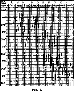
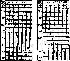
Figure 3. In paratyphoid A, the rash is finer, and hemorrhages are more frequently observed. The appearance and fading of roseolae have a wave-like character. Nervous system disorders in paratyphoid B are observed less frequently than in typhoid fever, merely due to the smaller number of severe forms of the disease. In the latter, not only is the status typhosus well expressed, but sometimes phenomena of depression appear especially brightly, and more rarely violent delirium. According to literature data, nervous system disorders in paratyphoid A are less pronounced. Regarding the gastrointestinal tract, the most pronounced phenomena (nausea, vomiting, abdominal pain, loose and sometimes malodorous stools) are noted in paratyphoid B. In this same form, more often than in others, the stool acquires a colitic character (frequent urges, mucus, blood, indicating primary involvement of the large intestines). The reaction of the cardiovascular system, blood, and excretory organs does not differ from that usual for typhoid fever. The number and severity of complications in both forms of paratyphoid are significantly less than in typhoid fever. This applies especially to the most formidable complications of the latter, which depend on severe damage to the lymphatic apparatus of the mucosa of the small intestines—namely hemorrhages and perforations; both of these are observed very rarely, especially perforations, with those in paratyphoid A even undescribed (Vogralik). This undoubtedly reflects the aforementioned differences in the properties of the pathogens of typhoid and paratyphoid. For the same reason, complications in the form of metastatic purulent foci in the cartilages, periosteum, joints and urinary tracts, muscles, and sometimes in the brain, etc., are more frequently observed in paratyphoid B. Septic forms caused by paratyphoid microbes proceed either by the type of acute septicemia, or septicopyemia, or pyemia lasting for years. The most vivid examples of the course of all these forms are represented by diseases caused by paratyphoid bacilli, for which the names fixed in the USSR are: N1 and N2 (Ivashentsov and Rapoport) = Ca and O1 (Weisman), N1 Erzindjan (Neufeld/Neirkih), paratyphoid J, etc. The septic properties of this type of paratyphoid microbes are so sharply expressed that they prompted S. V. Kulesh to give them the name Bact. septico-pyaemicus hominis. Forms caused by other paratyphoid microbes differ in the degree of symptom expression. The characteristic features of hyperacute paratyphoid sepsis are: marked jaundice, rashes (roseolous or hemorrhagic), extreme dryness of the tongue, sharply expressed diarrhea sometimes preceded by constipation and accompanied by phenomena of hemorrhagic colitis, significant enlargement of the liver and its tenderness (to a lesser extent this applies to the spleen), mental impairment (clouding of consciousness, delirium), often a sudden drop in cardiac activity, sometimes with a paradoxical preservation of good well-being. Bacteremia is unusually sharply expressed; bacilli are detected even in blood smears, or over 5,000 colonies are cultured from 1 cm3 of blood. Complications with meningitis are frequent; in these cases, the cerebrospinal fluid is turbid and contains a large number of pathogens. The entire disease lasts up to a week and ends lethally. Septicopyemic forms, differing in the lesser expression of all the listed symptoms, drag on significantly longer, often bearing the character of a typhoid form, and are distinguished by the formation of various purulent foci. Yielding a very high percentage of lethality, these forms can transition into forms of protracted pyemia and end in recovery. The variety of purulent foci and at the same time the primary involvement of bones, cartilages, and joints is manifested especially clearly in precisely such protracted forms, which sometimes develop even without a preliminary picture of tumultuous septicopyemia. The diagnosis of the paratyphoid nature of the disease is significantly assisted by the examination of the blood of patients for the agglutination of paratyphoid strains. Blood cultures are sterile. The decisive diagnostic fact is the isolation of cultures from material obtained by puncture. Among internal organ lesions, the most frequently noted are splenic abscesses, cholecystitis, cystitis, and kidney abscesses. The most constant phenomena accompanying protracted pyemic forms of paratyphoid infection boil down to erratic temperature rises, a yellowish-earthy coloration of the skin, stubbornly persisting diarrhea occasionally alternating with stool retention, progressive anemia, exhaustion, and various neuralgias and neuritis.
G. Ivashentsov. Diagnosis. Until the recent past, the bacteriological diagnosis of paratyphoid diseases was based on inoculating the initial material either directly onto appropriate differential nutrient media or after preliminary enrichment. At present, it is customary to combine the direct inoculation method with the enrichment method. The initial material can be any object suspected of containing the corresponding pathogen. In human pathology, such objects include blood, feces, duodenal juice, urine, pus, and others. The contents of the gallbladder are also examined in corpses. The method for obtaining hemocultures is the same as for typhoid fever. As large a volume of blood as possible (up to 20 cm3 obtained from a vein) is inoculated into ox bile or 10% bile broth, followed by plating onto differential media. Results are no worse when blood is inoculated into distilled water according to Klodnitsky-Gildemeister. Liquid feces are inoculated per se, and formed stool after emulsification in physiological saline or peptone water. Inoculation is performed sequentially on 3-4 Petri dishes over the surface of differential media (Endo, Conradi-Drigalski, Gassner, Padlevsky, Bitter, and others). Simultaneously, inoculation is carried out on an enrichment medium (Lentz-Titz medium, Müller) followed by plating onto the above-mentioned media. The procedure is similar for other initial materials. Further processing and identification of cultures are carried out according to generally accepted rules in bacteriology. The new method proposed by Wilson and Blair for isolating the typhoid-paratyphoid group using bismuth-sulfite medium deserves attention. Only cultures meeting the basic requirements for microbes of the paratyphoid group are additionally studied serologically. If cultures exhibit an unusual character on media or in serological respects, one must not lose sight of the fact that the observed deviations may depend on the "impurity" of the obtained culture. By the method of repeated purification of the culture, through passages via Petri dishes with subculturing from colony to colony, it is often possible to establish the mixed nature of the primary inoculation. Errors of this kind in diagnosis have been noted not infrequently in recent times (Seligmann, Clauberg). As additional methods of differential diagnosis for the coli-typhoid group, it has been proposed to use synthetic media with ammonium chloride, ammonium sulfate, or ammonium phosphate as the sole source of nitrogen nutrition, combined with various sources of carbohydrate nutrition. The growth results of various representatives of the paratyphoid group are presented in Table 11. Table 11. Cultures Medium with ammonium salt with the addition of arabinose rhamnose succinic acid malic acid Bacter. paratyphi B Schottmülleri . . . Bact. enteritidis Bres- Bacter. paratyphi C Glässer-Voldagsen . Bacter. paratyphi C Kunzendorf .... Bacter. paratyphi C hu- + + + + + + .+ + + + + . The second scheme (Pesch and Maschke) proposes using for the same purposes water agar with ammonium chloride (1.63%) as the sole source of nitrogen nutrition, combined with three carbon sources (Table 12). Table 12. Cultures Medium with ammonium chloride with the addition of grape sugar lactic acid rhamnose Bact. typhi abdominalis . . . Bact. enteritidis Breslau . . Bact. Gartner-groups I and II + + ± + + + + + ' + i i Serological diagnosis of paratyphoid diseases has the same practical significance as in typhoid fever. Widal's reaction is set up with various representatives of the paratyphoid group (typhoid, paratyphoid A, paratyphoid B Schottmüller type, Breslau type, Gartner's bacillus, and N2). Serum dilutions are taken from 1 : 50-90, 1 : 800 and higher. The result is recorded after 2 hours of standing in an incubator at 37° and after 24 hours. Group and paradoxical reactions occur frequently and complicate differential diagnosis. The method proposed by M. K. Fischer—the use of hypertonic NaCl solutions suppressing group agglutination—is quite appropriate in such cases. By setting up the agglutination test at common salt concentrations equal to 1,500 or 3,000 millimoles (8.77% and 17.54%), it is possible to show that Schottmüller type serum agglutinates only the homologous microbe with sharp suppression of the agglutination of the Breslau bacillus. Conversely, Breslau type serum at these concentrations agglutinates both microbes, in a somewhat lower titer, but to the same degree. By a more complex route, the specific nature of agglutination can be revealed by Castellani's saturation method. Due to the diphasic nature of paratyphoid B cultures, diagnostic difficulties may arise if, in diseases caused by a microbe in the specific phase, cultures represented in their nonspecific phase are accidentally used to set up Widal's reaction. In such cases, Widal's reaction may be completely absent. The introduction of the euglobulin fraction of agglutination sera into serological practice (Fischer) simplifies differential serological diagnosis.
O. Hartoch. Clinical diagnosis. Abdominal paratyphoid is diagnosed similarly to typhoid fever. Its differential diagnosis includes all forms that must be distinguished from typhoid fever. The distinctions between typhoid fever and abdominal paratyphoid are so conventional and ill-defined that distinguishing them in the clinic in each individual case proves very difficult. Speaking in favor of the paratyphoid form are a more acute onset, early appearance of roseolae, their abundance, in particular on the hands, large size, brightness, elevation; presence of diarrhea; and in severe cases, sharply pronounced mental disturbances. Early diagnosis is of not only academic but also practical interest in view of the indicated differences in prognosis. The issue is resolved only by bacteriological examination (see above). For the diagnosis of gastroenteritic forms, see Meat poisoning. The diagnosis of septic forms is based exclusively on the results of blood culture or pus from metastatic foci. Treatment: see Typhoid fever and Sepsis. Prophylaxis. The preventive measures for all paratyphoid diseases are determined by the etiology and epidemiology of each of them. All sanitary-hygienic measures, general public improvements, especially water supply, sewerage, cleaning of populated areas, public and household latrines, cesspools, the fight against flies, compliance with personal hygiene rules, etc., which form the basis of the fight against typhoid fever, retain their significance in relation to abdominal paratyphoid as well. As for paratyphoid B, nutritional conditions, especially public catering—the shortcomings of which are the main source of mass gastroenteritic forms of paratyphoid diseases—acquire even greater importance for it than for typhoid. The strictest supervision is necessary over the quality of meat released from slaughterhouses (see Meat poisoning), the order of its transport and storage, methods and conditions of its processing (sausage production, canned goods, etc.). No less attention should be paid to the manufacture and storage of all products consumed raw, cold dishes and appetizers, ice cream, as well as dishes not consumed immediately after their preparation, which allows for the accumulation of introduced infectious agents in them after boiling or other processing (salads, jellied dishes, soups prepared for several days, etc.); the fight against rodents, especially rats, is also of no small importance. Methods of specific immunoprophylaxis boil down to vaccination with paratyphoid A and B cultures, analogous to typhoid vaccination and usually carried out simultaneously with the latter through the use of a divaccine (typhoid bacilli 1 billion and paratyphoid A or B 500 million in 1 cm3) or trivaccines (typhoid fever 1 billion and paratyphoid A and B 250 million each in 1 cm3). The significance of vaccination against paratyphoid in parallel with typhoid vaccination was clearly demonstrated during the war by the example of the French army: the incidence curve of typhoid forms, which showed a decline after vaccinations with Eberth's cultures, showed a new significant decline after paratyphoid vaccination. The equivalence of enterovaccination (as well as regarding typhoid fever) has not yet received universal recognition.
G. Ivashentsov. Paratyphoid in children, like typhoid fever, occurs at all ages. However, there is exact, exhaustive statistics on the frequency of its spread by years, especially in young children. This is explained by the features of the course of this disease in children, difficulties in diagnosis, and as a consequence, incomplete accounting. It has been established that children are more frequently affected by paratyphoid B than A; cases of mixed disease have also been described. Infection with paratyphoid can occur even in the antenatal period from a mother sick with paratyphoid. In these cases, children are most often born dead or in a severe condition with phenomena of hemorrhagic septicemia (Schmidt, Yamada). Cases of infection during childbirth have also been noted, which also present a severe septic course. The greatest opportunities and conditions for infection in cases of a paratyphoid environment are found in infants and small children. Here, a decisive role is played by close contact with adults caring for the child, primarily the mother, and defects in care and feeding. The child is easily infected when using a shared bed, contaminated linen, diapers, a shared baby bath, toilet, when crawling on the floor, through a dirty pacifier, toys, when washing with dirty hands, through a towel, etc. The child's food - milk, vegetables, later - meat, prepared and stored without observing the sanitary minimum, are also frequent sources of infection. All these conditions are specific to the old family way of life, closed and individual upbringing of the child against the background of general sanitary neglect. A reflection of this is the so-called family epidemics. The clinical picture of paratyphoid in children differs even less from the clinical picture of typhoid fever than in adults. The younger the child, the more enteric paratyphoid in children differs in its course and symptomatology from enteric paratyphoid in adults. It proceeds especially atypically and polymorphically in infants and small children. At the age after 8-10 years, the course is almost analogous to that in adults. The main features of paratyphoid in the first years are a generally milder course, shorter duration of the disease, polymorphism, and a large percentage of outpatient and abortive forms. Along with this, there is also a significant number of severe diseases, especially in infancy, which, according to some authors, predominate here. However, in this case, it must be borne in mind that in infants, many forms of paratyphoid, especially mild and atypical ones, are undoubtedly overlooked and not accounted for. Therefore, the predominance of severe forms of paratyphoid in infants should be taken with great reservation. The incubation period is usually characterized by rather vague symptoms. In infants, refusal to breastfeed, refusal of food in general, a sharp degree of apathy, and sometimes pronounced restlessness, especially at night, are noted. Older children complain of headache, fatigue, become drowsy, stop being interested in their surroundings, abandon their usual activities, and prefer to stay in bed. In cases of infection through meat, an acute picture of meat poisoning may develop during the incubation period, which gives this period a very severe character. In the case of a favorable outcome of this paratyphoid toxemia, sometimes after a short (a day or two) clear interval, symptoms of enteric paratyphoid proper develop (observations of the State Scientific Institute for the Protection of Motherhood and Infancy). The onset of enteric paratyphoid in the overwhelming majority of cases in children is acute and accompanied by a rapid rise in temperature, often chills, vomiting; in sympathomilics and neuropathic children - tonic and clonic convulsions. Sometimes herpes labialis is observed at the same time. The further development of the disease also proceeds rather quickly. All symptoms characteristic of enteric paratyphoid appear in a shorter period than is the case in adults. The termination of the disease is also rapid, with a critical drop in temperature, but there are also cases of a slow lytic termination (observations of the State Scientific Institute for the Protection of Motherhood and Infancy), dragging on for several days. There are also some features in the development of individual symptoms. The temperature, reaching high figures within 1–2 days, subsequently often gives the type of an intermittent curve, often resembling a malarial one. Following the drop in temperature, during the recovery period, relapses are noted, evening temperature rises often lasting for a week or more. Some authors have also described an afebrile state. Parallel to the temperature, the development of phenomena from the nervous system also proceeds. In young children, restlessness is particularly often noted, accompanied by crying, sometimes prolonged, sometimes short, occurring periodically. Along with this, there is drowsiness, loss of consciousness, delirium, unmotivated movements of the hands, grasping at the air, picking at the lips, and in the stage of highest development of the disease - deep stupor. From the gastrointestinal tract, constipation most commonly occurs at first, and then diarrhea follows, varying both in frequency and character of the stool. In infants, the stools are dyspeptic in character, sometimes from the very beginning taking the type of a cold diarrhea or bloody dysenteric stool. In some cases, one type of stool replaces another. Along with this, rather frequent regurgitation occurs. Older children complain of pain in the ileocecal region, which gives rise to suspicion of appendicitis. The spleen is as a rule enlarged and palpable; the latter is not achieved only in the case of pronounced flatulence, quite often encountered in enteric paratyphoid. A large enlargement of the spleen is accompanied by pain in the hypochondria. In infants, enlargement of the spleen may be absent. Skin changes in children are significantly sharper than in adults. A roseolar rash appears quite early, on the 2–3rd day of the disease. The roseolae are larger in size, brighter, and erupt not only in specific places - the abdomen, back, chest - but spread throughout the entire body, including the face. Along with roseolae, sometimes, especially in severe cases, petechiae appear. Frequently, the rash has a patchy measles-like character or finely punctate, scarlatiniform. Vesicular and vesiculopapular eruptions have been described. Along with this, especially in infants, the skin is often dotted with miliaria crystallina. In very young infants, there is often no rash at all. During the recovery period, significant peeling is observed, especially following an abundant preceding rash. Mucous membranes are altered from the first days of the disease. The lips are dry, in severe cases covered with crusts and bleeding cracks. The gums are loosened, swollen. On the soft palate, arches - red patchy eruptions, sometimes ulcers. The tongue is coated with a thick grayish coating; in severe cases, dry, cracked, the coating takes on a brownish, sometimes black color. The nasal cavity is mostly clear. Sometimes only bleeding is observed. In the nasopharynx, phenomena of hyperemia, inflammation, a lot of mucus. The larynx presents more serious changes. Here, laryngitis, laryngospasm, and the pseudo-croup symptom are observed. Diffuse bronchitis is usually determined. The heart suffers to varying degrees depending on the severity of the case. The pulse is usually frequent, almost corresponding to the temperature. Slowing and dicrotism are usually not observed. The blood picture in paratyphoid A shows clear leukopenia, a drop in neutrophils, and an increase in lymphocytes, proceeding parallel to the development of the disease (Walterhofer). The number of eosinophils drops or they are completely undetectable at the beginning of the disease and appear only in the last stage. In paratyphoid B, leukopenia is not a constant symptom. Leukocytosis is frequently present; the shift to the left is less significant than in typhoid. The number of eosinophils is normal or slightly elevated. From all the diversity in the course of enteric paratyphoid, three most striking forms can be distinguished. The typhoid form, in which nervous typhoid phenomena are in first place and the main symptoms described above are expressed in various degrees and combinations, in young children often gives particularly pronounced meningeal phenomena - Kernig's sign, Brudzinski's sign, neck stiffness, convulsions, dermographism, and sometimes strabismus, nystagmus (obs. State Scientific Institute for the Protection of Motherhood and Infancy). The cerebrospinal fluid in these cases is only under high pressure and does not give any other pathological changes. All cerebral phenomena disappear parallel to the drop in temperature and improvement of the general condition. The gastrointestinal form gives a picture of intestinal intoxication - diarrhea, vomiting, and varying degrees of disturbance of the general condition. In severe cases, the disease can proceed according to the type of infant cholera with profuse diarrhea or according to the type of acute enterocolitis, dysentery, reproducing the entire complex symptom-complex of this severe lesion. The septic form is encountered more frequently in young children and newborns and proceeds as diversely as sepsis in the first months of life. Complications in enteric paratyphoid in children are numerous and diverse. In infants and small children, purulent lesions are often noted - otitis, antritis, pyoderma, parotitis, purulent serous arthritis, abscess of the liver, thyroid gland, muscles. In the oral cavity - severe thrush, aphthous stomatitis, noma. Purulent meningitis is of particularly great importance. The diagnosis of enteric paratyphoid is the more difficult, the younger the child.
Therefore, in any severe febrile illness, one should not forget about typhoid-paratyphoid diseases. It is especially easy to confuse abdominal paratyphoid with summer infant diarrheas, pyelitis, undiagnosed pneumonia, meningitis, sepsis, etc. The question of the true nature of the disease in all cases can be resolved on the basis of bacteriological and serological data. At the same time, it must be borne in mind that the Widal reaction in children often appears late and quickly disappears. Therefore, decisive importance belongs to the finding of paratyphoid bacteria in the blood and discharges of the patient—urine, feces, pus. It is also necessary to take into account epidemiological data—incidence in the family, etc. The prognosis for abdominal paratyphoid in children is generally favorable. The cited mortality figures fluctuate within the limits of 3-5%. Small children fall severely ill, as well as children who have previously suffered from intestinal disorders. The gastrointestinal form is particularly severe in them, yielding up to 50% mortality. Treatment, just as in adults, consists primarily in organizing the most careful nursing and proper nutrition, and in the rational and timely use of symptomatic remedies. Prophylaxis. The fight against paratyphoid among children must take first place among measures for the eradication of paratyphoid epidemics in general in the USSR. The reorganization of daily life, a wide network of children's collectives, the organization of proper care, and public children's nutrition provide all the prerequisites for overcoming the factors causing the spread of paratyphoid among children and the population in general. It is only necessary, simultaneously with general health-improving measures, to fight for the proper organization and establishment of work in children's institutions and the adoption of a number of special preventive measures. Here, first of all, it is necessary to subject all working personnel to a particularly thorough examination for bacillary carriage. Furthermore, constant observation of children, especially those falling ill with diarrhea, is needed in this same direction. Suspects and carriers must be isolated. Children from families where there are suspect patients are also subject to segregation. If it is a matter of an infant whose mother is seriously ill, it is better to wean this child in the very first days of the illness—this provides greater assurance against the child's infection. Children who have had paratyphoid may be admitted to the collective only with a negative result of an examination for bacillary carriage. Until then, special care must be organized for them. Special attention must be paid to the storage of food products, primarily milk, vegetables, and meat. The children's kitchen must be kept in the strictest order. Great attention must also be paid to the maintenance of baths and potties, where the principle of individual care must be carried out especially strictly. The children's institution must carry its regimen and sanitary minimum into the child's family, from whence everything that promotes the spread of epidemic diseases and hinders their liquidation must be banished.
Related articles
Mentioned in
Cite this page
“Paratyphoid.” Soviet Medical Encyclopedia. English translation of Bolshaya Meditsinskaya Entsiklopediya, 1st ed. (Moscow, 1928–1936), ed. N. A. Semashko. https://sovietmedicalencyclopedia.pages.dev/article/paratyphoid/