Spondylitis
Historical document, translated for reference. It reflects medical knowledge of the 1920s–30s and is not medical advice.
Summary
This article from the first edition of the Great Medical Encyclopedia covers the history, statistics, and pathology of spondylitis, with a primary focus on tuberculous spondylitis (Pott's disease). It details the epidemiological data, age distribution, localization, and metastatic nature of the disease in the Soviet population of the 1930s.
Encyclopedia article (1928–1936)
SPONDYLITIS, spondylitis (from Greek spondylos—vertebra), literally inflammation of the spine. In fact, however, the term spondylitis denotes a wide variety of diseases of an inflammatory and non-inflammatory character. Diseases of the intervertebral joints are singled out into a special group of spondyloarthritis (see) in contrast to spondylitis proper—lesions of the vertebrae themselves. Furthermore, by analogy with joint diseases, where a division into arthritis and arthrosis is made, it is customary to single out from the general mass of spondylitis spondylosis (see) as chronic dystrophic, deforming processes of the spine developing on the basis of metabolic disorders, endocrine disorders, rheumatism, prolonged intoxications and other harmful influences, including thermal and mechanical occupational hazards and trauma. In contrast to these diseases with an unclear etiology, spondylitis in the proper sense of the word refers to acute and chronic inflammatory processes in the spine caused by specific bacterial agents, the most characteristic feature of which is the primary destruction of the vertebra itself by the infectious process, followed by deformation of the spine. Tuberculous spondylitis (spondylitis tuberculosa), caries of the spine (caries vertebrarum, spondylartrocace), Pott's disease (malum Potti) belongs to the same category of infectious processes, but quantitatively predominates so much over all the above-listed groups that the term spondylitis itself has become established mainly for tuberculous lesions, whereas lesions of another etiology, constituting less than 5% of the former, are more often called otherwise: vertebral osteomyelitis, vertebral syphilis, vertebral actinomycosis, etc. Tuberculous spondylitis has been known since ancient times; even Hippocrates associated the formation of a hump with the destruction of the spine by some chronic process (phymata), as did Galen, who knew about spinal curvature "from internal causes." In the Middle Ages, Avicenna spoke of the destruction of vertebrae by pus, while the undoubted connection of cold abscesses with the carious process was proven in 1731 by Ledran in two autopsies. Finally, a completely accurate description of the most characteristic sign of tuberculous spondylitis—humpback formation—was given by the English physician Percival Pott (1779), after whom the disease itself came to be named, although the true cause of the latter was clarified later by Nélaton, Delpech, Laënnec, and in particular by the studies of Rokitansky, Broca, and Virchow, who proved the tuberculous nature of the carious process. Later studies by König, Volkmann, and especially the capital works of Lannelongue and Ménard finally clarified the etiology, pathogenesis, pathological anatomy, and clinical picture of these severe and at that time completely hopeless afflictions. Statistics. In terms of frequency, tuberculous spondylitis occupies the first place among all other forms of surgical tuberculosis, constituting from 30% to 40% of the latter in outpatient material and up to 50% in inpatient material. Thus, according to Tikhov's cumulative statistics, tuberculous spondylitis constituted 33% of all osteoarticular lesions, according to Moscow data by Bulashevich—36.7%, and according to the data of the Institute of Surgical Tuberculosis in Leningrad (LISHT), out of 10,816 registered patients, spondylitis accounted for 35.7%, coxitis—18.9%, gonitis—16.7% (Kornev). Consequently, more than a third of all osteoarticular lesions are spondylitis, which makes it possible to determine the annual total incidence of the population as approximately 3 per 10,000, based on the constant coefficient established by Kornev for Leningrad of 8 per 10,000 regarding the incidence of all forms of osteoarticular tuberculosis. The total number of all spondylitides ("affliction rate") should be approximately triple the number of primary patients per year, i.e., about 1 per 1,000 inhabitants, and consequently for the entire Union not less than 150,000. The male sex is affected slightly more often than the female (52:48). Regarding age, spondylitis is the earliest disease, affecting, according to Kornev's data, in 52.4% of all cases the first five years of life (coxitis 24.6%, gonitis 21%), in 13.3%—the second five years, in 17.6%—the second decade, and in 18.4%—all other ages older than 20 years. Consequently, two-thirds (66%) of spinal diseases fall in the first decade (according to Tikhov even 78%) and five-sixths (83.3%) in the first two decades. Most often, small children at the age of 2–3 years fall ill, and their seeking help is significantly delayed. Regarding localization, the maximum incidence falls on the lower thoracic vertebrae. Next in descending order of frequency are the cervical vertebrae and finally the sacrum. In general, the thoracic region accounts for 60%, the cervical and sacral 5% each, and the lumbar 30%. Most often, two to three vertebrae are affected, less often one and four, but the process can involve 10 or more vertebrae. Tuberculous spondylitis is a metastatic disease caused by the tubercle bacillus, carried into the vertebra by the bloodstream from a primary focus, or the so-called
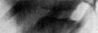
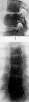
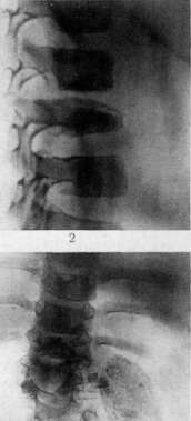
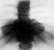
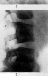
Figure 1 and 2—Initial roentgenological signs in tuberculous spondylitis: Fig. 1, narrowing of the intervertebral fissure with a limited focus in the body of the vertebra; Fig. 2, destruction of the body of the vertebra... 1 Narrowing of the fissure and indistinct change in the adjacent vertebra. Fig. 3. Prevertebral descending cold abscess in tuberculosis of a thoracic vertebra: spindle-shaped shadow. Fig. 4. Roentgenogram of a lumbar cold abscess with calcification in tuberculous lesion of the lumbar vertebrae. Fig. 5. Intrathoracic cold abscesses: paravertebral shadows with lowering of the lower thoracic vertebrae. Fig. 6. Sign for polyps... [Article continues]
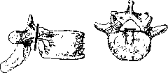
Fig. 1. Blood supply to the vertebrae.
mere introduction of the bacillus into the tissues is not yet sufficient for the development of the disease; there must be additional factors that weaken the resistance of local tissues, which are: 1) special virulence and massiveness of the infection brought into one or another section of the skeleton (embolism according to König and Volkmann); 2) anatomical features of blood supply that contribute to the retention and greater accumulation of bacilli (Lexer); 3) functional load and exceptional activity of one or another section of the organs during their development (Menard). The latter factor apparently has the greatest significance in relation to the early manifestation of the disease (maximum in the 2nd–3rd year), when greater demands are placed on the spine than on the extremities (Kornev). Trauma, significant or slight, in and of itself is not the cause of the occurrence of tubercular spondylitis, since 1) a direct connection with trauma is determined according to all statistics in no more than 30% of all cases (secondary nature of the etiological moment); 2) the frequency of tubercular bone lesions after significant trauma is no higher than in other children (Broca), and 3) experimentally in animals infected with tbc, trauma does not cause spondylitis (Lannelongue and Achard), whereas according to Kornev's data, when infection is introduced into the spleen without any trauma, it is possible to obtain a caseous lesion of the vertebrae (experiments by A. Chistovich at the Leningrad Institute for the Study of Occupational Diseases). But nevertheless, trauma is of great importance in revealing covertly proceeding processes in the spine and in the exacerbation of calmly proceeding processes. The transfer of acute infections by children has the same significance, with those diseases that proceed with pulmonary complications—measles, whooping cough, and especially pneumonia—having the worst effect on the course of the local process, which themselves can affect the deterioration of the primary focus and contribute to the dissemination of the process. That is why spondylitis is frequently revealed in children who already have lesions of small bones (spina ventosa) or isolated subcutaneous nodes (tuberculomas) that appeared after transferred pneumonias or pneumonia complications of various childhood infections. The significance of trauma can also be understood in such a way that if a child has tubercular bacteremia, trauma creates a locus minoris resistentiae in the sense of the localization of microbes at the site of trauma. Heredity has no direct influence on the occurrence of spondylitis, but one must keep in mind the inherited reduced resistance of the organism to tbc in general and the possibility of early contact infection from bacillary parents. From this point of view, socio-household factors have even greater significance, not only as general conditions influencing infectivity by tbc and the state of the organism's resistance against tbc, but also on the development of the process itself (see below - prophylaxis). Finally, immuno-biological factors determine first of all the isolation of the process in the bones. If one adheres to Ranke's classification, the moment of the infection of the vertebrae itself belongs to the second stage—generalization, associated, on the one hand, with the appearance of various metastatic foci, and on the other hand, with such a tension of the allergic state that delimits the process in one place, and from this time one can consider the transition of the process to the third stage—isolated, organ tbc, which usually guarantees against the appearance of new foci, unless the latter already existed in a hidden state, revealing themselves by external signs after a more or less prolonged time. In the vast majority of cases, we have an isolated lesion originating from a single vertebra; double and triple lesions, as well as combined lesions of large bones and joints (5–8%), are rarely observed (1–3%). Pathological anatomy. As was said, the tubercular process usually develops in the anterior sections of the spine—in the bodies (spondylitis anterior)—and posterior spondylitis (spondylitis posterior)—lesions of the arches and processes—is very rarely encountered. The lesion of the bodies can have the character of osteoperiostitis—superficial ulcerations with detachment of the longitudinal ligaments (spondylitis superficialis)—which in its pure form is encountered very rarely and predominantly in adults; in the overwhelming majority of cases, deep tubercular ostitis or osteomyelitis develops. The latter in children from the very beginning has a more diffuse character, causing the melting of the vertebra very early, whereas in older children and especially in adults, a more localized, nested process is more often observed. The tubercular bacillus brought with the bloodstream into the spongy substance causes the formation of a specific granuloma, which, as an active living tissue developing in the bone marrow between bone trabeculae, causes the resorption of the latter, which with the slow development of the process leads to the formation of bone cavities of greater or lesser magnitude—cavities containing grayish-red granulations that subsequently undergo caseous degeneration and frequently contain remnants of trabeculae—"bone sand." In addition, the proliferating granuloma frequently undergoes caseous degeneration very early and causes the necrosis of the bone trabeculae enveloped by it, which leads to dry necrosis of certain sections of bone that gradually begin to delimit, separate in the form of sequestra and melt, and also form cavities. Such necroses, appearing on sections in the form of yellowish bloodless foci, are usually considered bone infarcts—the consequence of the blockage of terminal arteries by a tubercular embolus (König, Volkmann, Lexer) or localized tubercular endarteritis (Orth, Heil, Oberst), which in the light of recent research (Randerath, A. Chistovich) has not received confirmation. In the necrotic sections, the framework of the destroyed granuloma is preserved in the form of a dense argyrophil network determined by the deep silver-plating method (Bielschowski, Foot). Located usually in the center of the bone, such necrotic foci can remain in a hidden state for a very long time without causing clear pain symptoms or deformations, which is observed mainly in adults, in whom the strength of the cortical layer is an obstacle to the spread of the process outward and prevents the collapse of the vertebra, from which sometimes almost a shell remains, which during excessive load or trauma can crush or crack according to the type of pathological compression fractures with all the consequences of the latter. In children with their soft bones, the collapse of the vertebra is observed as a rule and depends on the more diffuse character of the process and the involvement of the outer sections—the periphery of the vertebra. In such cases, the central foci approach the anterior section, soften (destroy) it, and thereby contribute to the collapse of the vertebra, with the involvement of neighboring ones—upper and lower (Fig. 2), producing such frequent lesions of three vertebrae. When the primary focus is located closer to the discs, the latter as a rule and very early are involved in the process, are destroyed relatively quickly, and involve the adjacent surface of the neighboring vertebra [see separate table (p. 375–376), figures 1 and 2], causing its subsequent contact destruction. Although it is generally accepted that the intervertebral disc itself is never affected, this position is currently contested by some authors. Thus, the primary lesion of the intervertebral discs by the tubercular process has been described (Doub, Badgley). This phenomenon becomes possible as a result of the ingrowth of vessels into the elements of the intervertebral disc from the intercostal arteries passing through the anterior ligament. Congenital defects of the cartilaginous plates—the preservation of capillaries and juice canals—favor the primary lesion of the intervertebral discs. This is also promoted by the presence of Schmorl's nodes, in which the erosion of the cartilaginous plates provides the possibility of vessel ingrowth into the intervertebral disc. The possibility of a primary lesion of the intervertebral discs is especially probable at a young age, when the intervertebral discs are sufficiently vascularized (Ognev). Cartilage itself is never primarily affected. Dis—
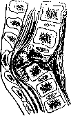
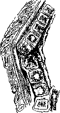
Figure 2. Destruction and collapse of vertebrae at the height of the disease. Compression of the spinal cord by caseous masses. Formation of a prevertebral abscess and detachment of the anterior longitudinal ligament.
—spread of the specific process, its breakthrough through the boundary barrier—the cortical layer—causes the development of perifocal reactive changes in the surrounding soft tissues, determining the severity of clinical phenomena, primarily spinal cord disorders caused by the disruption of lymph and blood circulation. The localization of the breakthrough on the surface of the vertebral body predetermines the further development of the process. Thus, the breakthrough of the anterior surface frequently leads to significant detachment of the anterior longitudinal ligament and secondary lesion of the exposed vertebrae; lateral—to the spread of cold abscesses in the soft tissues; posterior—to the detachment of the posterior longitudinal ligament or to the passage onto the dura mater.
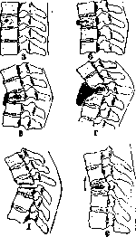
Fig. 3. Mechanism of destruction and collapse of vertebrae (scheme): a, b, c, d—development of the central focus, with destruction and displacement of the anterior section, formation of a cold abscess and involvement of two neighboring vertebrae; e—contact destructions of two neighboring vertebrae and cartilaginous disc; f—formation of a button-like protrusion during the inclination of the overlying healthy vertebra.
and finally, the localization of destruction on the upper or lower surface of the vertebra produces a contact lesion of the disc and adjacent vertebrae. The most characteristic sign of Spondylitis is the formation of a hump (gibbus), an angular curvature of the spine. The exclusion of an individual segment from the general column usually leads to flexion-rotation around the articular processes of the overlying vertebra as a two-armed lever, the anterior arm of which—the body—tilts forward and downward, while the posterior—the spinous process—rises upward, increasing the diastase and the protrusion of the tip of the process, which is the first sign of wedge-shaped collapse according to
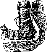
Figure 4. Old quiescent spondylitis with major deformation. Such a protrusion may also depend on the posterior displacement of the entire arch together with the wedge-shaped remnant of the vertebral body, which is displaced dorsally by the pressure of the collapsing vertebrae. The faster the vertebra is destroyed, the sharper the angle; with slower destruction and the involvement of a larger number of vertebrae, the hump acquires a more
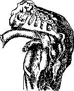
Figure 5. Doubling of the curve with major deformation. Side view of the previous specimen (Fig. 4).
uniform arcuate bend (kyphosis) with the formation of compensatory lordoses above and below. With significant destructions, when many healthy vertebrae participate in the curvature, complete collapse leads, as it were, to a doubling of the spine (Figs. 4 and 5). In rare cases, large displacements lead to true pathological dislocations, where the upper vertebra usually slides first posteriorly and then anteriorly. With uneven destruction of the bodies, lateral curvatures—scolioses—develop, which are observed mainly in the lumbar region. In the thoracic region, where the spinous processes, lowered obliquely downward, overlap each other and the vertebrae themselves normally form a kyphotic bend, the curvatures reach their greatest degrees, especially in the upper part of the thoracic region. In the lumbar region, with its physiological lordosis and horizontally positioned spinous processes, kyphoses are observed much less frequently; more often one deals with the flattening of the normal lumbar lordosis and direct collapse—settling of the vertebrae. Approximately the same picture is observed in the cervical region. The described anatomical-pathological changes undergo further evolution in connection with the stage of the disease and the age, or rather the growth, of the child. The height of the disease is characterized by maximum destruction and intensity of reactive inflammatory phenomena, while the stage of subsidence is accompanied by a decrease in these non-specific perifocal processes, a halt in destruction, and the appearance of reparative phenomena, which are manifested by greater bone strength, their clear contour, more complete collapse of healthy vertebrae and consolidation of diseased ones, often with the formation of lateral bony bridges, periosteal growths, etc. In rare cases, complete fusion of two or three vertebrae into a common bone mass is observed, which usually indicates the elimination of the process.
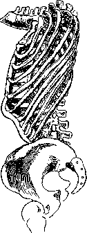
Figure 6. Deformation of the chest in upper-thoracic spondylitis.
Concurrently, ankylosis of the intervertebral joints and ossification of the posterior longitudinal ligament are observed, which ultimately creates even greater stability for the affected area of the spine (Turner). This is also facilitated by secondary changes—the formation of supra- and infra-gibbar lordoses, which level out the mechanics, and the descent of the ribs until they touch the pelvic bones. The destroyed vertebrae do not recover and do not grow, and therefore lag behind in growth from the rest of the skeleton, due to which the deformations formed in the active period increase sharply thereafter. The earlier the disease manifested itself, the more significant the subsequent deformations caused by it. Large curvatures of the spine lead to significant secondary changes in the entire skeleton, and first of all, the chest is deformed. The higher the lesion, the lower the ribs drop, the flatter and longer the chest becomes,
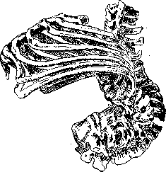
Figure 7. Deformation of the chest in thoracolumbar spondylitis.
and the more the sternum approaches the spine. The lower the lesion, the higher the ribs rise, and the sternum assumes almost a horizontal position (Figs. 6 and 7). Changes also undergo the pelvis, which in lower lesions tilts backward, the limbs become disproportionately long compared to the trunk ("monkey arms", reaching down to the knees and lower) and even the skull stretches in the occipito-mental direction. Skeletal deformations lead to compression and significant displacement of internal organs, which particularly affects the aorta, which, being embedded in pathologically altered tissues and following the curvature of the spine, sometimes forms such folds, bends, and outright duplicatures that cannot fail to affect the blood circulation and nutrition of the lower limbs. The second most important sign of tuberculous Spondylitis is the formation of cold abscesses, or, as they are often called, cold abscess formations—that is, accumulations of cold tuberculous pus, possessing the ability to migrate and spread along intermuscular spaces and neurovascular bundles, sometimes over very significant distances (e.g., to the thigh and even into the popliteal fossa in lesions of the lower thoracic vertebrae). Containing no pyogenic bacteria and possessing no enzymatic-proteolytic action, a cold abscess can remain in the organism indefinitely without causing special disorders, but at the same time it is always a source of three kinds of complications: toxic, mechanical, and secondary infection. Toxic phenomena can depend not only on the absorption of the contents of the abscesses, but also on the very spread of the process as a secondary tuberculous focus in the soft tissues, since the formation of a cold abscess is not a passive process of accumulation of liquid decay released by the carious focus, but an active spread of tuberculous granulation through the soft tissues, as Lannelongue pointed out as early as 1881, which has now been confirmed in the works of Licht (A. Chistovich and Vinogradov). The danger of mechanical complications depends on the compression of certain important organs by large accumulations (esophagus, spinal cord, etc.). Finally, a cold abscess can break through either internally—into cavities and hollow organs, which is observed extremely rarely, or it can break through outward through the skin, which leads to the most frequent and most dangerous complication—the formation of a fistula and secondary infection. Pyogenic bacteria entering from the outside drastically change all the properties of "cold" pus, making it "hot," polynuclear with proteolytic action, which leads to increased destruction-melting of tissues, a sharp inflammatory reaction of the surrounding tissues, atypical spread, and general septic poisoning of the organism—fever, emaciation, and amyloidosis of internal organs. The pathways of spread of closed cold abscesses are determined by the presence of loose connective tissue layers—spaces (spatium) in intermuscular spaces, under aponeuroses, along neurovascular bundles, etc. A simple spreading (rupture) of pus does not occur; Figure 8. Intrathoracic cold abscesses (after Schauffler's scheme): a - initial accumulations with displacement of the aorta and esophagus; b - large accumulations with involvement of the aorta, compression of the lungs, and penetration into the spinal canal. a rapidly developing granulation with decay in the center forms a sac that gradually pushes tissues apart and, by virtue of hydrostatic pressure and partly gravity, moves further.
In high cervical lesions, the cold abscess spreads either posteriorly between the occipital muscles or laterally into the submandibular region, but more often the accumulation of pus is determined on the anterior surface of the bodies in the form of a retropharyngeal abscess, which can cause difficulty in feeding and respiration. In lesions of the middle cervical vertebrae, cold abscesses form in the lower lateral cervical triangle behind the sternocleidomastoid muscle above the clavicle and higher, and in rarer cases they can descend along the spine into the posterior mediastinum.
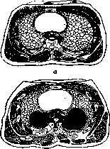
In lesions of the thoracic vertebrae, cold abscesses form either in the form of "swallow nests" sitting on the lateral surfaces of the thoracic vertebral bodies, which can increase to significant spherical accumulations (Fig. 8), determined only radiologically, or they can spread along the ribs laterally and in any place penetrate to the outer surface of the chest, or finally, the cold abscess can detach the longitudinal ligaments—periosteum—and spread up and down along the exposed vertebral bodies, forming longitudinally extended flat accumulations visible on X-ray images in the form of fusiform shadows (see separate table, pp. 375–376, Fig. 3) extending far beyond the limits of the destroyed vertebrae upwards (Fig. 9).
In rare cases, a cold abscess breaks through posteriorly, immediately to the side of the spine on the back. Through the diaphragm along the vascular pathways, mediastinal abscesses do not descend (Löffler) and only in lesions of the lower thoracic vertebrae Fig. 9. Pneumoabscess, outgoing cold abscess. Detachment of the anterior longitudinal ligament can penetrate under the diaphragm and spread along the course of the psoas muscle. The psoas major muscle originates from the lateral surfaces of the bodies of the lower thoracic and lumbar vertebrae, which is why lesions of the latter as a rule lead to the spread of cold abscesses downwards along the course of this muscle, inside the aponeurosis enveloping it. Stopping above Poupart's ligament, most often
intervertebral discs. Pathological fracture of the spine.
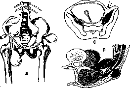
Figure 10. Scheme of the spread of psoas abscess: a - spread and formation of iliac, femoral (on the right), iliac and gluteal (on the left) abscesses; b - cross-section at the level of the upper sacral vertebra - ilio-psoas abscess; c - formation of a lumbar abscess - breakthrough of the psoas abscess into the sheath of the quadratus lumborum muscle and through Petit's triangle.
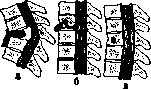
these cold abscesses are most often felt in the iliac fossa, and moving lower under Poupart's ligament, they accumulate on the antero-internal surface of the thigh. From here, the cold abscess can spread even lower along the course of the vessels - to the popliteal fossa or, rounding the thigh, emerge posteriorly under the gluteus maximus muscle (Fig. 10). In adults, cold abscesses often penetrate posteriorly into the sheath of the quadratus lumborum muscle and from there emerge into the lumbar region through Petit's triangle. Finally, in lesions of the lumbosacral region, the cold abscess can emerge through the greater sciatic foramen and accumulate above the greater trochanter - above, under the gluteal muscles, or descend downwards, below the trochanter. Cold abscesses, as secondary foci developing in soft tissues, go through the same stages of development as primary lesions in the vertebrae: onset - formation, height - spread, and subsiding - delimitation, stabilization and reduction or, as they say, resorption, unless a complication - a breakthrough - has occurred. However, such a seemingly spontaneous cure turns out to be as imperfect as in the bones themselves, since the descended, condensed contents, surrounded by a dense capsule with calcareous deposits, remain in the body for an indefinitely long time, being the cause of such frequent relapses. Finally, the third cardinal sign of spondylitis is spinal cord disorders, observed in approximately 10-15% of cases and depending on the involvement of the adjacent parts of the spinal cord and roots in the process. The direct transition of the tubercular process to the substance of the spinal cord - tuberculous myelitis - is observed extremely rarely; in the overwhelming majority of cases, these changes depend on perifocal spinal cord vascular inflammatory changes - local edema of the spinal cord (Schmaus, Kahler) and less often on mechanical causes - compression of the spinal cord. The latter may depend on the protrusion into the spinal canal of caseous masses, granulation proliferations, and cold abscesses, which usually detach the posterior longitudinal ligament without destroying it, but sometimes pathological masses penetrate to the dura mater, which is either simply compressed by them or itself becomes involved in the process (pachymeningitis externa), thickens, sometimes with germination onto the inner surface, where plaques are formed (Sorrel). Even more rarely, compression is caused by bone sequestra - the posterior displacement of fragments of destroyed vertebrae that have lost connection with the arches, or sharp protrusions of the posterior sections at the site of significant curvatures and displacements observed during the rapid development of the process or pathological fractures and dislocations (Fig. 11). Usually, however, curvatures that occur slowly, regardless of their magnitude, not only do not lead to a narrowing of the spinal canal, but rather to its expansion in this section. Most often these changes are observed in the thoracic region, where the spinal canal is relatively narrower than in the cervical, and especially in the lumbar regions, where in addition the cauda equina begins. Emerging through
Figure 11. Scheme of compression of the spinal cord (after Calot): a - by the remnant of a destroyed vertebra (sequestrum); b - by caseous masses or a cold abscess; c - by thickening of the meninges.
the intervertebral foramina, the roots are also involved in the process according to the same type of vascular changes; they themselves are exceptionally rarely affected by the tubercular process. Finally, the spread of the process to the antero-lateral surfaces of the vertebral bodies can involve the sympathetic border trunks, ganglia, and communicating branches passing here (Sorrel-Dejerine). Course and symptomatology. As with any bone-joint tuberculosis, lesions of the spine usually go through a sequential cycle of development. The onset of the disease can for a long time proceed latently or with very vague symptoms. Children become more sluggish, less mobile, sometimes complain of pain, cry out
at night. Then the pain
Figure 12. Muscle hypertonia in spondylitis. "Reins" (Korneev's symptom) upper, often radiating
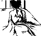
pains radiating into the side, abdomen, or legs (depending on the level of the lesion), a forced posture, difficulty in movement, and finally more clear signs are discovered: local pain upon pressure and load, local limitation of mobility, and protrusion of the spinous processes determined during a detailed examination of the spine (see Spine, research methods). Of the greatest importance is the sign of persistent limitation of mobility, depending on the reflex contracture of the back muscles, which sometimes causes hyperextension - throwing the torso back ("proud" gait), and in some cases - with lesions of the lower thoracic and lumbar vertebrae - a special muscular hypertonicity, manifested during movements or when tapping with a small hammer on special muscle cords diverging at an angle from the affected vertebra to the inner edge of both scapular spines (Kornev's "reins" sign) (Fig. 12). Adults for a long time, sometimes for years, are treated for neuralgia, lumbago, sciatica, until a clear anatomical cause is discovered. An early sign of cervical lesions is torticollis and the patient constantly supporting the head with hands (Fig. 13). The height of the disease is characterized by clear anatomical changes - deformation of the spine, cold abscesses, and spinal cord disorders. The deformation of the spine, initially manifested by the so-called button-like protrusion of a single spinous process without changing the general line of the spine, subsequently acquires the character of greater or lesser curvatures, which are more often angular (gibbus), less often arched (kyphoses) and trapezoidal. Lateral curvatures can be angular, stepped (displacement along the horizontals) and torsional with rotation around the vertical axis. Finally, primary lordotic curvatures are observed extremely rarely and only in the cervical region, but as secondary ones they are encountered as a rule above and below kyphotic curvatures, which depends not only on compensatory adaptability to restore disturbed statics, but also on reflex parabibbar muscular contractures, giving very early, more often lower, less often higher than the lesion, a flat depression as it were, which in the presence of persistent limitation of mobility is one of the early and sure signs of spinal cord lesion. Due to increasing pain, the patient stops walking without support, persistent contractures of the dorsal musculature lead to great stiffness of movements, and developing contracture of the ventral musculature leads to greater kyphotic curvature, which can also develop in the supine position of the patient when the weight of the overlying part of the body is eliminated. Subsequent subsidence and limitation of movements depend already on the degree of anatomical destruction and scar changes. In adults, as has been said, curvatures do not reach such degrees as in children, but in them pains and stiffness of movements are even more sharply manifested. The formation of cold abscesses is usually accompanied by slight increases in temperature, an increase in neutrophilic leukocytosis, and an intensification of local inflammatory changes. In particular, when an abscess forms in the region of the psoas muscle, a contracture of this muscle often develops with moderate adduction of the leg (the possibility of flexion with the impossibility of extension, in contrast to coxitis, where all movements are difficult). Subsequently, the cold abscess begins to be clearly palpable in the depths, first in the form of an infiltrate, then fluctuation appears, and upon resorption it is felt in the form of a dense wide cord. Mediastinal abscesses are determined only radiologically [see separate table (art. 375-376), Fig. 5]. When the cold abscess has formed (Fig. 14), usually the reactive changes indicated above decrease, but nevertheless the toxic effect can manifest itself for a long time, especially with high pressure on the walls. Increasing in size, gradually separating the muscles and pushing apart tendon stretches, the cold abscess approaches the skin and can cause changes in the latter: tension, pallor, dilation of veins, local increase in temperature, and then cyanosis, redness, and the formation of a fistula. Fistulas usually initially have a benign appearance, but then, as a secondary infection enters, the pus becomes thicker, more uniform, greenish, the edges of the fistula become inflamed, pockets begin to form, new fistulas open, fever appears, and the general condition worsens. The longer the fistulous tract, the more severe this complication, while with shorter tracts healing occurs sooner. Ulceration of the skin with exposure of caseous-altered tissues is often added to the fistulas: The presence of a secondary infection at best delays the process, but more often transfers patients into the category of hopeless fistulous chronics with subsequent amyloidosis. In those cases where it is possible to prevent secondary infection, the fistula can close relatively quickly, which also coincides with the liquidation of the cold abscess. Spinal cord disorders are usually very early expressed in an increase in knee reflexes, which with the development of the process gradually intensify to spastic states of the musculature of the lower extremities with clonus of the foot and patella and can turn into spastic paralyses-paraplegia.
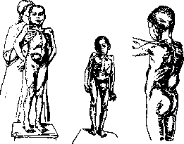
a
б
в
Fig. 14. Types of external cold abscesses: a - iliac; б - femoral; в - lumbar. Motor paralyses are usually accompanied by greater or lesser sensory disturbances up to complete anesthesia below the level of the lesion (with a zone of hyperesthesia at the level or slightly above the lesion), and in more severe cases are complicated by trophic changes - bedsores and disorders of the pelvic organs, urinary retention and defecation, subsequently replaced by incontinence. The faster paralyses develop, the more often these severe, often fatal complications manifest. Superficial bedsores on the sacrum in bedridden patients with loss of sensitivity are observed quite often, and they depend on insufficiently attentive care for the patients; extensive ones, however,
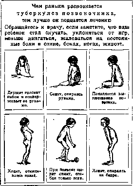
Figure 13. Early signs of spondylitis (poster by P. T. Kornev).
Strictly progressive bedsores involving the bone as well are a consequence of deep neurotrophic disorders that occur upon the rapid and complete loss of sensitivity; such bedsores can lead to severe inflammatory changes in the surrounding tissues, with the formation of phlegmons, tracking abscesses, and a septic state. Paralysis of the urinary bladder is dangerous due to the possibility of developing purulent cystitis and fatal ascending pyelonephritis, the cause of which lies in the infection of the distended bladder during prolonged catheterization used for emptying the bladder when there is initially complete urinary retention. In lesions of the cervical vertebrae, the paralysis may also involve the upper extremities, as well as the intercostal and abdominal muscles, whereby respiration, supported solely by the diaphragm, is rendered extremely difficult. Conversely, in lesions of the lumbar region, where the spinal cord ends and the cauda equina begins, the described disorders are encountered significantly less frequently and manifest as root-type lesions with a predominance of sensory disturbances and a reduction in tendon reflexes; if paresis and paralyses develop, they are flaccid in character. The earlier spinal cord disorders manifest, the faster they develop, and the more intense the motor disorders are, the more dangerous they are for life, but at the same time, the sooner and more completely they disappear (on average within 8-12 months), whereas slow and late development with an incomplete lesion syndrome poses less threat to life, but has a greater tendency to transition into a chronic state (Sorrel). In the course of the development of spondylitis, there is usually a sequence of phenomena ranging from initial signs of deformity to cold abscesses and paralysis, but frequently this sequence is disrupted in the direction of an earlier manifestation of paralysis or cold abscesses. In general, the active period of the disease—or as it is called, the period of height—lasts about 1.5 to 2 years, gradually transitioning to a calmer course and a subsiding of the process. The period of subsidence is clinically characterized by the disappearance of inflammatory phenomena—pains, muscular contractures, the resolution of cold abscesses (fistulae) and spinal cord phenomena, as well as an improvement in the general condition, stabilization of body temperature, and the approximation of biological reactions to normal. Most frequently, this coincides with the formation of permanent deformations due to the collapse of healthy vertebrae at the site of destruction, and the stability of the subsidence is often related to the degree of collapse and the strength of the bony fusion of the vertebrae. Radiologically, signs of bone repair appear—greater distinctness of the edges, greater bone density, and sometimes incipient bony fusions. In rare cases, two vertebrae fuse into one [see separate table (p. 375-376), Fig. 6]. Outcomes. This period of subsidence also lasts for 1-2 years and leads to a state of stable subsidence, but unfortunately, not to a cure, which occurs only in exceptionally rare cases and after many years. Usually, however, in a latent form, localized foci of 'slumbering' infection remain, threatening exacerbation and relapses. In addition, cases of a protracted course of the disease are frequent, when the usual cyclicity is disrupted either towards longer periods or towards a wave-like course with exacerbations and complications. This is observed more frequently in adults and particularly in the elderly, less frequently in children with an abnormal constitution, general complications, and especially with fistulae. Social and living conditions, as well as timely and full treatment, are of decisive importance. The diagnosis of spondylitis includes clinical, radiological, and laboratory research methods to determine the location, degree, and nature of the lesion. Clinical research methods (see Spine, research methods) boil down to determining the degree of inflammatory changes—pain, functional disorders—limitation of mobility, anatomical destructions—curvature, spinal cord and trophic disorders, cold abscesses, fistulae, changes in internal organs, and the general condition. The degree of manifestation of these symptoms determines the degree of clinical burden—the severity of the disease, which according to Kornev's method of 'unitary accounting' can be expressed in certain numerical values-indices. Radiological studies refine and complement clinical ones regarding the localization of the lesion, the number and degree of destruction of the vertebrae, the stage of this destruction and the degree of repair, as well as regarding the recognition of hidden cold abscesses. X-ray images must be taken obligatorily in two projections. On the anteroposterior view, the exact count of the affected vertebrae by the ribs, the degree of narrowing of the intervertebral discs and the bodies themselves are well determined, and cold abscesses are also well determined—mediastinal in the form of spherical, pear-shaped, and elongated shadows along the spine, and iliac, determined by the expansion of the outer boundaries of obliquely running shadows of the psoas muscle [see separate table (p. 375-376), figure 4]. On the lateral view, the details of destruction, the degree of collapse, the clarity of contours, and especially surface lesions and ulcerations on the anterior surface of the vertebral bodies not determined on the anteroposterior image are precisely determined. Cold abscesses are usually not visible on the lateral image. In addition, the comparison of both images gives an idea of the density and rarefaction of the vertebral bodies, the presence of focal changes—cavities, periosteal proliferations, and bony fusions. It must be remembered that radiological signs of repair lag significantly behind clinical and biological manifestations of subsidence and are distinguished by significantly less clarity than in joint lesions. Biological reactions rather reflect the degree of activity of the process and the immuno-biological state of the organism. The height of the disease and the particular severity of the latter are characterized by neutrophilic leukocytosis, accelerated erythrocyte sedimentation rate (ESR), and reduced lipase content; subsidence and improvement—by an increase in lymphocytosis, slowing of the ESR, and normal lipase content. The degree of intensity of tuberculin reactions (Pirquet, Mantoux, etc.) does not resolve the question of the nature of the disease, indicating only the presence of the tubercle bacillus in the organism and the allergy caused by the latter. For etiological diagnosis, greater importance attaches to the isolation from pus of a pure culture of the tubercle bacillus, determined in approximately 70% of cases (Sovetova). Differential diagnosis of tuberculous spondylitis in far-advanced cases with a clear classical triad—gibbus, cold abscess, paralysis—is simple and leaves no doubt. In earlier stages with less brightly expressed phenomena and especially when one or another sign is missing, recognition becomes difficult, and the diagnosis has to be made based on the aggregate of all data by way of exclusion. Thus, the most characteristic and constant sign—the deformation of the spine, the gibbus—by itself can be the consequence of the most diverse causes (see Spine, Kyphosis, Lordosis, Scoliosis). Among congenital developmental anomalies, one has to bear in mind: asymmetric fusion of hypoplastic vertebrae (see Spine, developmental defects), hypertrophic spinous processes observed in the cervical, thoracic, and lumbosacral regions, platyspondylia (congenital flattening of the vertebrae described by Putti, widespread or localized). These deformations are painless, do not cause muscular contractures, and do not produce spinal cord symptoms. Painless deformations of the same character (kyphoscolioses) are produced by the consequences of rickets; kyphoses at the height of rickets in small children are always arch-shaped, painless, and unstable, and are corrected under pressure. Arch-shaped curvatures of a large radius are observed in juvenile epiphysitis (see Spine, acquired deformations on the basis of non-inflammatory changes), as well as in deforming spondylosis (see) and spondylarthritis (see). More limited angular curvatures are observed in Kümmell's disease—see Spine, acquired deformations on the basis of non-inflammatory changes, as well as in certain compression fractures and their consequences, which may be accompanied by pain and spinal cord disorders, but without cold abscesses. Finally, all destructions of an inflammatory nature—osteomyelitis, syphilis, etc., as well as neoplasms, especially metastatic ones, can lead to the collapse of vertebrae and angular curvatures. Chronic osteomyelitis of the vertebrae usually proceeds as fistulous tuberculous spondylitis and is distinguished from the latter with difficulty only by the anamnesis, the presence of osteomyelitic foci in other, mainly long, tubular bones, higher neutrophilic leukocytosis (15-20 thousand and higher), and greater hyperosteal reaction, sclerosis, and periosteal lateral proliferations determined on X-ray images, although identical changes may occur in the reparative period even with old, especially fistulous, tuberculosis. Isolated closed chronic osteomyelitis of the vertebrae is a great rarity and is determined mainly bacteriologically—by the presence of pyogenic infection in the pus of abscesses accessible to incision, the formation of which may resemble cold abscesses, but without a tendency to significant migration. Spinal cord disorders are also observed here, but rarely and do not lead to complete paralysis, as in tuberculosis.
Vertebral syphilis is an exceptionally rare disease and may be suspected in a syphilitic patient with lesions of individual vertebrae, particularly the upper cervical ones, wherein proof is provided by the success of specific treatment. Cold abscesses and paralyses are almost never observed here. Post-typhoid spondylitis, in which the cartilage is primarily affected, proceeds with pain and restricted mobility, but without angular curvatures, since the vertebral bodies are not destroyed, but are fused together by symmetrical brackets that are clearly visible on X-ray photographs. Actinomycosis affects the vertebrae secondarily, usually at the end of the disease, spreading from the submandibular region or the lungs, and presents no particular difficulties for diagnosis. Difficulties in diagnosis are presented by spinal cord disorders when they are not accompanied by visible anatomical changes, as is observed in spinal cord tumors, vertebral tumors growing into the spinal canal, and echinococcal cysts growing into the same; recently, the possibility of such same disorders with changes in the intervertebral discs has been pointed out. In connection with this, it is necessary to note changes in the intervertebral discs, which are acquiring ever greater significance in the pathology of the spine, in particular the so-called Schmorl's cartilaginous nodes (Schmorl), i.e., the penetration of elastic cartilaginous masses into the vertebral body through a damaged boundary (so-called limiting) plate, forming a kind of herniated protrusion of the nuclei pulposi. Such nodes, according to Schmorl, are found on autopsies after 20 years in almost 30% of all cases and cause a rarefying process in the vertebrae, which is the cause of post-traumatic weakness of the spine (insufficientia vertebrae, according to Schanz), and possibly of traumatic spondylitis of the Kümmell type and even juvenile kyphoses. Therefore, in cases of persistent pain in the middle sections of the spine, with comparatively limited stiffness, sometimes even with certain spinal cord disorders that developed after a longer or shorter period following spinal trauma, but without clear anatomical changes, one must keep in mind these peculiar diseases, usually treated as covertly progressing tuberculous spondylitis. Treatment. Tuberculous spondylitis, as the most severe form of bone lesions, requires for its treatment with special insistence a combination of general and local measures as two sides of one and the same system. Underestimation of one or the other side is a major mistake and a source of poor treatment results. General treatment consists in eliminating external harmful factors and raising the general condition of the organism, increasing its stability and resistance. This goal is achieved, on the one hand, by carrying out broad preventive measures, and on the other hand, by creating conditions of the greatest physiological favorability for the organism, best achieved during sanatorium treatment (which can also be carried out at home), combining three main elements: a correct regimen, proper nutrition, and systematic air-and-light treatment associated with a mandatory daily stay in the fresh air for at least 2-3 hours. Heliotherapy is a powerful factor in raising the general condition of the organism, but must be precisely dosed so as to eliminate the danger of overheating, which causes an exacerbation of the process. Artificial light sources—the mercury-quartz lamp, Sol-lux, and others—are auxiliary means that to a certain extent replace solar treatment. Such auxiliary means are also intramuscular injections of iodine-iodoform according to Gotz-Grekov and calcium therapy, the significance of which is ultimately very modest. Tuberculin therapy is usually not used for spondylitis. Local treatment has as its task the provision of complete rest to the affected section of the spine through immobilization and unloading, which is achieved first of all by putting the patient to bed for a long period—a task difficult not so much in a therapeutic regard, but mainly in an organizational one, since this is associated with incessant skillful care of the patient, taking him out into the fresh air, etc. The bed must be comfortable, with a firm, non-sagging mattress (hair or double felt) placed on a solid wooden board or a solid base. Spring mattresses, mesh frames, and feather beds are inadmissible. It is more convenient to have a bed on wheels for transporting the patient for air treatment or a portable one in the form of a wooden board with narrow sides, installed on a wooden stand according to the French model. The patient must gradually, over the course of several days, become accustomed to lying in bed so that in the future he can carry out this lying down in the strictest manner, under no circumstances getting up, sitting up, or turning over; he must lie in one position all the time, on his back, with only a small pillow under his head, so that he is turned twice a day when remaking the bed and wiped with camphorated alcohol; in the case of ulcerations or bedsores on the apex of the hump or sacrum, the patient can be allowed to rest in a prone position. In children, such bed confinement is possible with the help of special fixing corsets of the Rollier type, embracing the shoulders, torso, and in especially restless children, the thighs (Fig. 15). In the recumbent position, elementary rest is created and a certain unloading is achieved, which is however insufficient to overcome the contracture of the dorsal and especially ventral musculature, which is one of the main causes of progressive kyphotic curvatures. Therefore, additional unloading is necessary either in the form of traction by the head (raising the head of the bed) using a Sayre head halter in lesions of the cervical and upper thoracic regions, or in the form of artificially enhanced lordos.
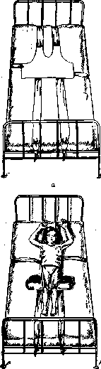
Figure 15. Fixation of the patient on the bed: a—cloth corset-bra; b—fixation of the patient with the bra.
ing of the spine, the so-called reclinat. (Fig. 16). The latter is achieved in the supine position by placing hard pillows of various sizes, stuffed with sand or even better linseed, under the hump, thanks to which the anterior sections of the vertebral bodies—first of all the healthy vertebrae above and below the hump—are moved apart, consequently the general deformation of the spine is corrected to a certain extent, and at the same time the pressure in the affected vertebrae themselves is reduced. Trying at all costs to push apart the collapsed vertebrae is both an unattainable and irrational task; forced straightening can only exacerbate the process, increase muscle contracture, and lead to even greater deformation. Therefore, very great caution and gradualness are needed here. With more quiescent and subsiding lesions in the thoracolumbar and lumbar regions of the spine, reclination is achieved
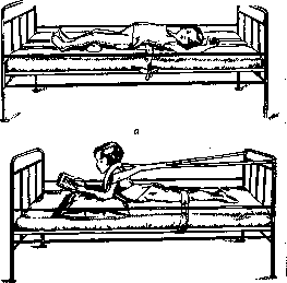
Figure 16. Reclining positions: a—in the supine position with a pillow placed under the hump; b—in the prone position—lordosing.
also by the patient lying on the stomach with a triangular pillow placed under the chest for lordosing. More perfect fixation is achieved in plaster beds (according to Lorenz) embracing the head, torso, and thighs, which are especially useful in the home-outpatient treatment of children and taking the latter out for air-and-sun treatment; they are also necessary in more acute processes and in poorly disciplined patients. By applying the beds to patients in a prone position with rolls placed under the pelvis and clavicles in a state of maximal lordosing, we also achieve a certain reclination, which can be quite safely enhanced by the gradual placement of strips of pressed cotton or gauze under the hump (Finck). Only careful observation of the skin and especially of bedsores on the apex of the hump is necessary. Practically every sick child up to 8–10 years old must be provided with a well-made, papered, convenient, and durable bed with two or three replaceable covers for it (Fig. 17). Such treatment in the recumbent position must continue throughout the entire active period of the disease, i.e., 2–3 years, and then with complete quiescence of the process, the patient can gradually
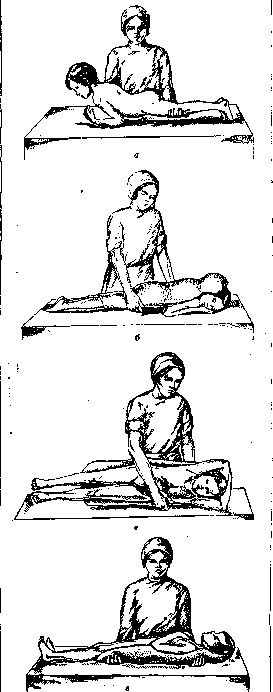
Figure 17. Diagram of using a plaster bed: a—correct position of the child before applying the bed; b—applying the bed; c—turning with the bed; d—correct position in the bed.
for half a year, learning to stand and walk by adding 1 minute a day, as Sorrel advises, but obligatorily in a corset, first a solid plaster one, and then a removable gelatin-formalin or celluloid one (preferably from non-flammable celluloid, i. e., emalinite or aeroplane lacquer), applied also in the maximally straightened and reclineated position (suspension in a special frame in the "flying bird" position) (Fig. 18). In lesions of the upper half, starting from the eighth thoracic vertebra and above, the corset must have a collar to support and fix the head. In adults, it is better to make an orthopedic Hessing-type splint-leather corset with crutches. The patient must wear the corset for a very long time, at least 3 years, and adults practically for the rest of their lives. A mistake is the outpatient treatment of spondylitis with corsets in the active, especially acute, period. The treatment of cold abscesses should be reduced mainly to the prevention of fistula formation. The faster the cold abscess forms, the closer it approaches the integuments and the greater its tension (pressure on the walls), the sooner a breakthrough occurs. Therefore, timely evacuation of the liquid contents of a progressive abscess by puncture is the most rational preventive intervention, provided it is carried out under strictly aseptic conditions, with a long (8-10 cm), sufficiently thick (2 mm) needle or trocar, from healthy overlying obliquely shifted tissues. Direct punctures at the apex of the protruding cold abscess, and even from altered tissues, are just as gross a mistake as an incision of a closed cold abscess with its open introduction. The danger of puncture is the possible infection of tuberculous pus and the possibility of damage to large vessels (especially the femoral vessels in ilio-femoral cold abscesses), the peritoneum and internal organs. The danger of infection is especially great during punctures through the mouth of a retro-pharyngeal abscess, which is why it must be freed by a puncture from the lateral side of the neck (according to Calot), guided by the transverse process of the third cervical vertebra, pushing anteriorly the sternocleidomastoid muscle and the neurovascular bundle. In those rare cases when punctures fail to eliminate a threatening cold abscess, the latter can be removed surgically along with the capsule, but obligatorily with layered wound closure (Kornev - 20 cases). Intrathoracic abscesses, if there is reason to think they depend on persistent phenomena of spinal cord compression, can also be emptied by punctures either paravertebrally (Schede, Finn and others), which is far from safe in the sense of vessel and pleural damage, or through the intervertebral foramina, entering the spinal canal (Calvé's technique), which is more complex and requires special instrumentation. The costo-transversectomy operation proposed for the same purpose by Ménard—a lateral approach
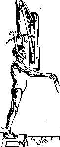
Fig. 18. Plaster corset. to the cold abscess with the removal of the transverse process and the head of the rib (Fig. 19)—is nowadays used only in exceptional cases. Laminectomy (see) has been completely abandoned. This operation is safer in quiescent cases with old paralyses, but here it brings little benefit. The treatment of paralysis, apart from the above-listed, very rarely used interventions, is as a rule strictly conservative and consists in the particularly careful and meticulous implementation of immobilization and unloading methods. Paralyses are only a symptom of the underlying disease, usually a manifestation of perifocal nonspecific changes, and therefore the correct treatment of the primary focus in the vertebrae, the alleviation of inflammatory phenomena as a rule entails an improvement in spinal cord disorders, which with systematic treatment are eliminated in approximately 70-80%. Finally, it is necessary to mention fistulas, which are easier to prevent than to treat when they have opened, and especially when they have infected. Fresh, newly opened fistulas must be most carefully protected from infection by careful handling of tissues and the implementation of the strictest asepsis (gluing the bandage to the skin). When infected, one has to act differently: in severe cases with abundant discharge—frequent dressings; in case of tracking—incisions; in case of infiltrates—compresses; in milder, but protracted cases—careful instillations of iodoform-glycerin, Calot's fluid, camphor-naphthol, and in the most stubborn cases—forced treatment with the instillation of 1-3 cm3 of a 10-12% solution of CuSO4 + ZnSO4 (Franke)—a method that is not safe and requires experience. But probably the most important thing that has the best effect on the course of fistulas is, once again, careful treatment of the underlying process—the strictest immobilization and unloading, which is the first condition for the treatment of all complications—cold abscesses, fistulas, and paralyses. Operative fixation of the spine. In general, the treatment of spondylitis must pursue two main goals: the elimination of the tuberculous process itself in the vertebrae and the prevention of spinal deformities. Unfortunately, both of these goals are still rarely achieved in full, and only early diagnosis and early full-fledged treatment can increase the stability of cure and reduce the degree of deformative changes. The classical conservative method set forth above, which is the basis of all our measures and is aimed at providing complete rest to the affected vertebrae, despite all its value and importance, still does not solve this problem completely, since it cannot prevent constant traumatization associated with respiratory movements, with inevitable movements during coughing, sneezing, repositioning, physiological acts, etc. These moments contribute both to the spread
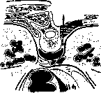
Fig. 19. Costo-transversectomy (Ménard's operation).
as if squeezing out the process (frequency of cold abscesses), and to the intensification of destruction, and consequently the collapse of the vertebrae—deformity. Therefore, the desire naturally arose to achieve more perfect immobilization by operative fixation of the posterior sections of the vertebrae. Proposed by Chipault (Chipault, 1897)
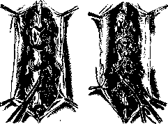
a
b
Figure 20. Hibbs' operation—plastic surgery of the spinous processes and lamches: a—formation of flaps from laminae; b—overturning of the chiseled spinous processes. tying the spinous processes and laminae with silver wire was gradually replaced by osteoplastic methods, the purpose of which is complete bony fusion of the posterior sections of not only the affected, but also the neighboring healthy vertebrae. Such fusion of the spinous processes like a bony bridge, first of all, perfectly immobilizes the vertebrae, prevents their vertical (in particular respiratory) movements. At the same time, by sealing the outer ends of the two-armed levers and finally closing the movable hinges on the articular processes, the same bony bridge can, on the one hand, unload the affected sections, transferring the center of gravity of the overlying vertebrae to the underlying healthy ones, bypassing the affected ones, and on the other hand, to a certain extent prevent the further collapse of the vertebral bodies. All the numerous methods proposed for this purpose can be divided into two main groups of operations that achieve the fusion of the spinous processes either by means of plastic surgery of the spinous processes themselves or by means of free bone transplantation. The first method, proposed by the American Hibbs (Hibbs, 1911), consists in chipping the spinous processes and laying them on top of each other in the manner of shingles in anticipation of subsequent bony fusion, which is reinforced by bone bridges knocked out of the laminae and overturned towards each other (Fig. 20). Numerous modifications of this basic method consist in the method of splitting the spinous processes and laying them, and all of them have the disadvantage that fusion may occur after a more or less prolonged period. Therefore, the second method, proposed in the same year and also by the American Albee (Albee), became more widespread—the transplantation onto the spinous processes of a plate taken from the tibia, which immediately and definitively fixes the spine. Numerous modifications of this method concern either 1) the method of transplant transplantation: intraspinally—into the longitudinal split of the processes (Albee, 1911), subspinally—under the chiseled processes onto the exposed laminae (Haldstead, 1915), paraspinally—laying the transplant on the sides (Henle, 1911), or finally in the form of rafters for oblique unloading in lesions of the lumbar vertebrae (Vreden); 2) the place from where the transplant is taken: from the scapula (de Quervain, 1912), from the rib (Hoessly, Shamov), the iliac crest (Fromme), the fibula (Bachlectmer); 3) the property of the material itself: autoplastic, as in all the above-described methods, homoplastic—from parents to children, heteroplastic—from boiled bovine bone (Kleinberg, Turner) and finally alloplastic—from celluloid plates laid on the side of the spinous processes (Lange). The original Albee method has acquired the greatest distribution, in which 1) the best mechanical conditions are created, thanks to the deep insertion of the
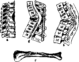
Figure 21. Methods of osteoplastic fixation
spine: a—Albee; b—Halstead-Vreden; c—Kornev "staple"; d—formation of a staple from the tibia. 2) the best conditions for the nutrition and ingrowth of the graft, placed entirely in the bone bed, and 3) minimal trauma, since the entire ligamentous-muscular apparatus is preserved. In the technique of the operation, which must be careful and sparing, it is necessary to pay attention to the precise identification of the location and number of affected vertebrae, in order to fix all affected vertebrae and two healthy vertebrae above and below the sick ones, precise longitudinal splitting of the spinous processes over their entire length down to the arches themselves, precise cutting of a graft (with periosteum) from the tibia and its deep insertion into the formed cleft followed by suturing the edges of the latter over the graft. Kornev attaches special importance to the transplantation of a strong and solid (without fractures) graft, which with greater or lesser significant curvatures of the spine appears to be a difficult task and can be facilitated by cutting out from the upper, widest part of the plenum tibiae a graft to measure, in the form of a "staple" (Kornev, 1928) with wide flat branches and a solid strong median rod knocked out from the entire thickness of the tibial crest, taken together with a part of its outer surface (Fig. 21). Indications for the operation are isolated, uncomplicated lesions of one to three vertebrae with small curvatures and a general good condition; contraindications are multiplicity and extent of the lesion, poor general condition, especially with tuberculosis of internal organs, completed cases with large deformations and fistulas. Cold abscesses are best eliminated before the operation, but in exceptional cases residual, stable, deeply lying cold abscesses are not an absolute contraindication to the operation, as well as limited mediastinal abscesses, if only they do not have a creeping character in the form of spindle-shaped ascending shadows detectable on X-ray images, indicating significant detachment of the longitudinal ligaments and exposure of overlying, seemingly healthy vertebrae. In the latter case, the operation rarely brings benefit. Paralysis is also better eliminated earlier, but in some stubborn cases, a technically well-performed operative fixation quickly shifts the process in a favorable direction. One should not operate in the acute period of the disease, with unstable temperature, disturbance of biological equilibrium (blood, ESR); it is more profitable to calm the process by conservative measures, and some authors recommend operating only with complete subsidence of the process, considering the operation as an additional orthopedic fixation aid (Henle, Biesalski). Most modern orthopedic surgeons recognize the value of these operations, and some (Sorrel, Calvé et Galland, Henle, Langenskiöld) use it only in adults, while others consider it possible to operate on children as well, mainly of older (up to 12 years) and middle (up to 7–5 years) age, and some consider age in general only a relative contraindication, operating depending on indications in individual cases even in early childhood, from 3 to 5 years (Albee, Gorres, Johansson, Kornev, Vreden, Waldenström).--Along with adherents of the operation, there are also opponents—relative (Biesalski, Haglund, Turner, Krasnobaev) and absolute (Bier, Lexer, Ludloff, Löffler), who do not recognize the value of this method. In any case, it must be remembered that operative fixation is not a radical, but only a therapeutic and auxiliary measure, essentially conservative-orthopedic, but achieved by operative means. Therefore, surgical intervention should be regarded as one of the links (greater or lesser central) in the chain of all therapeutic measures—both preparatory, aimed at raising the general condition, calming the process, possible reclinatorily and eliminating complications, and successive ones, covering not only the immediate postoperative period (8–12 weeks of lying down), but also a long subsequent period, when the patient must wear a removable corset for 2–3 years and be in an appropriate environment under the supervision of a physician. Consequently, the basis of treatment should be the sanatorium-orthopedic conservative method, in which the operative method is not opposed, but rather complements it, being applied according to certain indications in certain periods of the disease as an integral part of the general treatment plan. The immediate results of treatment of spondylitis in various sanatorium institutions give a relatively high percentage of favorable outcomes—70-80%, while the long-term ones are far from comforting. Thus, with conservative treatment, mortality with long-term observation up to 5 years reaches 40% (Seemann, zur Nedden, Langenskiöld), and in this respect the published long-term results of surgical treatment are significantly better, giving 50-70% favorable outcomes and no more than 25% mortality. As for the immediate results of surgical treatment, with correct indications, good technique, and proper preparatory and sequential treatment, it gives a percentage of favorable outcomes not lower, but higher than conservative ones. Thus, at the Leningrad Institute for Surgical Tuberculosis and Bone-Joint Diseases over 12 years, surgical intervention was used in 368 patients with a total percentage of favorable outcomes of 87.5 (42% improvement and 45.5% persistent subsidence) and 4% mortality, whereas out of 332 conservatively treated patients, the mortality percentage was 17, with 62% favorable results (Kornev, Molchanova). Although these outcomes largely depend on the selection of patients, nevertheless, on such a relatively large material, it is obviously revealed that surgical interventions do not entail greater danger than strictly conservative treatment. The advantages of the former are obvious, since, on the one hand, undoubtedly operative fixation in a more perfect way leads to the strengthening of the affected part of the spine and thus improves the degree of cure, and on the other hand, significantly shortens the periods of hospitalization of patients without prejudice to the quality of treatment. Prophylaxis of tuberculous spondylitis cannot be separated from the general prophylaxis of tuberculosis in general and must be carried out through broad health-improving measures and special anti-tuberculosis measures, so widely carried out in our Union and which have already given definite positive results in reducing the incidence of tuberculosis. Regarding bone forms, in particular spondylitis, the center of special attention should be shifted to the detection of early forms, which, as noted above, most often appear in early childhood, at the age of 2–5 years. Therefore, special importance is attached to the familiarity of micro- and macro-pediatricians with the basics of recognizing the initial, usually latently running forms of tuberculous spondylitis, to identify these forms in maternity and infancy protection centers and children's health centers, in children's prophylactic outpatient clinics, preschool and school institutions, so that at the first suspicious symptoms children could be sent to special institutions for bone and joint tuberculosis, which with special persistence must master the methods of early diagnosis. The treatment itself should have a prophylactic bias in the sense of providing preferential assistance to early and fresh forms, with special attention paid to the identification of complications. Early treatment and timely elimination of complications are the best guarantee of success in the fight against those severe consequences that untreated or poorly treated tuberculosis of the spine entails—disability and the transfer of patients to the category of hopeless chronics. Such timeliness and usefulness of assistance is possible only with the planned implementation of all measures aimed at covering all patients, by combining hospital and outpatient care into one general system, by establishing the examination and treatment of these patients at home with the provision of all types of social assistance—patronage and general dispensary registration of these patients with long-term observation of them. It is necessary to take care of producing X-ray images for these patients (necessarily lateral ones as well), supplying them with plaster and removable orthopedic apparatuses, taking care of training qualified cadres of doctors in bone and joint tuberculosis, which are not available now, as well as the deployment of special institutions, which are insufficient. The fight against tuberculous spondylitis is a hard struggle to protect the younger generation from physical disability, to which public attention should be drawn to a greater extent than has been the case up to now.
Related articles
Mentioned in
Cite this page
“Spondylitis.” Soviet Medical Encyclopedia. English translation of Bolshaya Meditsinskaya Entsiklopediya, 1st ed. (Moscow, 1928–1936), ed. N. A. Semashko. https://sovietmedicalencyclopedia.pages.dev/article/spondylitis/