Tuberculosis
Historical document, translated for reference. It reflects medical knowledge of the 1920s–30s and is not medical advice.
Summary
Tuberculosis is an infectious disease caused by a specific microorganism discovered by R. Koch, affecting various organs but most commonly the lungs, intestines, bones, and joints. The article provides a historical overview of tuberculosis knowledge from ancient times through the 18th century, including its recognition in different cultures, evolving understanding of its pathology, and early treatment approaches.
Encyclopedia article (1928–1936)
74 VII. Immunoprophylaxis and immunotherapy... 9 6 Tuberculosis (from Latin tuberculum - tubercle), an infectious disease affecting humans, animals, and birds, caused by a specific microorganism discovered by R. Koch (see below), apparently widespread everywhere at present, with diverse and prolonged courses, affecting various organs but most frequently the lungs, intestines, bones, and joints. The manifestation and severity of the disease, besides features of localization, massiveness of infection, and character of the biological reaction of the organism, are decisively influenced by the working and living conditions in which the patient finds himself. This closest connection of T. with social conditions gave rise to calling T. a social disease, a proletarian disease. K. Marx in 'Capital' characterizes T. as an inevitable consequence of the capitalist economic system. I. Historical sketch. T. has been known since ancient times, but until the 19th century, when Laennec proposed this name, it was known under other names: phthisis, phthise, consumptio (burning up), tabes, Schwindsucht, Auszehrung, consumption; its local manifestations for a long time went under various names: scrofula, lupus, tabes mesaraica and others. Although before the discovery of the causative agent of the disease T. was confused with various diseases similar in symptom complex, still even in the earliest medical works that have come down to us, the characteristic signs of T. are quite distinctly outlined, and we are able to form a fairly clear idea of the views of physicians of past epochs on T., its pathogenesis, clinic, and treatment. The earliest indications of T. are found in the code of Hammurabi (2250 years before the modern era), where in §§ 148 and 149 a very vivid description of the pulmonary form is given and the right to divorce a woman who has developed consumption is regulated. In the book of laws of Manu (1400 years before the modern era), pulmonary consumption and lesions of the lymph glands of T. are recognized as a 'dirty, incurable' disease, and judging by the prohibition of marrying women from families where such diseases occur, the observations of that time had already noted the transmission of the disease from one family member to another. The data of Egyptology allow us to consider T. in Egypt as little widespread among the ruling nation (landowners and slave owners) and more widespread among the oppressed nations ('Semitic disease'). It is quite certain that pulmonary T. was known in China: the treatise on medicine by Wang-Shu-Ho (6 centuries before our era) even gives prognostic indications (slow pulse, soft-favorable sign, tense, frequent-bad prognosis). Deserves attention the advice to treat scrofula and T. of the lymph glands with alkaline waters and seaweed (iodine).
Ancient Greek medicine, in its early development, under the influence of Egyptian, gives us many valuable indications allowing us to assert that even at that time physicians had noted the connection of T. with constitution, the influence of hereditary predisposition and even its contagiousness. Fragments of the treatise of the Cnidian school allow us to establish that Greek physicians of that era knew how to recognize outbreaks of the process, accompanied by softening of foci, the cirrhotic nature of the process, rapidly confluent and miliary forms, and a number of complications accompanying pulmonary T. (spontaneous pneumothorax, hydropneumothorax, Hippocratic splash, empyema, amyloidosis, etc.). The semiotics of these complications is set forth very vividly and accurately. Therapeutic measures (removal of pus by opening the chest cavity with subsequent drainage and pouring of a spirit-oil emulsion), as well as the prognostic evaluation of cold-tuberculous and hot-septic pus leave no doubt that physicians of that era had a fairly clear understanding of the clinical picture. A number of indications on symptoms characteristic of tuberculous patients-sweats, constantly cold feet and hands, diarrhea, loss of appetite, lesions of the larynx, pleura, coin-like sputum-very vividly illustrate the great experience of Greek physicians in recognizing the manifestations of T. With a pessimistic view of the curability of T., given that only severe forms of T. were known to physicians due to the state of diagnostics, therapeutic measures come down to hygienic-dietary regimen [rest, warm compresses, intestinal hygiene, expectorants; with improvement or transition to chronic course-moderate physical exercise, abundant but non-irritating food, milk (preferably female, donkey, and goat)]. Among the Romans, who mainly used Greek physicians or slaves trained in Greek schools, we find nothing essentially new. The most original works are those of Aulus Cornelius Celsus, who emphasizes the connection of T. with meteorological conditions, the importance of age (consumption of the transitional period) and includes in the arsenal of therapeutic measures long sea voyages. In the works of Galen the principles of the Greek school find a brilliant interpretation. In the following epochs until the end of the 16th century we note the transmission of the principles set forth by the Greek schools in Arab medicine (Rhazes-10th century, Maimonides-12th century) and in medieval Christian medical schools. Patho-anatomical study of T. dates only from the middle of the 17th century, precisely from the time when scientists began more or less systematically to perform autopsies on deceased patients. Researchers of that time describe sometimes found in the lungs of corpses nodules, which they denote as tubercula or as skirrhos. However, the connection of these nodules with the clinical picture of 'pulmonary consumption' was first established by Sylvius de la Böe (Sylvius de la Boë, 1614-72), who claimed that consumption begins with the appearance of these nodules, which subsequently increase, soften, and ulcerate. Sylvius de la Böe, as well as a number of subsequent scientists (Morton, Cullen, Portal, Kortum, Hufeland), considered that tubercles represent enlarged lymph nodes of lung tissue, invisible in normal conditions. Their increase occurs due to stagnation of lymph or, according to Portal, due to deposition in them of a special product in the form of tuberculous or scrofulous matter (obstruction theory of the origin of consumption). Incidentally, the authors of that time found much in common between the nodules present in the lungs in consumption and the changes in the lymph glands related to the so-called scrofula, which is why the designation of tubercles as scrofulae (German Skrofeln) and the attribution of changes in the lungs in consumption to scrofula is found very often in the works of scientists of the 17th and 18th centuries. The view of tubercles as enlarged lymph nodes lasted quite long despite doubts expressed about this in 1766 by Morgagni and in 1785 by Reid; the latter author claimed that tubercles, or 'granulations' represent not lymph nodes, but a product of exudation. Reid also objected to the combination of tubercles with the concept of scrofula; in his opinion tubercles and manifestations of scrofula are completely different diseases. In this respect Reid can be considered the founder of the so-called dualism in the evaluation of tuberculous changes (see below). Morton's phthisiology (end of 17th century) is the first monograph summarizing the experience and knowledge about T accumulated by previous centuries. Morton attempts to establish the etiology of T. and quite correctly notes the provoking influence of a number of diseases (typhus, scurvy, scarlet fever, measles, smallpox, etc.), depressive mental states, overwork, damp foggy climate. In the development of the process he notes on the basis of autopsies the phases: serous infiltrate, reaction of lymph nodes, pneumonia and cavernization. The description of symptoms, especially the characterization of the instability of mental reactions in tuberculous patients, is very vivid and accurate. The therapeutic and preventive measures recommended by Morton fully coincide with the indications of the Greek schools. The idea of the infectiousness of T. in the 18th century is documented by the law of 1751 issued in Spain and later similar regulations in Naples (1782) and other Italian states. These laws prescribed a number of measures for disinfection of dwellings and personal belongings of consumptives. The treatise by Auenbrugger published in 1761 on percussion (see) gave a great impetus to the development of precise diagnosis of lung diseases and in particular of consumption. A great step forward in the development of the patho-anatomy of T. was made by the Englishman Baillie (1793); he gave a good description of the tubercle and other changes in pulmonary consumption and established that 'tubercles', 'scrofulae', nodular changes and diffuse caseous infiltration represent different morphological expressions of the same morbid process. An important merit of Baillie is that he also discovered in other organs besides lungs the formation of tubercles that could merge into large caseous nodules. In addition he categorically rejected the interpretation of tubercles as enlarged lymph nodes. The recognition by Baillie of the pathogenetic unity of tubercles and caseous, 'scrofulous' infiltration forces us to recognize him as one of the founders of unitarism in the evaluation of tuberculous changes. The dissertation published in 1797 and which went through a number of editions and translations into French and English by Hufeland gave a brilliant analysis of the semiotics and clinic of tuberculosis of the lymph glands and chronic tuberculous intoxication (scrofulous diathesis) and the connection of these phases of the tuberculous process with bone T., T. of the mesenteric glands and others.
An attempt to further clarify the nature of changes in T. was made by the French scientist Bayle (1774-1816). He was the first (1810) to introduce the term 'miliary gray tubercle,' or 'miliary granulations'; Bayle traced the evolution of miliary tubercles from initial dense formations to their caseation and softening, and expressed the view that they are the basis of all tuberculous changes. T., according to Bayle, is not a disease only of the lungs, it is a general disease, at the basis of which lies a special diathesis; tubercles appear in one organ or another as manifestations of this diathesis. Bayle gave a differential diagnostic characterization of non-tuberculous lung lesions and pulmonary T. that has retained its significance to the present day. However, Bayle greatly complicated the concept of 'consumption' by recognizing six forms of it: 1) phtisie tuberculeuse; 2) phtisie granuleuse; 3) phtisie avec melanose; 4) phtisie ulcereuse; 5) phtisie calculeuse; 6) phtisie cancereuse. All these uncertainties were eliminated by the capital works on the pathological anatomy of T. by one of the greatest French scientists, Laennec (1781-1826). He united under the concept of 'tuberculosis' various changes that had been excluded by some from the categories of tuberculous changes, in particular he clarified the essence of scrofula as T. of the lymph glands. On the other hand, he purified the concepts of 'tuberculosis' and 'consumption' from the superfluous forms that complicated them. Laennec considered Bayle's classification incorrect, in particular in his opinion the division of consumption into tuberculous, granulosis, melanotic, and calcareous forms was illogical; the ulcerous form, as belonging to gangrene of the lung, and the cancerous form, as belonging to another disease—cancer—should be excluded. Thus, being a representative of unitarism with respect to tuberculous changes, Laennec recognized only two types of initial tuberculous changes: 1) isolated 'yellow' tubercles and 2) tuberculous infiltration; these formations first have a yellow, translucent appearance, then become dry and opaque; next comes their softening, suppuration, and in case of the rejection of purulent masses—the formation of ulcers and cavities. Thus, for the first time the dynamics of tuberculous changes were precisely outlined. As for the essence of tuberculous changes, Laennec, like Bayle, spoke against their inflammatory origin and assumed their closeness to the process of tumor formation. Laennec's research had enormous influence in the first half of the 19th century on the concepts of patho-anatomical changes in tuberculosis. Laennec checked and critically evaluated all methods of physical diagnosis used before him and by introducing auscultation with the help of a stethoscope gave these methods the necessary completion. By studying autopsies of patients whom he observed in the clinic, Laennec was able to compare clinical data with changes revealed at autopsy, and this ensured his descriptions exceptional accuracy and vividness, which has preserved the significance of his research to the present day. Like Bayle, Laennec dealt mainly with severe hospital material, and this explains their pronounced therapeutic pessimism and the absence of any substantial new therapeutic indications. In the following years, up to the beginning of the development of histology of tuberculous changes, almost no additions were made to Laennec's doctrine. One can only point to the discussion about the essence of tuberculous changes: while Louis defended Laennec's point of view on the tumorous origin of the tubercle, Andral expressed the view that tuberculosis is a product of a special tissue secretion, Brisseau, Rokitansky and others insisted on the inflammatory nature of tubercles and tuberculous infiltration, on the fact that they represent a product of inflammatory exudation. To the same time belongs the discovery in the lungs of cattle of changes similar to those encountered in human consumption. The development of physical diagnosis after the powerful impetus given by Laennec continued. Piory introduced the plessimeter, Winterlich—the percussion hammer. A stethoscope for simultaneous listening with both ears was proposed, and finally in 1839 Skoda (Vienna) gave for the first time an precise acoustic analysis of percussion and auscultation data, which has retained its significance almost in full to the present day. With the development of biochemistry, attempts began to study the structure of tuberculous foci by chemical means. The first detailed histological studies of tubercles belong to Lebert (1849), who established that tubercles consist of special round bodies (corpuscule, globule tuberculeux) separated from blastema; however, the simultaneous research of Henle and Reinhart, who discovered these same bodies in pus, did not confirm the specificity of them for T. that Lebert had proposed. Incidentally, Reinhart, on the basis of his microscopic research, discussed the question that interested researchers of that time about the difference between gray and yellow tubercles. In contrast to Laennec, who put forward the idea that gray and yellow tubercles belong to different stages of the same process, Reinhart began to assert that these are two completely different formations: the gray tubercle is a focus of exudation with organization of the exudate, while the yellow tubercle is simply thickened pus. Thus Reinhart revived the dualistic point of view on the essence of tuberculous changes. The research of Rokitansky (1855) on the histology of the tubercle, belonging to the same time, is interesting in that he was the first to see that in the tubercle among mononuclear cells, large multinuclear elements are often encountered. The most significant significance in the middle of the 19th century had the research on tuberculosis by Virchow. At first (1847) Virchow followed the path outlined by Laennec, but later (1856) he took a categorical stand on the dualistic point of view. He distinguished tuberculosis as a new formation originating from connective tissue, by nature close to lymphosarcoma, he well described its cellular structure, the absence of blood vessels in it, its outcome in scarring or in caseous softening, ulceration. In contrast to the tubercle of scrofula and diffuse caseous infiltrates, according to Virchow, they have an exudative origin, belong to the outcomes of non-specific inflammation and represent a manifestation of a special pathological constitution, a special vulnerability of tissues. The authority of Virchow was the basis of the fact that his dualistic view of the changes found in consumptives became widespread among both pathologists and clinicians (Niemeyer and others). It became a common saying: 'The greatest danger for a consumptive is to get tuberculosis.' The assertion of dualism in the doctrine of T. was facilitated by histological research of a number of French researchers; thus, Jaccoud came to the necessity of distinguishing tuberculous and caseous (pneumonic) consumption, Robin began to draw a sharp line between gray granulations, never undergoing caseation, i.e. 'tuberculation,' and yellow tubercles with their outcome in softening, and Empis emphasized this even more sharply, separating small gray tubercles with the special name 'granulie' and calling the disease with the eruption of a mass of gray nodules as 'maladie granuleuse.' It was pointed out that Bayle and Laennec, not having the method of microscopy, apparently knew little about the true miliary tubercles, i.e. Empie's granulie, and described mainly large (in the lungs—pneumonic) nodules and therefore inclined to unitarism. The further development of the pathological anatomy of T. proceeded along two paths. On the one hand, there was a further deepening of anatomical and especially histological information about tuberculous changes, and on the other—clarification of the nature of changes in experimental T. From patho-anatomical research, the research of Buhl (1857 and 1873) deserves mention; in the first work he established that the eruption of miliary tubercles is observed only in the presence of a caseous focus, from which by blood occurs the spread of a special product of decay appearing in such a non-specific focus; in the second work Buhl developed in detail the histology of caseous pneumonia, noting the prominent participation of alveolar epithelium in the exudate, which distinguishes this pneumonia from banal inflammations. The research of Langhans (1868) on the histology of tuberculosis was of extremely great significance, who was the first to describe giant cells in tubercles and evaluated them as something very characteristic of T. (hence the name of these cells 'Langhans' giant cells'). He also outlined two possibilities for the formation of these cells (growth of one cell during division of its nuclei and fusion of several cells). The research of Koster, Bazin, Klebs, Wagner, Schiippel developed the histology of tubercles to a large extent; in particular Schiippel (1871) very accurately describes three types of cells that make up the tubercle—1) giant cells with numerous nuclei, 2) large plate-like cells resembling epithelium and which might be called 'epithelioid' cells, 3) small cells of lymphatic type—and also correctly indicates the topographical relationship of these cells in the tubercle. Schiippel also convinced himself that from tubercles in lymph glands, a continuous caseous (scrofulous) mass can subsequently form.
However, despite the accuracy of information about the structure of the tubercle, the question of the origin of the cells comprising it and the question of the essence of the tubercle were resolved discordantly. Wagner classified the tubercle as a lymphoid neoplasm (lymphadenome tuberculeux); Köbs derived the cells of the tubercle from the endothelium of lymphatic vessels; Schüppel initially thought about the origin of the elements of the tubercle from blood leukocytes, but later inclined toward the endothelium and even expressed the assumption about the beginning of the development of the tubercle inside a blood vessel, etc. It can also be noted that the pathological anatomy of pulmonary T. was very thoroughly developed by a number of researchers. The experimental work on T. dates back to the well-known studies of Villemin ["The tubercle from the point of view of its localization, development and nature" (1862), report at the Medical Academy (5/XII 1865) and monograph "Studies on tuberculosis"]. The works of Virchow on the cell as the morphological expression of life and the works of Pasteur, which refuted the idea of the spontaneous generation of microbes and created the foundation for the study of the etiology of infectious diseases, had a decisive influence on the formation of Villemin's views. Being a talented and inquisitive researcher, Villemin managed to provide brilliant arguments in favor of the infectious origin of T. and confirm his considerations with convincing experiments. Rejecting the influence of diathesis, constitution, heredity (e.g., mass diseases of T. in the French army among soldiers selected from the most physically fit and healthy people), Villemin points to a number of circumstances that force one to accept an infectious etiology for T.: a) T. is observed in all climatic conditions; b) T. is little spread where the population density is small (mountaineers, nomads), and, conversely, is most often observed where people live particularly closely (large cities, barracks, prisons, etc.); c) T. is rare among livestock living on pastures and is observed when kept in stalls, it is unknown among wild animals and affects domesticated ones living among people, in zoos. All these moments lead Villemin to draw a complete analogy between T. and glanders or typhus, the infectious nature of which was already obvious. To confirm these considerations, Villemin conducted 17 series of experiments with the inoculation of tubercular tissue, blood, and sputum from humans and animals to animals. Thus, he inoculated human tubercular material to rabbits, guinea pigs, dogs, cats, sheep, birds. He also inoculated tubercular material taken from cows, from rabbits to rabbits; in addition to the expressed tubercular tissue, he inoculated material from "scrofulous" foci. The success of his experiments was facilitated by the original method of introducing the test material into the trachea. Thanks to this, all his experiments gave a positive result. Based on his experiments, Villemin comes to the inevitable conclusion about the parasitic etiology of T. The works of Villemin caused a number of objections from his contemporaries. They reproached him for taking for experiments animals that were allegedly capable of "spontaneously" contracting T., the success of his experiments was compared with the failure of a number of experiments preceding Villemin's works (Corthum, Lepelletier, etc.) and with the experiments of Cruveilhier, who obtained changes in organs similar to tubercular nodules upon injection of mercury; these comparisons were used as objections against Villemin's arguments. Even Virchov's considerations about the possibility of changes similar to tubercular foci with non-tubercular etiology were used against Villemin.
The discovery by Koch of the causative agent of tuberculosis—the tubercle bacillus—settled the question of the unitary etiology of its manifestations. It would seem that the experiments cited above should have completely abolished the dualistic viewpoint regarding the etiological difference between tubercles and diffuse caseous infiltrates and returned researchers to the unitarianism of Laennec. However, in reality the authority of Virchow and the leading pathologists and clinicians who adhered to his views (Aufrecht, Wildenburg, etc.) was such that their dualism in Germany persisted for a very long time, despite the very weighty objections from major pathologists such as Baumgarten and Congheim, who held a unitarian viewpoint. In France, the dualistic viewpoint ceased to exist much sooner thanks to the acceptance of unitarianism in tuberculosis by such major scientists as Trench, Lepin, Charcot, and others, although some hint of dualism among the French still appears to this day in their tendency to separate Laennec's yellow tubercle from Bayle's gray tubercle. In England, the unitarian viewpoint spread through the works of Widson and Fox, in Italy through Sgalli. After Koch's discovery, the etiological dualism of Virchow and Büll finally collapsed. Through brilliant works by Koch, Baumgarten, and Congheim, which represented a synthesis of bacteriological and pathological-anatomical research, it was established that both tubercles and any diffuse cheesy infiltrates had the same etiological origin. Despite the fact that etiological dualism ceased to exist, some researchers began to preach a dualism of a somewhat different kind, namely in the sense of two variants of the morphological expression of the tuberculous process. The founder of this new dualism must be considered Orth. The latter insisted that in tuberculosis there are changes of two types: 1) a productive process in the form of tubercle formation and 2) an exudative process in the form of exudate secretion. The basic difference between these two processes is that in a tubercle there are no clear manifestations of exudation, whereas in exudative changes, in an exudate always rich in fibrin, there are no phenomena of proliferation. Baumgarten, Frenkel, and others objected to this viewpoint, pointing out that in tuberculosis productive and exudative changes always combine with each other; in particular, in a tubercle one can find fibrin between the cells, and in exudative processes there is always proliferation, for example in the lungs in the form of multiplication of alveolar epithelium. In 1901-02, a lively discussion on the aforementioned topic took place between Orth and Baumgarten, partly at meetings of the German Pathological Society and partly in the press. Subsequently, Kaufmann, Tendeloo, Marchand, and others put forward the position that tuberculosis in all its expressions is an inflammatory reaction and as such always represents a combination of alterative, exudative, and productive changes. Therefore, in tuberculosis there cannot be purely exudative and purely productive changes. Despite this, Orth's views on the fundamental difference between exudative and productive forms of tuberculosis gained considerable popularity, particularly in the teachings of Aschoff and in the classifications of Beitzke, Nicolaï, and others. The discovery of the causative agent and further works by Koch on the bacteriology and epidemiology of tuberculosis, the development of the doctrine of immunity, advances in pathological histology, pathophysiology, and biochemistry, and in recent decades, radiology and pulmonary surgery (artificial pneumothorax and other surgical interventions), along with the emergence of a whole system of preventive and therapeutic tuberculosis institutions, have sharply accelerated and deepened the development of theoretical and practical knowledge about tuberculosis. The literature on tuberculosis has grown to enormous proportions, and the number of scientific studies continues to grow. Among the most significant stages in the development of knowledge about tuberculosis in the last 50 years, it is necessary to mention the works of Behring and Ehrlich (1903-04) on the immunization of animals and humans against tuberculosis, which laid the foundation for all subsequent attempts that led to the widespread application of the BCG vaccine (see below—causative agent of tuberculosis); the works of Pirquet (1902, 1907) on allergy and the significance of allergic reactions in the clinical picture of tuberculosis, which served as the starting point for numerous and still continuing research; studies on the routes of entry of tubercle bacilli into the body (the doctrine of dust and droplet infection, developed by Flügge, Nierre, Lange). The works of Cornet (1888) and Hohn (1912) on the regularities observed in the primary localization of the tuberculous virus in the body (primary affect, reaction of regional lymph nodes), which led to a series of studies that culminated in the brilliant research and generalizations of K. E. Ranke (1916-22), who established the connection between the primary phase of the development of the tuberculous process (primary complex) with subsequent phases of generalization and organ lesions (isolated phthisis) and outlined the regular interrelationships between morphological changes and allergic reactions of the body. The modern works of Asmann, Redeker, and many others on the manifestations of the tuberculous process that directly progress to pulmonary phthisis (the doctrine of the infiltrate; see below—clinical picture) and the works of Grau (1916-19) on the hematogenous forms of tuberculosis, along with numerous pathological-anatomical and radiological studies of various forms of tuberculosis, have provided a powerful impetus for the revision of clinical methods for recognizing pulmonary tuberculosis at a time when timely treatment methods can give a rapid and lasting effect. Finally, the method for treating pulmonary phthisis by artificial pneumothorax, proposed by Murphy and Forlanini in 1882, and the surgical methods for treating tuberculosis (thoracoplasty, etc.) subsequently introduced into clinical practice, as well as physical methods for treating other manifestations of tuberculosis, the modern development of which in sanatorium-climatic conditions began with the works of Bremer and his closest students and collaborators, have sharply increased the effectiveness of our therapeutic measures in tuberculosis.
A- Abricosov, V. Khoatsman. P. The causative agent of tuberculosis. The tubercle bacillus (MB), discovered by R. Koch in 1882, belongs to the group of acid-fast (resp. acid-resistant) microbes [see separate table (art. 19-20), fig. 1 and 2], which, in addition to MB, includes the leprosy bacillus, as well as a whole series of acid-fast saprophytes widely distributed in nature. Acid-fastness consists of two properties: difficulty of staining and non-decolorization under the action of acids, alkalis, and alcohol. MB, like other acid-fast microbes, are stained only under prolonged action of the dye on cold, or when staining with heating, or finally when enhancing the dyeing effect by adding a mordant (carbolic acid, etc.) to the dye. However, once stained, acid-fast microbes are not decolorized (or are decolorized with difficulty) when treated with diluted acids, alkalis, or alcohol. In the group of acid-fast microbes, MB possess acid-fastness to the greatest degree; typical MB are not decolorized, no matter how long acids or alkalis act on them. The question of the cause of acid-fastness cannot be considered settled; the opinion that such a cause is the presence in the bacillus of certain substances (fatty or waxy according to Much'y, Goris'y, etc., chitin-like according to Ruppel'io) is opposed by the viewpoint of researchers who see the cause of acid-fastness in physicochemical factors (Auclair, Paris). MB, like other acid-fast microbes, are not decolorized when stained by Gram's method; this property (iodophilia) is expressed very sharply, and even prolonged stay in a mixture of acetone with alcohol does not decolorize Gram-stained MB. Tubercle bacilli are immobile; accordingly, they lack flagella. Morphologically, MB are rods 1.5-3.5 m in length; longer bacilli of 5-8 m are also encountered, as well as (rarely) thread-like forms. The rods are either solid or granular; in the latter case, one, two, or more coccal-like formations (granules) are embedded along the length of the rod, or the rod itself consists of a chain of such granules, resembling a streptococcus. The granules (see separate table, fig. 4, 5 and 8) are round or (rarely) oval formations; their size is not uniform; along with large granules with a diameter exceeding the cross-section of the rod, very small granules are encountered; the number of granules varies; often there are 1 or 2, and in such cases they are usually located at the poles; rods with 3-4 or more granules are also encountered; rarely are there more than 6-7. Apparently, the granules represent a constant constituent part of MB, since rods that are solid when stained by Ziehl-Neelsen's method are found to be granular when stained by Gram's method (see separate table, fig. 11). The causative agent of T. is characterized by pronounced polymorphism (pleomorphism), and in addition to typical forms, atypical forms are encountered. This atypicality goes in various directions. First, the granules that make up MB can also exist independently, and thus, in addition to acid-fast rods, the existence of acid-fast granules must be accepted. But the atypicality of MB also goes in the direction of their loss of acid-fastness. Along with acid-fast forms, non-acid-fast forms exist, and some of these, while not being acid-fast, retain the ability to not be decolorized by Gram's method (Much's forms - see separate table, fig. 3 and 12), while others lack both acid-fastness and the ability to be stained by Gram's method. The diversity is further increased by the fact that the granules can also be non-acid-fast; theoretically, the existence of six forms of the tubercle virus is conceivable: acid-fast rods and acid-fast granules, non-acid-fast Gram-positive rods and similar granules, non-acid-fast Gram-negative rods and similar granules; practically, non-acid-fast rods and granules are usually Gram-positive (Karwacki). The transformation of typical acid-fast forms into non-acid-fast forms has most often been observed in MB cultures and in animal experiments; in cultures, such transformation occurs under the influence of the most varied conditions (aging, frequent transfers, cultivation on nutrient-poor media, on media with the addition of harmful substances, organ extracts, other microbes, etc.). In pathological products and organs, non-acid-fast forms were not often found, which is largely due to the difficulty of their differentiation. Non-acid-fast tubercle forms are described differently by various authors; most often, diphtheroid rods (see separate table, fig. 6, 7 and 10) (Sweany, Duffy, Karwacki, etc.), streptobacilli, but also yeast-like microbes (Reenstierna, Vaudremer, Maher), sarcina-like forms (Kirchhoff), etc. (see separate table, fig. 5 and 9) have been observed as such forms. A special form of the tubercle virus is the filterable virus or tubercle ultravirus (Calmet). Under this name are understood the forms of the tubercle virus discovered by Fontes, capable of passing through a porcelain filter. These forms are invisible in the microscope at ordinary magnifications and do not grow on media used for cultivating typical MB. The only method for recognizing the presence of ultravirus is the experiment on a guinea pig, which consists in injecting this animal under the skin with a filtrate of MB culture, secretions, or organs containing MB; if the filtrate contains ultravirus, the guinea pig reacts with a specific disease: it becomes cachectic, it develops enlargement of the lymph glands (mainly tracheobronchial) and enlargement of the spleen; histologically, atypical tuberculous changes in the form of hyperplasia of the reticulo-endothelium and scattered epithelioid cells are found in these organs. The latter, when combining, can form limited epithelioid nodules. The disease caused by ultravirus usually does not result in the death of the animals: after 1-11/3 months all phenomena disappear. In the enlarged lymph glands, upon careful examination of smears, acid-fast rods that do not cause typical T. in the first passage in the guinea pig can sometimes be found. Research by the French school showed that, by injecting a guinea pig infected with ultravirus with an acetone extract of tubercle bacilli (2 times a week, 1 cm3 each) for 6-7 weeks, one can obtain considerable multiplication of acid-fast bacilli and enhancement of their virulence up to the ability to cause generalized T. The rapid appearance of acid-fast bacilli from ultravirus is obtained as a result of introducing the filtrate into the abdominal cavity of a guinea pig, which 1-2 days before had been injected into the same cavity 2.5 cm3 of a suspension of precipitated calcium phosphate (0.5 cm3 of 5% calcium chloride + 2 cm3 of 5% disodium phosphate). Already 1-2 days after the introduction of the filtrate into the abdominal cavity, numerous acid-fast bacilli are found. For the French school headed by Calmet, ultravirus is a special form of the tubercle virus, sharply contrasting with classical MB and capable of causing an atypical tuberculous process. For most researchers, the tubercle ultravirus is one of the forms of existence of the tubercle virus, capable of passing through a porcelain filter; most likely, this is about the smallest tubercle granules. According to Plotz, Jousset, and others, individual low-virulence MB pass through the filter, giving in the guinea pig a picture of benign atypical tuberculosis. The polymorphism of the causative agent of T. is not exhausted by the fact of the existence of its non-acid-fast varieties. There are numerous data indicating the possibility of MB transforming into actinomycetes (resp. streptothrices). In addition to old data, rich material on this question has been collected recently by Karwatsky, who managed to transform all the strains of tubercle bacilli at his disposal (a total of 80 streptothrix strains) into Streptothrix. Similar observations on the relationship between MB and actinomycetes belong to V. I. Kedrovsky, M.V. Trius, E.I. Politova. All these facts with full definiteness prove the relationship between MB and actinomycetes (resp. streptothrices) and explain the term Mycobacterium tuberculosis, long given to tubercle bacilli by Lehmann and Neumann. Types of MB. There are three types of true MB: the human tuberculosis bacillus (typus humanus), the bovine tuberculosis bacillus (typus bovinus), and the avian tuberculosis bacillus (typus gallinaceus, s. tuber-# culosis avium). Morphol. differences between these -&/ if f ^ I ."1 \ J '! Mi ** &* :'
£.0
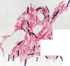
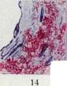
Fig. L More typical view of TB. Fig. 2. Predominantly large grains, Fig. 3. TB culture («risks» on Chistukhin). L and ieK is. r»V 11" f HII >fl! :icpiia. M*IC. "p. 11 f реи^ЛЛДЛ И pT tIP acid-fast aTHItliMHI-IC i-i .iiP'iKii. Fig. 4. Acid-fast and individual diphtheroid rods, Ptrc Г. Many diphtheroid rods - 1>нс. K. ^ерлв; actinomycetes, Fig it. \:ими......(. forms !Ж [e colonial cultures and lp,)г Рис, IU- Lылсьнлныс кисло- тоупорти лерка v лифтерондние палочки, |*ис, 11. ПК a чо кроте. (Fig. 2 10 - TB culture on -.-1.- i-.i medium, fig. 1, 2. '1 11 - stains by -' Iclil - \i 1.1--,и,y. j Fig. 12. TB in sputum {stain by _4uclihy.] Fig. 13. TB in urine: phagocytes Ex. II. TB h tissue. K ст. Тубфхумх", "IЫ", "EJHC IS. Types of TB colonies on different media, and fig. i u инле cy\«i'i чиршииш: шн пленен ни плоскости; Ь-гладкие колонки; c - growth in view: wrinkled film with нгыеитнропдннин рл^рдстяинеч
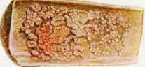
Fig. 1. Typical view of tuberculosis. Fig. 2. Predominantly large grains. Fig. 3. TB culture («risks» on Chistukhin). L and ieK is. r»V 11" f HII >fl! :icpiia. M*IC. "p. 11 f реи^ЛЛДЛ И pT tIP acid-fast aTHItliMHI-IC i-i .iiP'iKii. Fig. 4. Acid-fast and individual diphtheroid rods. Fig. 5. Many diphtheroid rods. Fig. 6. Actinomycetes. Fig. 7. Various forms of colonial cultures. Fig. 8. Acid-fast rods and diphtheroid rods. Fig. 9. TB in sputum. (Fig. 2-10 - TB culture on medium, fig. 1, 2, 3-11 - stains by Zielh-Neelsen. Fig. 12. TB in sputum {stain by Ziehl-Neelsen.]. Fig. 13. TB in urine: phagocytes. Fig. 14. TB in tissue. To st. Tuberculosis, "IЫ", "EJHC. Types of TB colonies on different media, and fig. i u инле cy\«i'i чиршииш: шн пленен ни плоскости; Ь-гладкие колонки; c - growth in view: wrinkled film with нгыеитнропдннин рл^рдстяинеч
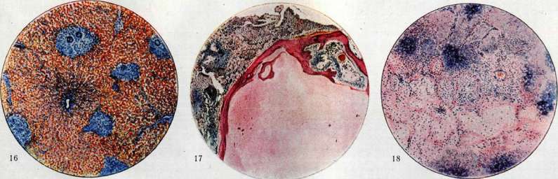
Fig. 1. Typical view of tuberculosis. Fig. 2. Predominantly large grains. Fig. 3. TB culture («risks» on Chistukhin). L and ieK is. r»V 11" f HII >fl! :icpiia. M*IC. "p. 11 f реи^ЛЛДЛ И pT tIP acid-fast aTHItliMHI-IC i-i .iiP'iKii. Fig. 4. Acid-fast and individual diphtheroid rods. Fig. 5. Many diphtheroid rods. Fig. 6. Actinomycetes. Fig. 7. Various forms of colonial cultures. Fig. 8. Acid-fast rods and diphtheroid rods. Fig. 9. TB in sputum. (Fig. 2-10 - TB culture on medium, fig. 1, 2, 3-11 - stains by Zielh-Neelsen. Fig. 12. TB in sputum {stain by Ziehl-Neelsen.]. Fig. 13. TB in urine: phagocytes. Fig. 14. TB in tissue. To st. Tuberculosis, "IЫ", "EJHC. Types of TB colonies on different media: a - growth in the form of a film on the surface; b - smooth colonies; c - growth in the form of a wrinkled film with нгыеитнропдннин рл^рдстяинеч
in the center: dry, rough tubercle columns. Fig. 16. Tubercles in the rarely fatty liver ('goose liver' in tbc). Fig. 17. Exudative affection of the lung; a wedge-shaped mass of affection is surrounded by a bony capsule. Fig. 18. Exudative affection of the lung: wedge-shaped necrosis of the exudate and underlying lung tissue. See Tubercles. The types are small and have relative significance. M. humanus is longer than typ. bovinus; the length of the bovine T. bacilli on average is 1-2 μ, these bacilli are thick, little granular. The bacilli of avian T. are relatively long, thin, often (in cultures) form swellings and sometimes take on the character of threads. The temperature optimum of these bacilli is 40-43°, whereas bacilli of typ. humanus and typ. bovinus grow best at t° 38°. In regard to growth on artificial nutrient media, the following differences exist. M. humanus grows faster than typ. bovinus and gives more luxuriant growth (eugonic growth of English authors). When sown in a flask with glycerin broth, after 5-6 weeks typ. humanus forms on the surface of the medium a thick, wrinkled film, rising up the walls of the flask. M. bovinus grows slower than typ. humanus and gives meager growth (dysgonic growth). Its colonies are smaller than those of typ. humanus, and on glycerin broth after 5-6 weeks of growth a thin, discontinuous film is obtained, representing in places warty thickenings. M. gallinaceus grows significantly faster than typ. humanus. On glycerin agar, growth in the form of a colorless, moist coating is obtained already after 4 days, whereas typ. humanus and typ. bovinus grow in the form of dry colonies appearing 10-14 days after sowing (typ. bovinus even later). All the listed signs have relative value and cannot serve for differentiation. The relative value is also the different relation to glycerin; addition of glycerin to the medium favors the growth of typ. humanus and sometimes has a retarding effect on the growth of typ. bovinus. The only criterion allowing to determine the belonging of BK to one or another type is their different pathogenicity for different animal species. M. gallinaceus is highly pathogenic for birds and causes in them generalized T: with the formation of nodules; in rabbit, rat and mouse this type causes tuberculous bacteriemia without formation of nodules or with formation of microscopic nodules. Guinea pig and man are practically not susceptible to infection with BK typ. gallinaceus. Typ. humanus is pathogenic for man and guinea pig and little pathogenic for rabbit. M. bovinus is pathogenic for cattle, man, guinea pig and rabbit. The different pathogenicity of typ. humanus and typ. bovinus for rabbit serves as the method which is most often used for differentiation of these two types. Good results are given by the method of Park and Krumwiede, consisting in that to rabbits weighing 1,500-2,000 g is injected into the ear vein an emulsion of BK—two doses of 1 mg and two doses of 0.01 mg. If the bacilli belong to typ. bovinus, the rabbits, even those receiving 0.01 mg, die within 2-3 months with signs of generalized T. The rabbit infected with typ. humanus does not die in this time; when killed, it gives on autopsy no changes or limited changes, mainly in the lungs.
The question of the interrelationships of the types of BK cannot be considered solved. For a time there were reports of the existence of transitional strains between typ. humanus and typ. bovinus; on further study they proved to be mixed. In recent years facts have appeared bringing closer together the bacilli of the three types. From a filtrate containing tuberculous ultravirus, using the acetone method, cultures of acid-fast bacilli (by sowing organs) can be obtained. The strains obtained in this way are not homogeneous (Valtis and van Deinse), but represent a mixture of dissociants—smooth S-forms and rough R-forms with predominance of the former. Such a strain has properties bringing it closer to typ. gallinaceus. With successive passages through the organism of guinea pigs, the R-forms come to the fore, and the strain approaches the original typ. bovinus from which the ultravirus was obtained. From these and similar observations the school of Calmette concludes that tuberculous ultravirus represents the true tuberculous virus, single and identical for all animal species, whereas the type arises only with the further passage of the virus through the organism of this or that animal. In recent years Valtis and others have described transitional strains between typ. bovinus and typ. humanus. Acid-fast saprophytes. Belonging by some researchers to the so-called true tuberculous bacilli, the tubercle bacilli of cold-blooded animals apparently belong to acid-fast saprophytes, widely distributed in nature and entering from there (from water, from soil) into the organism of fish, frogs, snakes, etc. Penetrating into the animal organism in large quantities, these bacilli can cause changes simulating tuberculous. Since the discovery of BK by various researchers a large number of acid-fast saprophytes have been described. Such are the bacilli of smegma, bacilli found by L. Rabinovich in sputum in gangrene of the lungs, bacilli of nasal mucus (Laabs, Karlinski), earwax (Bienstock, Gottstein), bacilli of Timofeev's grass (Moeller), bacillus of feces (Moeller), bacilli of milk (Petri, Rabinowitsch, Korn, Tobler and others) and oil, bacilli of dust, water, mucus of wind instruments, water faucets, etc. Acid-fast saprophytes are grown without difficulty on ordinary nutrient media at t° from 15° to 38°. On liquid media a film is formed; on solid media—dry or moist coating, often a pigment (red, yellow) appears. The bacilli are acid-fast, but this property is expressed less sharply than in true tubercle bacilli. The question of the interrelationships of acid-fast saprophytes can at present be considered solved. Recent research has established (Frey, Hagan) that these microbes are found everywhere in soil and water (Eichbaum). One can therefore think that there exists only one species of acid-fast saprophytes, but, settling in various objects, these microbes acquire properties somewhat differing from each other.
Chemical composition of BK. In chemical respect for BK is characteristic the high content of fatty and fat-like substances (Hammerschlag—26.2-28.2%, Schweinitz, Dorset—39.29-40.8%, Kressling—39.69%, Baudran—36.0-44.0%, Goris—38%, Johnson, Brown—43.5%, Model—18.8%). In qualitative relation to these substances belong neutral fats (complex esters of glycerin and fatty acids), waxes, sterols, phosphatides (complex esters of fatty acids and glycerin+phosphoric acid and nitrogenous base), sulfatides. According to Much, the composition of BK includes neutral fats, free fatty acids, lipoids and wax-like substances (in small amounts). According to data of American researchers (Sabin, Doan, Forkner), the lipoids of BK consist of substances insoluble in acetone and soluble in it. To the first group belong phosphatide A-3, soluble in alcohol and ether, and waxes (purified and soft), extractable with chloroform. To the acetone-soluble substances belong glycerides. American researchers established the fact (which however was already pointed out by Much) of different physiological action on the animal organism of various fat fractions of BK. Of greatest importance in this respect is phosphatide A-3, by itself manifesting the action characteristic of BK as a whole (ability to cause formation of nodules). The amount of carbohydrates in BK, as also of lipids, varies depending on the composition of the nutrient medium and the type of bacilli. Most carbohydrates are in typ. humanus. The capsule of the bacilli consists of cellulose (Hammerschlag), hemicellulose (Drieger, Marpmann), hydrocellulose (Auclair, Paris). Laidlaw and Dudley isolated from BK a polysaccharide precipitating the serum of immune animals in dilutions up to 1:6,000,000. According to Matius, the composition of the complex carbohydrate (polysaccharide) of BK includes mannose and arabinose, as also glucose, inositol and galactose (Chargaff, Anderson). The protein fraction of BK, according to Tamura, constitutes 57% of the weight of defatted bacilli; according to Johnson—84%. Most characteristic is the nucleoprotein entering into the composition of BK; the nucleic acid of tubercle bacilli was first obtained by Ruppel and named by him tuberculinic acid; Johnson and Brown from 436 g of defatted dry bacilli obtained 7.7 g of tuberculinic acid. Johnson refers the tuberculinic acid of BK to the animal type of nucleic acids. In qualitative relation for BK besides tuberculinic acid are characteristic globulins; albumins are little typical. Biologically the proteins cause neutrophilic leukocytic reaction, as also accumulation of histiocytes phagocytosing neutrophils. Water in BK (Hammerschlag) on average 85.9%; inorganic substances—6.75-6% of dry residue (Hammerschlag), 2.55% (Kressling), 1.92% (Dorset), 2.5% (Goris and Liot). Qualitative analysis gives the following:
Schweinitz Krauss and Goris and and Dorset Siebert
Phosphoric acid
55.23
51.25
0
0.84
0
0.60
10.10
9.18
5.27
26.55
6.92
3.22
9.03
2.17
9.7 Biology of TB. Nutrition. The best source of carbon for TB is glycerin. Glucose is an incomplete substitute for it: growth on glucose media occurs less intensively. Levulose, lactose, and maltose give even worse results. According to Long and Finner, there is a connection between the glycerin content in the medium and the amount of fatty substances in TB. Products of protein breakdown serve as the source of nitrogen for TB, with the greatest importance attached to simple amino acids, from which TB derive their amino group. Instead of amino acids, ammonium salts of inorganic and organic acids serve the same purpose (L. M. Model). Of the amino acids, asparagine gives the best results, as well as alanine and leucine. While needing glycerin and amino acids (resp. ammonium salts), TB are otherwise not particular in their choice of nutrient substrate. They give very good growth on synthetic nutrient media. From such media, the Levinstein and Pick medium can be mentioned: Asparagin 6.0, Ammon. lact. 6.0, Natr. phosph. (neutr.) 3.0, NaCl 6.0, Glycerin 40.0, Aq. dest. 1 000 cm³. The composition of the Soton medium-see Bacteria, Calmette-Guérin bacillus. As for mineral nutrition, TB require potassium, magnesium, sulfur, and phosphorus. TB are aerobes and are characterized by a greedy affinity for oxygen. Growth in relatively anaerobic conditions is possible, but extremely sparse (Calmette, Vodremer); under such conditions, atypical forms appear. TB multiply by transverse division. Along with this, under certain conditions, TB can arise from grains of both acid-fast and non-acid-fast types (Morton, Kahn). Some researchers also point to the possibility of TB multiplying by budding of rods from grains that make up the tubercle bacilli. TB are characterized by considerable endurance to various physical and chemical influences. Boiling kills them in 2 minutes; heating to 60°-in 1/2 hour. In a dried state, TB withstand 100° for 1/2 hour. Sunlight kills TB in an emulsion in 10 minutes; ultraviolet rays under suitable exposure conditions-in 2 minutes. In sputum under the influence of direct sunlight, TB remain alive for 6-8 hours; in diffused sunlight-for 1-6 days. In a dark room, TB can remain alive in sputum for 2-3 months or longer. A 1.5% phenol solution kills TB in 30 minutes, a 0.5% cresol solution-in 60 minutes, a 1%-in 50 minutes. In decaying substrates, TB remain alive for a long time. Methods of staining and cultivating TB. For staining, methods based on acid-fastness, alkali resistance, and iodophilia are used; methods of double staining are also employed. Of the methods based on acid-fastness, the Ziehl-Neelsen methods are most commonly used (see Ziehl-Neelsen method) and Spengler's method (see Sputum). Hermann's method is also good (staining before the formation of vapors in a mixture of three parts of a 1% solution of ammonium carbonate in distilled water and one part of a 3% solution of crystal violet in 95% methyl alcohol, decolorizing for several seconds in 10% nitric acid, treatment with 95% alcohol until the preparation is pale blue, additional staining with a 1% solution of eosin or safranin). In the Cepede method, the preparation is stained with carbolfuchsin, as in the Ziehl-Neelsen method; then simultaneous decolorization and additional staining with methylene blue in a solution of lactic acid (methylene blue in excess, pure lactic acid 20 cm³, Aq. dest. 80 cm³). The Gasis method is based on alkali resistance. The preparation is treated with a mordant for 1-2 minutes with heating; the mordant is prepared as follows: a piece of mercuric chloride the size of a lentil is dissolved with heating to boiling in a liquid of the following composition: crystal eosin-1 g, absolute alcohol-5 cm³, distilled water-95 cm³; the liquid is decanted. This is followed by decolorization in the following solution: caustic soda-0.5, KJ-1 g, 50% alcohol-100 cm³. The final step is staining for 2-3 seconds with the following solution: crystal methylene blue-1 g, absolute alcohol-10 cm³, hydrochloric acid-0.5 cm³, distilled water-90 cm³. Of the methods based on iodophilia, the modified Gram-Much method is most frequently used: 10 cm³ of a saturated alcoholic solution of methyl violet (B. N.) is mixed with 90 cm³ of a 2% solution of carbolic acid. Staining for 24-48 hours on cold or with heating to boiling. Treatment with iodine (1 g iodine, 3 g KJ and 300 cm³ Aq. dest.) for 5-10 minutes, washing with water, 5% nitric acid-1 minute, 3% hydrochloric acid-10 seconds, acetone-alcohol until complete decolorization, additional staining with an aqueous solution of safranin. Of the double staining methods, the Muhr-Weiss method should be noted: staining with a mixture of 3 parts carbolfuchsin and 1 part methyl violet used in Muhr's method, otherwise as in the Gram-Much method, excluding additional staining. To obtain a pure culture of TB, various methods are used: 1) the material is inoculated into a guinea pig, which is killed in the 3rd-4th week; the spleen is extracted, parts containing nodules are crushed, and the resulting mass is rubbed into the surface of an egg medium; the spleen can also be pre-treated according to Hon (see Sputum); 2) the material is mixed with 15-20% antiformin sa; the mixture is shaken, homogenized in an incubator, centrifuged, the precipitate is washed 3-4 times with sterile water, repeating centrifugation, the washed precipitate is rubbed into the surface of a solid medium (egg medium is best); 3) according to Petrov, the material (sputum) is mixed with a 4% solution of caustic soda aa, shaken, homogenized for 15-30 minutes in an incubator, centrifuged for 10 minutes, 1-2 drops of normal HCl are added to the precipitate, the precipitate is seeded on Petrov's medium with gentian violet; 4) of historical interest is the Kitasato method, consisting of washing a lump of sputum successively in 7-10 Petri dishes with sterile physiological solution; 5) the best results are obtained with the Levinstein-Sumiyoshi method, especially in the modification of Hon (see Sputum).--For media used for cultivating TB, see Nutrient media, special nutrient media. For obtaining pure cultures, egg media are of the greatest importance [see separate table (pp. 19-20), fig. 15] (Lubenaus, Petrovs, Hon, Petranjani, etc.); for maintaining cultures, glycerin potato, agar, broth, etc. are widely used. The Cumbari medium, used for obtaining non-acid-fast TB, is prepared as follows. A. Potato is left for 24 hours in a liquid consisting of Aq. dest., 6% glycerin, 3% ethyl alcohol, and ammonia until strongly alkaline reaction. The medium is prepared in the usual way. B. 400 g of potato pulp are boiled with 500 cm³ Aq. dest.; the mixture is filtered, strongly alkalized with ammonia, and boiled on a water bath until complete removal of ammonia (neutral reaction). For cultivation, medium A is used first, then the bacilli are transferred to medium B. For cultivating TB from blood, Levinstein proposed a medium of the following composition. To 150 cm³ of synthetic medium (monobasic potassium phosphate salt-1 g, sodium citrate-1 g, magnesium sulfate-1 g, asparagine-3 g, glycerin-60 cm³, distilled water-1000 cm³) are added 6 g of potato flour and 12 cm³ of glycerin; the mixture is boiled for 1/2 hour, left to stand for 1 hour at 56° until dextrin is formed, 4 eggs are added and then another one, shaken, 5 cm³ of sterile 2% solution of congo red or malachite green are added, filtered, and poured into tubes. Sweany and Ivanoff (Sweany, Evanoff) recommend an egg medium prepared on veal; veal is not infused with water but with milk. Particularly good results are obtained if 10% sterile cream is added to the medium instead of glycerin. Experiments on animals (see Sputum). The animal exclusively suitable for the experimental diagnosis of T. is the guinea pig. When infected with TB, it develops progressive weight loss; after 6 weeks-3-4 or more months the animal dies with signs of generalized T. When infected under the skin (the usual method of virus introduction), the picture of the disease consists of an infiltrate and ulcer at the injection site, enlargement and caseous degeneration of regional glands, similar lesions of other lymph glands, especially the nearest ones, lesions of the spleen (enlargement and nodules), liver (same), and lungs. With other methods of infection, the picture changes depending on the method of virus introduction. Smears of affected organs reveal the presence of TB [see separate table (pp. 19-20), fig. 14]; with prolonged existence of the process, bacilli may be few or absent altogether. Sources of infection with T.
The main source of infection with T. is a person sick with T.; taking into account that p/12 of all cases of human T. are pulmonary T., it must be recognized that sputum has the greatest importance as a source of infection. The question of how tubercle bacilli from sputum enter the body of a healthy person was answered differently over the last 50 years. Koch, based on his view (as is known, erroneous) of the existence of spores in the tubercle bacillus, came to the conclusion that dust infection plays the main role in the spread of T. Koch's student Cornet developed this idea into a coherent theory (the Koch-Cornet doctrine). According to this doctrine, sputum, drying and turning into dust, spreads T.; the tubercle bacilli in the dust retain their viability and, rising from the floor into the air (due to walking, drafts, etc.), become a source of infection. The danger of dust infection exists only in enclosed spaces; in the open air, the bacilli die (decay, the influence of light, etc.). In confirmation of the theory of dust infection, Cornet examined 147 samples of dust from rooms where tuberculous patients were located, and in 46 cases he found tubercle bacilli. Cornet's data were confirmed by other researchers, and in public places, tubercle bacilli were found only in railway carriages. Another confirmation of his theory, Cornet found in the experiment with the 'carpet'. A carpet was abundantly smeared with tuberculous sputum; the layer was dried and then the carpet was subjected to vigorous beating with brooms; guinea pigs placed in the resulting cloud of dust contracted T. Against the Koch-Cornet doctrine, Flügge appeared with sharp criticism. Relying on the work of his students, he showed that the conditions required by the Koch-Cornet theory for dust infection do not exist in everyday life. For tubercle bacilli from sputum to get into the air, the sputum must turn into the finest dust. This does not happen in practical life, because such a transformation requires several days, and during that time, even in the most uncultivated conditions, the sputum is removed from the floor one way or another. As for the experiment with the 'carpet', its conditions are such that they cannot be encountered in everyday life. According to Flügge, the main mode of infection is not dust, but droplet infection. Its essence is that when coughing, talking, laughing, sneezing, a person scatters around himself the smallest droplets of mucus containing tubercle bacilli in tuberculous patients. By placing slides at a certain distance from the mouth of a coughing tuberculous patient, it is easy to verify (staining, animal experiment) that in 80% of cases the droplets contain tubercle bacilli, sometimes in very large numbers (within 1/2 hour at a distance of 40-80 cm, more than 400). That droplets can cause T. is proven by direct experiments of Heymann (Neu-mann), who placed guinea pigs in cages in the immediate vicinity of the mouth of a coughing patient, and some of them (though a small number) contracted T. Droplet infection occurs only if the distance between the coughing tuberculous patient and a healthy person is not more than one meter. The theory of droplet infection quickly supplanted the Koch-Cornet doctrine. However, this theory could not stand up to the facts, and at present, in place of the theories of Koch-Cornet and Flügge, a new theory has emerged, synthesizing the first two; this new theory can be called the Chausse-Lange theory. The pioneer was Chausse, followed by Lange. The main objection raised by these researchers against Flügge's doctrine is that droplets usually have considerable sizes; in any case, they are much larger than 20 μ; meanwhile, only small droplets, smaller than 20 μ, can penetrate, as shown by the research of Chausse and Lange, into the lungs; thus, only in rare cases can droplets be the cause of pulmonary T. In contrast, dust particles have a very small size and are usually smaller than 20 μ. To be infected by dust is therefore incomparably easier than by droplets; however, Chausse-Lange dust is not the dust of Koch-Cornet. According to Chausse and Lange, the main source of dust is droplets scattered by the tuberculous patient. Separating from the patient, they settle on surrounding objects and, due to their size, quickly dry, turning into dust. Thus, the patient's linen, bedding, etc. pose the greatest danger. Chausse and Lange prove their doctrine with a whole series of very convincing experiments. Of these, only one can be cited: guinea pigs were placed in a patient's room at his bedside, but in his absence, during the time when cleaning and shaking out bedding was being done; some of the guinea pigs contracted tuberculosis. Thus, dust infection plays the main role in the spread of T., and the source of dust is droplets. Wet droplets play a secondary role in the spread of tuberculosis. Compared with sputum, other secretions of the tuberculous patient have limited importance as sources of infection. Tubercle bacilli can be found in women's milk; according to the research of Calmet and others, they are often present in faeces in the absence of intestinal T., because bile and faeces are normal secretions, using which the body gets rid of tubercle bacilli; urine can also in certain cases (not often) contain tubercle bacilli in the absence of kidney damage; finally, according to Piery, tubercle bacilli can also be present in the sweat of patients (30.76%, according to this author). It goes without saying that when the corresponding organs are affected, urine, feces, milk, pus, etc. can contain very large quantities of tubercle bacilli. Thus, all secretions of the tuberculous patient can be a cause of infection with T., but compared with sputum, all these sources have very secondary importance. Besides man, a source of infection with T. is cattle, because the milk of tuberculous cows very often contains tubercle bacilli. The question of what role cattle plays in the epidemiology of human T. is decided on the basis of how often typ. bovinus is the cause of T. in man. According to extensive research conducted in this direction in England and America (Griffith, Park, etc.), approximately one quarter of cases of T. in children from 0 to 15-16 years of age are caused by bacilli of typ. bovinus. According to Park, out of 1,038 cases, typ. bovinus was found in the age group 0 to 5 years in 26.5%, from 6 to 16 in 25%, and in older individuals in 1.31%. The forms caused by bacilli of typ. bovinus are mainly T. of abdominal organs, lymph glands, and others. The number of cases of pulmonary T. in adults caused by bacilli of typ. bovinus amounts to a few units. Thus, for children there is a real danger of contracting T. through infection with bacilli of typ. bovinus. This danger is particularly acute in some countries (due obviously to living conditions). Thus, in Scotland, according to Fraser, the percentage of infection of children with typ. bovinus reaches 70 and above. Bacilli of typ. bovinus enter the child's body mainly from milk; along with it, the role of other dairy products should not be underestimated; thus, according to Eber, in a large city, tubercle bacilli were found in milk in 10.5%, in butter in 12%, and in cream in 6%. Compared with cattle, other animals play a very small role in the epidemiology of human T. Pigs suffer from tuberculosis (typ. bovinus), but the danger they pose to man is very small, because swine T. is a closed and benign disease. In goats, T. is rare. Tuberculosis occurs in parrots (typ. humanus). There are also known cases of T. in cats. T. is often found in chickens. For man, this disease does not present great i 3» danger, because it is caused by bacilli of typ. gallinaceus, to which man is little susceptible. In a few (more than 10) cases, this microbe was the cause of T. in man, and apparently the source of infection was chicken eggs. The portals of entry of infection in man in T. Under the law of localization is understood the regularity according to which tubercle bacilli, penetrating the animal organism, cause specific changes at the site of introduction. In this form, this law does not correspond to reality. At present, it is well known that tubercle bacilli can penetrate the body without causing any changes either on the skin or on the mucous membranes; such penetration of tubercle bacilli into the body is rather the rule than the exception. In view of this, the law of localization should be understood in the sense that tubercle bacilli, penetrating the body, cause specific changes in the regional lymph glands nearest to the site of introduction. In this modified form, the law is essentially valid for guinea pigs, but even in these animals, not always (Oerskow), the introduction of tubercle bacilli is accompanied by damage to the regional glands. Virulence of the tubercle bacilli plays a large role in this regard. In relation to man, the law of localization has a conditional, limited significance. In view of this, from the fact of damage to one or another group of lymph glands, in most cases one cannot conclude that the bacilli were introduced in this particular place. A person's illness with T. can be the result of intra- or extrauterine infection. The existence of hereditary T. in the strict sense of the word cannot be considered proven.
There are essentially no data that would indicate the possibility of infection of the egg or sperm with tuberculosis. But even if we assume the possibility of infection of the sperm with the tubercle bacillus, there is every reason to think (Gartner) that the resulting fertilization by such a sperm would result in a non-viable fetus. Thus, when speaking of hereditary transmission of tuberculosis, what is meant is intrauterine infection of the fetus, with the bacilli passing from the maternal organism into the fetal organism through the placenta. It is impossible to deny the possibility of tuberculosis arising in this way. However, it can be stated that intrauterine infection with tuberculosis is a very rare phenomenon. If tuberculosis frequently arose through intrauterine infection, then congenital tuberculosis—the appearance of a child at birth with unmistakable clinical signs of tuberculosis—would be a very common phenomenon. Meanwhile, to this day in the literature there are only a few dozen cases of such congenital tuberculosis, and not all of these cases are reliable. The results of separating children from their tuberculous mothers immediately after birth (the Grancher system), as well as similar results of separating newborn calves (Bang), speak against the frequency of intrauterine transmission of tuberculosis. In both cases, the newborns are spared tuberculosis. The discovery of the tuberculous ultravirus has strengthened the position of supporters of placental transmission of tuberculosis. If the ultravirus passes through a porcelain filter, then there is reason to think that the placenta cannot be an obstacle to the tuberculous ultravirus. Indeed, soon after the appearance of works on the ultravirus, reports appear that both experiments and observations on humans allow one to think about the frequency of infection of the fetus with the tuberculous ultravirus (Calmette, Couvelaire and others). A doctrine is created that the congenital cachexia sometimes observed in newborn children is a disease similar to the disease that develops in guinea pigs after infection with the ultravirus. Under the influence of the doctrine of the ultravirus, some supporters of intrauterine infection (Lumiere) went so far as to deny the extrauterine origin of tuberculosis. Despite this, there is no reason to assert that the existence of the tuberculous ultravirus changes in any way our views on the rarity of intrauterine infection. As stated above, the tuberculous ultravirus in guinea pigs causes a disease that ends with complete recovery after 1-1½ months; thus, if the ultravirus frequently passed from the maternal organism into the fetal organism, the child would develop a quickly passing mild disease with an outcome of complete recovery. On the other hand, the placenta is by no means a filter that passes everything through it. According to physiological data, colloidal substances related to the proteins of the fetus pass easily through the placenta, and substances foreign to the fetal organism are retained. Antitoxin passes (Ramon, Grasset), but toxin does not pass (or passes in negligible amounts). Based on this, one can expect the passage of the tuberculous ultravirus into the fetal organism only if there is a large amount of it in the mother's organism. Such cases are not frequent, since a large amount of ultravirus is present only in the case of an active, progressive process. Thus—just as before the discovery of the ultravirus—it is necessary to adhere to the point of view that tuberculosis is a contagious disease arising through extrauterine infection. Intrauterine infection may occur, but its practical significance is very small. The portals of entry for tuberculous infection can be the skin, mucous membranes, and the alveolar pulmonary epithelium. Practically, the most important question is how pulmonary tuberculosis arises. Most researchers adhere to the point of view that pulmonary tuberculosis results from the penetration of the tubercle bacillus with the inhaled air into the pulmonary alveoli. The fact that the primary focus usually consists of a focus in the lungs and in the corresponding bronchial glands serves as proof. According to Ghon, the pulmonary focus is always older than the focus in the gland. In the experiment, the task of obtaining pulmonary tuberculosis by inhaling the tubercle bacillus appears to be easily solvable. In contrast to this point of view, Calmette believes that pulmonary tuberculosis arises as a result of alimentary infection. Calmette describes this process as follows. Penetrating the digestive tract, the tubercle bacillus passes through the mucous membrane without causing any changes at the point of entry. The bacillus, phagocytized by a neutrophilic leukocyte, enters a lymph vessel, then the mesenteric gland, the thoracic duct, and finally the lungs. Here it becomes stuck in a pulmonary capillary, whose lumen is smaller than the diameter of the leukocyte, and gives rise to a tuberculous focus. The fact that in the experiment it is possible to cause pulmonary lesions in an animal by feeding it the tubercle bacillus is usually cited as proof of the alimentary origin of pulmonary tuberculosis. Without denying this fact, established by the experiments of Calmette, Ort, and L. Rabinovich and others, it must however be pointed out that for such infection to be successful, very large amounts of bacilli are required (Findel, Reichenbach and Alexander), whereas the inhalation of very small amounts of them (Chaussée, Lange) is sufficient to cause pulmonary lesions. This fact is of great importance in resolving the question, since (Lange) humans are usually infected with small amounts of bacilli. The severe Lübeck experiment also serves as proof that when infected per os, tuberculosis of the abdominal organs usually develops, not pulmonary tuberculosis. (In Lübeck in 1930, 252 children instead of BCG vaccine received virulent tubercle bacilli per os in three doses, in total equal to 1-1⅛ billion live bacilli. 199 children became ill, and in all except one, only tuberculosis of the abdominal organs developed; in one child, along with tuberculosis of the abdominal organs, there was also pulmonary tuberculosis.) Other mucous membranes (the conjunctiva of the eye, the mucous membrane of the sexual organs, the palatine tonsils) can in certain cases be the site of entry of tubercle bacilli. Libin established the possibility of the introduction of tubercle bacilli through the pulp and canal of carious and gangrenous teeth. The significance of this route in the epidemiology of human tuberculosis, however, is not great. Immunity to tuberculosis. Natural congenital immunity (resistance) to tuberculosis is widespread among animals; there are species (horse, dog, rat, white mouse) that possess high, though not absolute, resistance to tuberculosis. Congenital resistance is also inherent in humans (Lange, V. Lubarsky). The following facts speak to this: 1) the relatively low incidence of tuberculosis in children despite the widespread presence of tubercle bacilli in urban living conditions; 2) the experiments of Seller, who vaccinated children with virulent tubercle bacilli in doses from 12 to 10,000 bacilli and never obtained the disease in them, etc. The degree of congenital resistance is not uniform. Acquired immunity to tuberculosis has the character of infectious immunity; it arises as a result of infection with tuberculosis and exists as long as this infection is present, i.e., as long as tuberculous changes and tubercle bacilli are present in the organism. According to Lange, the more active the tuberculous process and the more virulent the tubercle bacilli in the organism, the more valid and pronounced the immunity. According to Seller, immunity is most pronounced when there is a hidden, slightly active focus or a focus in the stage of healing in the organism. Finally, according to Calmette, for immunity to exist, it is sufficient for tubercle bacilli to be present in the organism, even if they do not cause specific changes (latent microbism). Experiments speak in favor of Lange's point of view. When there is a slightly active focus or only bacilli in the organism, immunity is present, but its degree is not great. Being associated with the presence of tuberculous tissue in the organism, immunity is at the same time associated with allergy, i.e., with increased sensitivity to tuberculin. An organism immune to tuberculosis usually reacts to tuberculin, and in any case, every organism that reacts to tuberculin also possesses a certain degree of immunity to tuberculosis. However, it cannot be denied that immunity can exist in the absence of sensitivity to tuberculin (Calmette); such cases are not common. An organism immune to tuberculosis is not susceptible to superinfection; thus, possessing immunity to foreign bacilli, the organism remains susceptible to its own bacilli that have penetrated it during infection. The highest degree of immunity exists in the case where the bacilli of superinfection are destroyed in the organism without causing any changes. If immunity is not pronounced, the bacilli of superinfection cause specific changes; however, these changes differ qualitatively and quantitatively from those that form during primary infection. The existence of acquired immunity to tuberculosis is proven by the experiment showing that vaccinating an animal (cattle, laboratory animals) with tubercle bacilli makes it unsusceptible to subsequent infection—of course, provided proper dosage, etc. The doctrine of acquired immunity, developed by Behring and Römer and bearing their name, retains its validity also in relation to humans.
Evidence of its existence in humans is provided by patho-anatomical data (the presence of tuberculous foci in almost all adult city dwellers), the results of determining tuberculin sensitivity (almost the entire adult population reacts to tuberculin), and others. Many are inclined to explain the different course of T. among peoples coming into contact with tubercle bacilli for the first time and among peoples among whom T. has existed for a long time. In the former, acute forms predominate with frequent outcomes in acute miliary T. and death ('normal' T.), while for the latter, isolated lung damage in the form of chronic pulmonary consumption without the lymphatic gland involvement obligatory in normal T. is characteristic ('modified' T.). Acquired immunity has a certain influence on the clinical picture of T., but an even greater influence is exerted by congenital resistance and especially social factors. In particular, pulmonary consumption can develop not only in an organism possessing acquired immunity to T., but also in an organism not having such immunity. The significance of social factors is revealed in the fact that acute forms of T. can occur in individuals immune to T. under unfavorable social conditions (V. Lubarsky and others), and on the other hand, chronic pulmonary consumption can develop under good social conditions in individuals not possessing acquired immunity to T. (Kleine). As the infection rate of the population with T. increases, the number of individuals immune to T. also increases ('natural immunization'). The tendency of some researchers (Sanarelli) to attribute the decrease in mortality from T. to it finds no complete justification in the facts, however, it is undoubtedly that natural immunization of the population is one of the factors reducing mortality from tuberculosis. Mechanism of immunity to T. The basis of congenital resistance to T. is the increased ability of the organism to destroy the VK penetrating into it. As shown by the research of Corper, Lurie and others, the animal organism possesses the ability to destroy VK, and a non-immune organism requires several months for this and not all bacilli are destroyed. In different organs, the process of bacillary destruction occurs with varying degrees of intensity and speed. In the liver and spleen, bacilli die sooner than in the lungs. In a T.-resistant organism, the death of bacilli occurs faster than in a non-immune one; moreover, all bacilli or at least the majority perish. The destruction of bacilli is apparently associated with the presence (or appearance) in the organs of substances (Lubarsky, Model) that inhibit the growth of VK. Along with the destruction of tubercle bacilli, the organism is freed from them by excreting them with bile and with faeces. In a resistant organism, this process occurs with greater intensity than in a non-immune one. In an organism possessing acquired immunity to T., antibodies (agglutinins, precipitins, etc.) appear (or may appear). Such an organism also becomes allergized. As for antibodies, they are not a factor of immunity. There is no parallelism between the content of antibodies and immunity; they may be absent in the presence of immunity and be in excess in its absence. The question of the relationship between allergy and immunity cannot be considered resolved. According to Krause (A. K. Krause), 'immunity is a function of allergy.' According to Calmet, allergy and immunity represent different and independent states. According to Maragliano, 'allergy is most closely connected with the evolution of the immune process.' According to Rich, a sharply expressed immunity corresponds to a weakly developed allergy and vice versa. Thus, at the present time, when neither the essence of immunity nor the nature of allergy is yet known, one can only speak of the connection of these states without deciding the question of their actual relationships. Reactions of immunity. Agglutination has no practical significance in the recognition of T., as it lacks strict specificity (it has been observed in pneumonia, typhoid fever, etc.) and moreover, it is obtained both in active and inactive forms of T., sometimes being negative in cases of active T. The technique was developed by Arloing and Courmont, who proposed for its production the so-called homogeneous culture (VK causing uniform turbidity of the broth). Precipitation also has not acquired practical significance. The existence of bacteriolysins is questionable; the serum in any case lacks lytic action. The complement fixation reaction has certain practical significance. Its success depends to a large extent on the antigen. Besredka's antigen is a 4-day culture of VK on its egg medium (see Nutrient media), killed at 115° and centrifuged. An emulsion of bacilli in a certain amount of physiological solution serves as the antigen. Boquet-Negre's antigen is prepared as follows. A six-week culture of VK on glycerin broth is sterilized for 30 minutes at 120° and filtered through paper; the bacterial bodies are washed on the filter with distilled water and dried. This should be followed by extraction with acetone (1 cm3 per 0.01 bacilli) for 24 hours, new drying and extraction with 99% methyl alcohol (1 cm3 per 0.01 bacilli) for 12 days at 37°. b. m. e. t. хххш. Shake. Filter. The liquid is the antigen. Petrov uses a glycerin extract of dried and powdered tubercle bacilli (a 21/2% dilution in physiological solution). Wassermann treated the bacilli with tetralin, washed them repeatedly with ether and dried. To the resulting yellow-white powder was added lecithin in the amount of 0.2%. With lecithin, the powder remains for 1 hour. This should be followed by centrifugation; the precipitate is the antigen. Witebsky, Klingenstein and Kuhn's antigen is obtained by treating dried VK with hot alcohol, extracting the precipitate with hot pyridine and purifying with acetone; the precipitate is weighed in a 10% solution of benzene; lecithin is added to the suspension. The diagnostic value of the complement fixation reaction is still small.
III. Pathological Anatomy. The pathological anatomy of T. at the present time, in the sense of etiological understanding of the basic changes in T., is strictly unitarian. It is clear to us that the basic changes, whatever their morphological expression, are associated with the introduction of the tubercle bacillus into the tissue. It is equally clear that the changes that occur when the tubercle bacillus is introduced into the tissue represent an inflammatory reaction of the tissue. This reaction, like any inflammatory reaction, is a combination of alterative, exudative, and proliferative (productive) changes in the tissue. However, the combination of alteration, exudation, and proliferation in this case varies greatly, which gives extreme diversity in the morphological and dynamic expression of the tuberculous process. This diversity is essentially based mainly on the predominance in individual cases of either the alterative, the exudative, or the productive factor; the reasons for this predominance of one or the other factor in the inflammatory tuberculous reaction are very complex, as they concern not only the peculiarities of the pathogen but also the diverse properties of the affected organism (see below). It is self-evident that such an assessment of the morphological expressions of tuberculous changes is far from both dualism and the primitive unitarianism of previous authors. If in the etiological assessment of tuberculous changes we adhere to a unitarian point of view, then in assessing all the pathogenetic conditions for the occurrence of one or another morphological expression of the tuberculous process, the modern point of view, in Pagel's apt expression, is synthetic. The pathological changes in tissue that occur when the tubercle bacillus is introduced into it may first of all present certain differences depending on the nature of the tissue. The structure of the organ and even which tissue of it has come into contact with the tubercle bacillus leaves its imprint on the type of reaction, i.e., on the morphology of the tuberculous process and its course. However, wherever tuberculous changes arise, they always initially show a tendency to express themselves as a limited nodular change, called a tubercle, a tubercle [see separate table (pp. 19-20), fig. 16; separate table (pp. 35-36), fig. 1, 2 and 5; separate table (pp. 111-112), fig. 4] (tuberculum, French follicule tuberculeux). To the naked eye, a fresh tubercle appears as a gray, semi-transparent, round nodule, usually slightly protruding on the surface of the organ and on its cut surface. The size of the tubercle can vary; the usual miliary tubercle (tuberculum miliare, French granulation miliaire) corresponds to the size of a millet seed (milium-millet grain), but tubercles can also be of smaller sizes, down to microscopic dimensions invisible to the naked eye. Tubercles larger than a millet seed are very rare; larger tubercles are distinguished by their central part having a yellowish tint and being opaque; at the same time, often, e.g., in the lungs, such larger tubercles are not round but angular. In the kidneys, tubercles are generally very poorly defined, appearing as pale spots. The development, or histogenesis, of the tubercle has been studied both experimentally and on human material by a large number of researchers and can be presented as follows. The tubercle bacilli, entering the tissue and multiplying in it, cause damage to the tissue elements (the alterative phase of tubercle formation), which can have varying degrees and extent; sometimes it is so insignificant that it is imperceptible under the microscope, but sometimes there is focal necrosis of the organ's tissue with disintegration of cells and intercellular substance, with karyorrhexis of nuclei. Very soon, hyperemia of small vessels is found at the site of damage and around it, exudate is secreted, and leukocytes and lymphocytes emigrate (the exudative phase), which in most cases is brief and weakly expressed. At the same time, the multiplication of local cells (the productive phase) begins, from which large, plate-like cells with homogeneous, light protoplasm and a bubble-like nucleus are formed; these cells, due to their similarity to flat epithelium, received the name "epithelioid" cells. In most cases, it is from these epithelioid (or according to Hubschmann "tuberculous") cells that the entire gray tubercle is formed; only at its periphery is there a varying number of small round cells of the lymphocyte, polyblast, rare plasma cell, and polymorphonuclear leukocyte type. The presence in the central part of the tubercle, among the epithelioid cells, of one or two multinucleated giant cells with numerous nuclei arranged at the periphery of the cell body is extremely characteristic of the tubercle. Because these giant cells with their peculiarities were first described in detail by Langhans, they are often called Langhans giant cells. Between the elements of the tubercle, the finest fibers (the "reticulum" of the tubercle) can be found, which were formerly attributed by some to the fibrin secreted between the cells, and by others to connective tissue. However, at the present time, based on good silver impregnation of these fibers, it has been finally established that they belong to argyrophilic connective tissue fibrils. The question of the origin of the elements of the tubercle cannot be considered fully clarified. There is no doubt that in the lymph glands, epithelioid cells are formed from the cells of the reticular tissue, in the liver (within its lobules) - from the endothelium and Kupffer cells of the capillaries; there are quite convincing data that the endothelium of small blood and lymphatic vessels actively participates in the formation of the elements of the tubercle; the same must be said about the fibroblasts of connective tissue. In the formation of tubercles among epithelial complexes, e.g., within the pulmonary alveoli, within the seminiferous tubules of the testicle, apparently the epithelium can also give rise to epithelioid cells. Finally, in recent times, the old view of Metchnikoff about the formation of all elements of the tubercle from wandering cells, macrophages, is being revived again; such a thought is expressed by Maximov, Timofeevsky, and Benevolenskaya on the basis of experiments with obtaining tubercles in tissue cultures; moreover, American authors (Sabin and others) have recently convinced themselves of the formation of tubercles from monocytes emigrating from the vessels. All this taken together forces one to conclude that the epithelioid cells of the tubercle can be formed not from one single type of cell, but from various cellular elements (reticular cells, endothelium, fibroblasts, macrophages, monocytes, epithelium); in other words, the formation of epithelioid cells is a specific form of reaction to the tubercle bacillus, characteristic of various cellular elements. - Giant cells are formed from epithelioid cells under the condition of amitotic division of the cell's nucleus without division of its growing body. This process, carefully traced by Herxheimer and Roth (Herxheimer, Roth), is associated with peculiar necrobiotic changes in the protoplasm of these cells, sometimes noticeable in the central part of the cell in the form of varying degrees of its staining, liquefaction or adsorption of small grains of lime. Giant cells can also be formed directly from the endothelium of capillaries (by their abortive budding).- The small round cells at the periphery of the tubercle belong to the manifestations of a banal inflammatory reaction. At the beginning of the development of the tubercle, in this marginal zone of it, there are full-blooded vessels and often phenomena of inflammatory exudation. Apparently this peripheral zone of the tubercle, which may be expressed to one degree or another, must be attributed to the manifestations of the so-called perifocal, collateral inflammation (see below). The reticulum of the tubercle represents partly preserved fibers of the pre-existing tissue; however, a significant part of the argyrophilic fibers is newly formed among the epithelioid cells. In general, the cellular elements of the tubercle and the reticulum represent organized tissue corresponding to granulation tissue; consequently the tubercle, tuberculum, represents a tuberculous granule-ma (see separate table, fig. 4), belonging to the infectious granulomas. The tubercle is usually characterized as an avascular granuloma; however, the avascularity of the tubercle is something relative. Indeed, in the part of the tubercle consisting entirely of epithelioid cells, vessels are as a rule absent; this is explained by the fact that the elements of the walls of capillaries that existed here earlier (perhaps also newly formed) participate in the formation of epithelioid and giant cells, and thus the vessels are as it were consumed, expended on the formation of the tubercle. In those parts of the periphery of the tubercle where there are few or no epithelioid cells, usually observed are so-
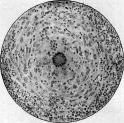
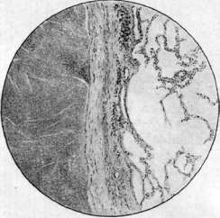
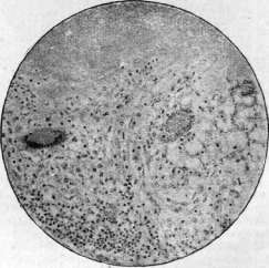
Figure 1. General view of tuberculous granulations; between the nodules - layers of fibrous-connective tissue. Figure 2. Tuberculous nodule; in the center - a giant cell; around it - epithelioid cells; on the periphery - lymphoid. Figure 3. Fibrous-connective capsule, delimiting the caseous focus in the lung. Figure 4. Productive tuberculosis; merging granulomas from epithelioid, lymphoid and giant cells; above - caseous decay.
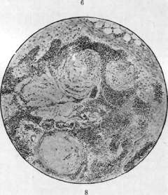
Figure 5. Tuberculous meningitis; Suggestive and diffuse infiltrates in the meninges; lymphoid infiltrates along the course of blood vessels. Figure 6. Chronic tuberculosis of the lung with exacerbation; sclerosis of lung tissue in the upper half of the specimen; exudative phenomena below. Figure 7. Tuberculous focus and pore of the adrenal gland. Figure 8. Healed hyalinized tubercles in a lymph gland. Figure 9. Healed primary affection of the lung: a - fibrous capsule around the affection (with coal deposits); b - trabeculae of BONE tissue with bone marrow (c); c - caseous center of the affection. S vessels; this is easily verified in cases where there is a group of closely located but not coalescing tubercles; in their coalescing peripheral zones there are always blood vessels, often very engorged with blood; the same is shown by experiments with injection of blood vessels. All that has been said above about the structure and development of tubercles refers to the most common and one can say typical type of tubercles, called epithelioid tubercles; they are characterized by the short duration and weak expression of the alterative and exudative phases, the rapid onset and predominance of the productive phase in the form of an epithelioid-cell granuloma. However, the course of development of the tubercle by no means always follows this path. In some cases, the first alterative phase is extremely sharply expressed and prolonged, for some time remaining in its initial state without the addition of subsequent phases, in particular without any hint of productive changes. Such tubercles are distinguished by the fact that they do not bulge in the form of grains, but appear in the form of miliary yellowish-gray spots. Under the microscope they turn out to be small foci of tissue necrosis with karyorrhexis of nuclei, embedded in normal tissue; the circumstance that here we are dealing with primary tissue necrosis, and not with secondary caseous necrosis of an already formed tubercle, is proved by the fact that the necrosis always uniformly captures the entire area without any hint of proliferation on its periphery, and especially by the fact that in the necrotic mass, especially when the specimen is impregnated with silver, the outlines of the tissue of this organ are clearly visible. Such necrotic, or toxic tubercles can be scattered in large numbers throughout all organs, which is characteristic of that form of generalized miliary T., which is distinguished under the name of tuberculous sepsis (see below). In other cases, after the alterative phase, which can be clear or barely noticeable, an effusion of serous or more often sero-fibrinous exudate with the accumulation of lymphoid cells and, less frequently, leukocytes is observed. In such exudative tubercles [see separate table (pp. 35-36), Fig. 6 and separate table (pp. 19-20), Fig. 18], which include those tubercles that were formerly called small-celled, or lymphoid tubercles, the productive phase is subsequently revealed (see separate table, Fig. 4); however, here it is expressed in the formation of a granuloma of characteristic structure (epithelioid cells, Langhans' giant cells, etc.), surrounding the area of exudation, which in turn turns into caseous necrosis. Along with these three types of tubercles, intermediate transitional types can also be observed. When sections are stained for tubercle bacilli, the latter are found in particularly large numbers in necrotic tubercles, and somewhat less in exudative tubercles. In epithelioid tubercles, bacilli are usually few. The bacilli lie both inside and outside the cells; there is no predominant localization in giant cells, as was usually mentioned earlier. From the above it is clear that tuberculosis, which represents the initial change at the site of introduction of the tubercle bacillus into tissue, can have different structure and course. In some cases, the productive phase predominates in it, in others the process as if freezes at the alterative phase (tissue necrosis), in others the exudation predominates in the process. The pathogenetic basis for such diversity in the structure and course of tubercles is not exhausted by any single cause. First of all, the possible assumption about the significance of the time of development of the tubercle must be rejected, i.e., that in a fresh tubercle alterative and exudative phenomena predominate, while in an older one productive changes occur. On the basis of the above description of the development of tubercles, it is clear that this is not so. Moreover, e.g. in cases of generalized miliary T., it is easy to see that one or another type of tubercle is not connected with the duration of the disease. In particular, Korteweg convinced himself that with exactly the same time separating the moment of death from the outbreak of generalized miliari-zation of T., the tubercles in some cases show a clear predominance of exudative manifestations, while in others they are everywhere purely productive, epithelioid. Much greater significance than the age of the tubercle is the nature of the place, the soil of its development, i.e., the structure of the organ and tissue in which the tubercles develop. E.g., it can be noted that sometimes when many tubercles are present simultaneously in the liver, those that form in the interstitial connective tissue bear all the signs of productive, epithelioid tubercles, while in those located inside the lobules, among the parenchyma, alteration in the form of necrosis of parenchymal elements predominates. In the lungs, due to the peculiarities of their structure (porosity, thinness of alveolar septa, thinness of capillaries), exudation occurs very easily. Because of this, purely productive epithelioid tubercles in the lungs are encountered relatively infrequently; usually exudation into the alveoli begins very early and often the tubercle turns out to be nothing more than a group of alveoli filled with sero-fibrinous exudate (miliary tuberculous pneumonia); productive changes can subsequently develop along the periphery of such a focus. However, the nature of the structure of the soil gives only a certain nuance to the structure and course of the tubercle and is not decisive in this sense. Again, in cases of generalized dissemination in generalized miliary T., it is easy to see that along with some difference in the type of tubercles depending on the organ, one or another variant of structure characteristic of all tubercles of this case can still be observed. In particular, in the lungs, in different cases, either productive tubercles or exudative ones in the form of foci of miliary pneumonia can be observed. Knowing that the tubercle is an expression of the inflammatory reaction, the picture of which always depends on the properties of the pathogen and on the properties of the macroorganism reacting to the pathogen (see Inflammation), it is easy to understand that this also applies to the tubercle. The idea that the main reason for the predominance in the tubercle of either productive or exudative changes lies in the properties of the pathogen originates from the experimental research of Baumgarten, Koch and others at the end of the 19th and beginning of the 20th centuries; it was established that the tubercle bacillus itself causes only a productive reaction, while its toxins cause alteration and exudation. Therefore, it was assumed that massive infection with virulent (toxic) bacilli gives predominantly an alterative or exudative process, while infection with a small number of bacilli that secrete few toxic products results predominantly in a productive process. This view was supported by the fact that when sections are stained for tubercle bacilli, the latter are found in the largest number in necrotic and (usually somewhat less) predominantly exudative tubercles; in productive epithelioid tubercles they are usually very few. The above viewpoint about the exclusive significance for the development of a tubercle of one or another type of infection is supported by some to the present time (Kaufmann, 1931); in particular, recently data have been put forward (Winn and others) that necrotic or toxic tubercles are caused by the introduction into the human body of bacilli of avian tuberculosis. On the basis of all data of the modern doctrine of inflammation, one cannot fully agree with the above viewpoint; if the nature of the infection undoubtedly has significance for the nature of the inflammatory process, nevertheless the reactivity of the organism is of no less importance. To one and the same cause, resp. pathogen, in quantitative and qualitative sense, two different organisms and even the same organism, but at different periods of its life, can react differently. All this is fully applicable to T. and to different types of tubercles. As early as 1904, Abricosov pointed out that in generalized miliary T. in weakened and cachectic persons (with severe anemias, cancerous cachexia, etc.) tubercles very often have a necrotic type. Saltykov notes that in persons with the so-called fibrous constitution T. as a rule has a sharply productive character and the tubercles are always epithelioid, with a tendency to rapid fibrosis. On the basis of all this one can think that the alterative (necrotic) type of tubercle, the predominantly exudative and predominantly productive types of it to a large extent represent reflections of different settings in the reactive manifestations of different organisms (for more details on this, see below).
The relationship between the number of tubercle bacilli and the nature of the process in the tubercle may be a sequential phenomenon; it is clear that in the necrotic mass and in the exudate, tubercle bacilli multiply much more quickly than among the cells of the epithelioid tubercle. The bacilli of avian tuberculosis probably have pathogenic significance for humans only in exceptional cases, and to explain all cases with necrotic tubercles as infection with avian tuberculosis would be a great stretch. The outcome of tubercles is largely associated with the type of their structure and development. Extremely characteristic for both tubercles and larger tuberculous foci is the outcome in cheesy, caseous degeneration (degeneratio caseosa), called for short caseation or caseification. This is a special necrobiotic process, expressed in the fact that the center of the gray tubercle becomes yellow, opaque, dry. When larger tuberculous foci are affected by caseation, a homogeneous yellowish-gray, dry mass appears, sometimes granular, resembling cottage cheese, sometimes more homogeneous, elastic or pasty, similar to certain types of cheese; hence the names cheesy, caseous mass, caseous degeneration, caseosis, etc. Upon microscopic examination, the cheesy mass appears as an amorphous, fine-grained protein substance with an admixture of small fat droplets; in fresh caseous mass, grains of chromatin (karyolysis of nuclei of former cells here) may be visible, whereas in old caseous mass there are no longer any signs of chromatin. When the preparation is stained for elastic fibers, the tissue skeleton of elastic fibers preserved in the amorphous cheesy mass can be revealed, and when the preparation is impregnated with silver, due to the preservation of argentophilic fibrils on the caseous mass, the outline of the previously existing tissue or the reticulum network of the tubercle becomes apparent. However, with prolonged existence of the caseous mass, these long-preserved tissue structures eventually degenerate and disintegrate. The area of caseation is surrounded by the preserved epithelioid tissue of the tubercle periphery, with the cells of this tissue, directly adjacent to the caseous mass, sometimes arranged radially, like a palisade [see separate table (pp. 35-36), fig. 3]. The course of caseation development in the tuberculous focus and the essence of caseous degeneration have not yet been fully elucidated. Virchow believed that the main essence of the process lies in the gradually increasing fatty degeneration of tubercle and exudate cells, ending in disintegration. According to Virchow, this necrobiotic process is a consequence of impaired nutrition due to the absence of blood vessels in the tubercle. Weigert and others attributed the caseation of the tubercle to manifestations of coagulation necrosis. Subsequently, it was found that cell obesity is not mandatory for caseation, that the process of caseification cannot be simply interpreted as a process of necrosis or necrobiosis, and that in it very important changes occur not in the cells, but between the cells. Pagel, Schleussing, and others believe that during caseification, a special transudate is secreted, which, under the influence of cell disintegration, especially chromatin, gives rise to precipitation-type coagulation (antigen-antibody reaction); some analogy is seen between the process of caseation and the deposition of hyaline and amyloid. Hübschman associates caseation entirely with exudation and believes that the basis of caseification is a special change (swelling) of fibrin, and the degeneration and disintegration of cells is a secondary process. Hübschman generally believes that caseation joins the exudative phase of the tubercle before productive phenomena; the latter as a rule follow caseation and represent as it were a reaction to caseation. Caseification of productive tuberculous tissue, e.g., epithelioid tubercle, according to Hübschman, is rare and, if observed, is always associated with a flare-up of exudation in the tubercle; the latter, for example, is expressed in the appearance of leukocytes among the epithelioid cells of the tubercle center. Hübschman's point of view that the tuberculous process always develops in the direction of exudation-caseation-productive reaction has not met with general approval; such a course of development, as can be seen from the above, is characteristic only of one type of tubercles (see above exudative tubercle). It can be emphasized that in the area of the tuberculous focus, caseation affects not only the exudate, but also all kinds of cells and the interstitial tissue of the stroma (e.g., alveolar septa of the lungs); from this it follows that the matter is not in exudation, but in a spreading change in the relationship between tissue and the tuberculous virus in all elements of the tuberculous focus; the fact that caseification, spread over the entire tuberculous focus and often simultaneously over several foci, sometimes occurs suddenly, as it were, abruptly, speaks to the fact that a violation of the reactive state of the body plays by no means the last role in the origin of the process of caseous degeneration. The moment of exudation undoubtedly greatly contributes to the onset of caseation, and it can be said that all predominantly exudative manifestations of the tuberculous process have caseification as their constant outcome. It has already been indicated above that necrotic and exudative tubercles as a rule undergo caseation before the onset of the productive phase (primary caseification according to Ranke); during this, the exudate cells disintegrate, fibrin turns into a fine-grained mass, elements of the interstitial tissue, e.g., alveolar septa of the lungs, die and disintegrate, merging with the general cheesy mass. The productive phase in this case is manifested only in the formation of a granulation wall around the cheesy focus. In the productive, epithelioid tubercle, the process of caseation occurs in the cell mass of the granuloma itself, i.e., it complicates the productive phase (secondary caseification according to Ranke); under the microscope, it is expressed in the fact that the cells of the central part of the tubercle undergo disintegration, the reticulum undergoes fibrinoid transformation and disintegrates, as a result of which the center of the tubercle turns into a granular amorphous mass. The cheesy mass can subsequently undergo softening or, conversely, thickens and becomes encapsulated. Softening sometimes occurs with the participation of leukocytes and macrophages that appear among the cheesy mass; in other cases, it occurs without the participation of cells by a special swelling and liquefaction of the cheesy mass. Most often, the process of softening first affects the peripheral zone of the cheesy focus, as a result of which the central, not yet softened parts separate, become sequestrated, and subsequently they too can undergo softening. Due to softening, the cheesy mass is transformed into a semi-liquid, pus-like ('pioid') mass or into a turbid liquid with a crumbly admixture. The process of softening is most susceptible to caseous masses of exudative origin with weak development of the productive zone. The process of softening itself, sometimes manifesting extremely rapidly, is apparently connected with a special violation of the body's reaction to the tuberculous virus. Softening is the main basis of tissue destruction; after softening, separation of softened sequestrated masses and the formation of cavities within organs can occur, and with involvement of one or another surface (skin, mucous membrane)-tuberculous ulcers. The outcome of caseation in encapsulation is connected with the fact that the cheesy mass thickens, and around it a connective tissue capsule develops. This connective tissue capsule can have a dual origin (Aschoff, Puhl, Pagel, etc.); it either develops due to fibrous transformation of the specific tuberculous granulation zone present around the caseous focus ('specific encapsulation'), or represents proliferation of ordinary connective tissue of neighboring parts ('non-specific encapsulation'). In addition, sometimes a kind of connective tissue, collagenous transformation of the cheesy mass itself is observed; this is possible either in the sense of organization of the dead cheesy substrate or in the sense that the always preserved fibrous substance among the caseous mass undergoes collagenization and compaction; there are also data that fibrin fibers can directly transform into connective tissue fibers through collagenization (Nageotte, Doljansky u. Roulet). In the encapsulated, dry cheesy mass, lime salts are usually easily deposited [see separate table (pp. 19-20), fig. 17 and separate table (pp. 111-112), figs. 2, 3, and 7]. This petrification is based on the adsorption of lime salts dissolved in tissue juices by the dead cheesy mass. Lime can be deposited in the form of a fine-grained substance diffusely throughout the caseous mass, which then turns into a white, easily crushable mass between the fingers (so-called omelotvorienie); in other cases, lime is deposited in dense concentric layers around one point (nucleus) in the cheesy mass, gradually capturing the entire mass; in this case, a true lime stone in a fibrous capsule is formed. Petrification is often followed by bone formation [see separate table (pp. 35-36), figs. 8 and 9]; usually it is preceded by the ingrowth of richly vascularized young connective tissue into the lime masses and the resorption of lime salts.
Along with this resorption, beams of true bone tissue with bone marrow between them appear on the inner surface of the capsule of the focus. Bacteriological and experimental research of petrified and encapsulated tuberculous foci shows that virulent tuberculous bacilli can persist in them for a very long time. The different outcomes of caseous foci (softening or encapsulation with petrification) are mainly associated with different states of the body's reactive manifestations. Productive epithelioid nodules also have an outcome in direct fibrous transformation^ fibrosis of the entire tubercle as a whole. The matter is that the granulation tissue of the nodule undergoes maturation up to mature fibrous and further scar connective tissue; epithelioid cells take on a spindle shape, arrange themselves in bundles, argyrophilic fibers between them increase in number, collagen fibers appear (the so-called fibrous nodule), with the increase of the latter and the decrease in the number of cells, the entire mass of the nodule transforms into a small roundish scar, which subsequently usually undergoes hyalinization. Large fibrous hyaline nodes, resembling grains of pearl, are formed in that form of T. of serous cavities, which is designated as pearl disease (see Pearl disease, Tuberculosis in animals); in humans this form is rare. -In relation to predominantly exudative nodules and generally tuberculous foci, two more possible outcomes must be indicated, which can occur before the onset of caseous degeneration. In some cases, an exudative focus, for example in the lung, may stop in its development, and the exudate undergoes resorption, just as in ordinary pneumonia; in these cases, no traces remain from the tuberculous focus or only a slight hardening of the tissue. In other cases, the exudate does not resorb but undergoes organization, which in the lungs gives the picture of carnification (see); this carnification can be non-specific (the exudate is replaced by ordinary connective tissue) or specific (the exudate is replaced by tuberculous granulation tissue); in the latter case, strictly speaking, there is not an outcome of the process but a change in its character: from exudative it turns into productive. Everything said above regarding tubercles as initial changes upon the introduction of tuberculous bacilli into tissue can be applied to the path. anatomy of the tuberculous process as a whole. All those variants of structure, course, and outcome that relate to nodules are also present in large tuberculous foci and in the tuberculous process as a whole. The difference between a nodule and an advanced tuberculous process is largely volumetric, and in order to imagine the dynamics of the entire tuberculous process, one must first recall the ways of its spread in the body. This spread can occur by contact, when the process directly involves adjacent tissue. This type of increase of the tuberculous focus can occur in different ways. Sometimes, starting from some place, the changes very quickly involve neighboring parts, which can simulate the appearance as if of a significant tissue lesion all at once. This happens, for example, in the occurrence of certain tuberculous pneumonic processes in the lungs (the so-called early infiltrate, lobar pneumonias). In other cases, there is growth of the tuberculous granuloma and its advancement onto adjacent tissue, while simultaneously in the center of the focus the area of caseation expands; a large roundish focus arises, caseous in the center and with a granulation zone at the periphery; such a tuberculous focus is called a solitary tubercle [see separate table (pp. 111-112), fig. 6 and separate table (pp. 119-120), fig. 6]. Similar foci can form in cases where an exudative process focus, for example in the lung, is successively surrounded by epithelioid granulation tissue; the latter subsequently undergoes growth in the outer layers, while in the parts adjacent to the caseous center, it itself becomes involved in caseification. In rarer cases, tuberculous granulation tissue sometimes grows to a considerable volume, without showing a tendency to caseation or with caseation but without softening of the caseous masses; this gives an o p u h o l e v i d n y i T. Large, isolated tuberculous growths are sometimes still designated as t u b e r c u l o m a. Diffuse growth of a granuloma from epithelioid cells without a tendency to caseation underlies that form of T. of lymph, glands, which is called large-cell hyperplasia. Diffuse growth of tuberculous granulation tissue is characteristic of tuberculous lesions of bone tissue. Finally, it happens that next to the initial nodule of one type or another new nodules appear, merging with the first; such tuberculous foci, designated as conglomerate-tubercles, usually have a scalloped edge, which causes the focus to resemble a clover leaf. In all these variants of spread by contact, there is growth of the initial small focus, the tubercle, and its transformation into a larger tuberculous focus that continues to grow. Such a tuberculous focus can represent the same variants of structure and course as those given above in relation to the nodule; it can be predominantly productive or predominantly exudative; undergoing caseation, it can become encapsulated, petrified, or undergo softening; when the softened caseous masses are sloughed off from it, a cavity or ulcer forms. In turn, from a cavity or ulcer the process can continue to spread to adjacent tissues. Spread by contact is also observed in the form of transition of the tuberculous process from bone to joint, from the spine or from a rib to the pleura, to the mediastinum, etc. By contact there also arises that accompanying change of the tuberculous process which is called perifocal, or collateral inflammation. This is a zone of exudative inflammation, often of a non-specific ordinary type, which sometimes surrounds the actual tuberculous focus, whether it be a tubercle or a larger tuberculous focus. Perifocal inflammation is explained by diffusion from the tuberculous focus of toxic products both those related to the life activity of tuberculous bacilli (tuberculin) and those associated with processes of decay in the tuberculous focus. Perifocal inflammation in T. can flare up and subside episodically, being a sign of increased tissue sensitivity; it is observed by no means in every tuberculous focus. Clinicians classify perifocal inflammation in the lungs as 'infiltrates'. Exudative inflammations of serous cavities of tuberculous origin have much in common with perifocal inflammation; here, in the presence of a tuberculous focus or tubercles, often productive, an exudative inflammation flares up in the tissue of the serous membrane (pleura, peritoneum, pericardium), which is in essence non-specific, ordinary, with the secretion into the serous cavity of serous, fibrinous, rarely purulent exudate. The second path of spread is along the systems of channels and tubes of the body, the so-called i n t r a c a n a l i c u l a r path. This includes the spread of the tuberculous process in the lungs along the bronchi, which is the usual basis for lesions in pulmonary T. of newer and newer parts of the lung (bronchogenic dissemination of the tuberculous process in the lungs). This same path is realized in the spread of the tuberculous process from the kidneys to the ureters and urinary bladder, from the epididymis to the vas deferens, seminal vesicles, and prostate. If in these cases it is a matter of secretory channels, usually the infection spreads in the direction of the flow of secretion. In some tubular systems, the so-called a u t o i n o c u l a t i o n, vaccination with infectious material in contact with one surface or another, is sometimes observed. Thus, sputum, excreted from the respiratory tract in pulmonary tuberculosis, often infects the mucous membrane of the larynx, pharynx, tongue, intestine, where a tuberculous process arises. Extremely important is the lymphogenous spread of the tuberculous process, i.e. along lymphatic paths; in this case, there is sometimes a lesion of lymphatic vessels in the form of the appearance of nodules in their walls, which gives a picture resembling a string of beads (tuberculous lymphangitis); in most cases, however, tuberculous bacilli move with the flow of lymph to the nearest regional lymph gland, where the tuberculous process arises. As a rule, lymphogenous spread occurs along the flow of lymph; however, with lymph stasis due to lesion of the lymph gland, the spread of infection can also occur in the reverse physiological direction of lymph flow (retrograde lymphogenous spread). From one gland the process can pass along lymphatic paths to other glands and finally reach the thoracic lymph duct, through which tuberculous bacilli directly penetrate into the blood (see below). Lymphogenous spread of the tuberculous process occurs mainly in primary T. in children (see below), and as a rule, first changes arise at the site of introduction of tuberculous bacilli (primary affect in the lung, in the intestine), and only after that does lymphogenous lesion of regional lymph glands arise. Only in rare cases does the tuberculous infection bypass the portal of entry, without causing changes in it and giving the first changes only in the lymph gland.
Sometimes the process shows a tendency to spread to many lymph glands, for example, the glands of the mediastinum, neck, retroperitoneal, and even the inguinal and axillary glands, which is referred to as lymphogenous generalization of the process. Such generalization most often occurs in the period of primary T. of childhood, but can be a manifestation of the so-called senile T. (see below). No less important is the hematogenous spread of the tuberculous process, which is a consequence of the presence of tuberculous bacilli in the blood. As has been established in recent years, in the presence of a tuberculous focus in the body, tuberculous bacilli are almost constantly present in the blood in one quantity or another; such tuberculous bacteremia, which in most cases is a consequence of individual tuberculous bacilli penetrating the lymphatic pathways and then through the thoracic duct into the blood, may not be accompanied by the appearance of metastatic hematogenous foci of T. Only with a corresponding change in the body's reactivity does the formation of hematogenous metastases here and there (for example, in the spleen, in the liver) occur in the form of tubercles of one type or another. In this case, it is often the case that the primary focus of T. is very small, has a hidden, 'latent' character, while the aforementioned metastatic focus undergoes proliferation and is clinically and anatomically identified as a tuberculous lesion of one organ or another. Previously, such cases were incorrectly attributed to primary, independent organ lesions. It is precisely cases of isolated bone and joint T., T. of the kidneys, sexual organs, adrenal glands, serous cavities, solitary tubercles of the brain that have such an origin. The above-mentioned hematogenous tuberculous lesions of organs and systems (the so-called organ T., German Organphthise) usually occur in the period of primary T. (see below), and there is a selective lesion of certain systems, often in accordance with certain physiological stages of development of these systems (lesion of bones during the growth period, sexual organs during the period of puberty, etc.). Hematogenous tuberculous lesions of some glandular excretory organs (kidneys, liver) are also classified as 'excretory tuberculosis' (German Ausscheidungstuberkulose) [see separate table (art. 19-20), fig. 16]. If tuberculous bacilli are present in the blood in significant quantities, then many miliary nodules appear in the organs, which is called miliary T. or mili-arization of T. [see separate table (art. 111-112), fig. 1]. Such miliarization can be local, for example when tuberculous bacilli penetrate a branch of the pulmonary artery and spread only in the area of its branching, and general if bacilli circulate in the systemic circulation; in these cases, when a large number of miliary nodules of one type or another appear in the most diverse organs (lungs, liver, kidneys, spleen, brain membranes, etc.), they speak of general miliary T., of general pros-ovid-naya bu-gorchka. In this case, if the rash of nodules is not particularly large and they have different sizes due to the different times of origin, this is characterized as chronic miliary T.; if there is a widespread rash of an abundant number of small tubercles of the same age, this is referred to as acute miliary T. As already indicated, the presence of a small number of bacilli in the blood can be a consequence of their penetration from a tuberculous focus into the lymphatic pathways and then through the thoracic duct into the blood. As for the appearance of a large number of bacilli in the blood at once, which is a necessary prerequisite for the development of acute miliary T., this can occur either in case of caseous T. of the thoracic duct itself, when the caseous mass with abundant tuberculous bacilli penetrates into the venous blood, or (which happens more often) there is a tuberculous lesion of the wall of a blood vessel and the rupture of the tuberculous focus directly into the blood. The importance for the development of miliary T. of such a 'vascular focus' was first noted by Weigert (1882) and subsequently confirmed by most researchers, who in careful autopsies of cases of acute miliary T. found such an initial vascular focus in 90-95% of them (Schmorl, Hartwich and others). On the basis of this, the doubt recently expressed by Hübschman as to the correctness of Weigert's theory and the opposition to it of a very complex assumption about the latent saprophytic presence of tuberculous bacilli in the blood and in the cells of the reticulo-endothelium until a corresponding change in the body's reactive abilities, as it were reviving these bacilli, was not successful. At the same time, it is clear that for the development of miliary T., in addition to the mechanical moment in the form of rupture of the tuberculous focus into the bloodstream, a special state of reactive abilities is necessary. This is proved by the fact that acute miliary T. in most cases occurs in subjects with a weakly developed primary tuberculous focus, while in a widespread tuberculous focus it is rare, despite the existing vascular lesions; this makes one think that the weak development of the primary focus gives corresponding immunobiological prerequisites for the possibility of developing miliary T. In addition, it has been noted that an outbreak of miliary T. often coincides with one or another episode that may have the consequence of increasing the body's sensitivity to the tuberculous virus. Most often, the tuberculous lesion of a vessel, which becomes the source of miliary T., occurs in the area of tuberculous lesion in the lung, and most often affects a branch of the pulmonary vein; either the transition of the tuberculous process to the outer wall of the vein is observed, or the tuberculous process arises on the inner membrane of the vessel (endangiitis tuberculosa). The type of nodules in acute miliary T. is different. Sometimes the nodules everywhere have a necrotic type; in these cases, the disease proceeds very severely, which serves as the basis for designating it as severe, or malignant tuberculous sepsis. In other cases, productive or exudative nodules with shades related to the peculiarities of the structure of organs predominate everywhere. Macroscopically, in miliary T., we see the lungs hyperemic and stuffed with a mass of tubercles, which when touched with a finger give the sensation of sand; in the spleen they appear as small grains on the background of hyperemic pulp; in the liver they are usually poorly visible and often are found only under the microscope; in the kidneys they look like pale, resorbing spots; in the brain in miliary T., tuberculous basilar meningitis usually occurs. The hematogenous dissemination of tuberculous bacilli from the primary focus to various organs underlies the hematogenous generalization of the tuberculous process, which, like lymphogenous generalization, is most often observed in the period of primary T. and in senile T. Concluding the description of the path-anatomical substrate of T., it should be noted that all the described tissue changes are extremely characteristic of T., but not strictly specific to it. Both the formation of nodules with epithelioid and giant (Langhans') cells, and the outcome in caseous degeneration and scarring are also found in other infectious granulomas (syphilis, leprosy, etc.). There is often a need for histological differential diagnosis between T. and syphilis, precisely because in the latter the picture of changes in the gummas can show much similarity with the changes in T. It is usually indicated that in syphilitic gummas, lymphoid and plasma cells predominate, in T.-epithelioid; in gummas, scarring appears very quickly, in tubercles-later; in gummas, vessels are easily opened, in T.-they are rare; caseous in gummas occurs only during the period of scarring, in T.-before scarring; in the caseous mass of gummas, the outlines of the former tissue are visible, in T.-this is not the case. However, these distinctive features are very relative and in some cases the question cannot be resolved without a number of additional data (bacteriological and serological research data, clinical picture, etc.).-The path-anatomical picture of T. can be greatly disrupted by the addition of a secondary infection by foreign microorganisms, which can especially easily occur in 'open' T., namely, when there are ulcers, cavities accessible to the influence of the external environment. In these cases, the introduction of catarral micrococci, pneumococci, staphylococci, streptococci, etc., gives rise to the addition of phenomena of banal inflammation and suppuration to the tuberculous processes; the addition of fusospirochetal infection causes gangrenous processes. The dynamics of the tuberculous process and its variants in connection with the reactive state of the body represent a problem that has been thoroughly developed in the last 20-25 years. This was greatly facilitated by the development of the doctrine of allergy, i.e., about that change in the reactive manifestations of the body that occurs after contact of a given virus with the body; such first contact of the virus with the body sensitizes the latter, due to which at the second contact of the same virus, an 'allergic' reaction is already detected, either in the sense of manifestation of increased sensitivity (hyperergic reaction) or decreased sensitivity (anergic reaction).
In tuberculosis, as in any inflammatory process, hyperergic and anergic reactions are expressed in different patho-anatomical pictures. The first to draw attention to this was R. Koch, who, experimenting with the causative agent of tuberculosis that he had discovered, became convinced that the second introduction of tubercle bacilli into an animal produces a different picture of the process than the first infection (the so-called Koch'sche Grundversuch). Subsequently, these experimental data were confirmed by a large number of researchers. In relation to human tuberculosis, the dependence of the dynamics of development of the tuberculous process and its variants on reactive abilities, i.e., on changes in the allergic state, was particularly clearly revealed by the work of Ranke, who first gave the corresponding 'typical scheme' of development of the tuberculous process (see below). The entire course of infection with tubercle bacilli and development of the tuberculous process can be imagined as follows. The introduction of tubercle bacilli into the human body in the vast majority of cases (90-95%) occurs through the respiratory tract; in 2-10% of cases, infection occurs through the digestive tract; other portals of entry (skin, sexual organs, tonsils, middle ear, congenital transmission through the placenta) are significant only in isolated cases. In European countries and the USA, the first infection with tuberculosis occurs in childhood; its frequency increases from early childhood to adolescence. The introduction of tubercle bacilli into tissue does not yet lead to the development of a reaction in the form of a pathological tuberculous process. Tubercle bacilli can remain in a latent state for a long time. According to some authors, the tuberculous reaction begins to develop only after the bacilli undergo dissolution, bacteriolysis, which is carried out with the help of bacteriolysins produced in the body; since in this production of antibodies some change in the body's reactivity, i.e., allergy, already manifests itself, then, as for example Lewandowsky and others assert, tuberculous changes from the very beginning of their occurrence are allergic. If tubercle bacilli for some reason do not undergo bacteriolysis, they remain to exist in the tissues as saprophytes. This point of view, however, still needs verification. In any case, tubercle bacilli sometimes at the site of their initial introduction into tissue (for example, in the lung, in the intestinal wall) may not cause any changes and pass along the lymphatic pathways to the regional lymph node without causing any reaction; only in this lymph node does the tuberculous process begin. However, such a beginning of tuberculosis is the exception; in most cases, the first changes begin at the site of first contact of the tubercle bacillus with the body tissues. Both the fact of the occurrence of the tuberculous process and its development depend on a number of conditions. Among them, it is necessary to note hereditary and constitutional factors that give so-called predisposition to tuberculosis; professional and domestic factors that change the reactive state of the body play a huge role; finally, the nature of the infection, its massiveness and repetition are also important. The first change that develops in the body, first coming into contact with tubercle bacilli, is observed as a rule in childhood. This so-called primary affection has the appearance of a small focus of tuberculous process, predominantly of the exudative type with rapid caseation of it; around the caseous center, a zone of productive process develops. Very soon, this is joined by tuberculous damage to the regional lymph node, which just as quickly turns into caseation. According to Ranke's teaching, this initial tuberculous change in the form of a focus together with the introduction of infection and damage to the regional lymph node, developing in childhood, is designated as the primary tuberculous complex and belongs to the first stage of the tuberculous process; Ranke characterized this stage as an expression of allergy I. At present, it is considered more correct to attribute the entire primary complex to a normergic reaction, since here we are dealing with the results of the first contact of the tuberculous virus with the body. The primary complex is observed in the lung in the vast majority of cases, much less frequently in the intestine (see above). Most often, the primary complex does not progress and undergoes healing; around the caseous focus of the primary affection (also called a Ghon focus, see) and in the caseous lymph node, specific and non-specific encapsulation develops, giving rise to the formation of a connective tissue capsule, lime is deposited in the cheesy mass, and bone tissue often forms on the inner surface of the capsule. Such healed foci of primary tuberculous affections in the lungs are found in almost all adult corpses (see below - patho-anatomical statistics). In a number of cases, however, the primary complex shows a tendency to spread, generalization; the latter can occur directly after the development of the primary complex (early generalization) or occurs later when encapsulation of the primary complex has already begun (late generalization). Ranke attributed generalization to the second stage of the tuberculous process, which is an expression of allergy II. From the modern point of view, this generalization in immunobiological terms coincides with the negative, anaphylactic phase of immunity and is characterized by increased sensitivity of the body to the antigen, in this case to the tubercle bacillus; therefore we now attribute generalization to manifestations of allergy of the hyperergic type. Generalization manifests itself differently and in particular as local spread of the process and as general dissemination. Locally, around the focus of the primary affection, perifocal inflammation of the exudative type develops, the focus increases by contact; in connection with exudation, the cheesy masses in the caseous focus also soften, which gives rise to disintegration, ulceration, formation of cavities. Spread also occurs intracanalicularly, for example in the lungs along the bronchi; finally, lymphogenous and hematogenous metastases develop (see above). The outcome of the generalization period can be different. Significant local spread of the tuberculous process, for example in the lungs, or acute generalization in the form of general miliary tuberculosis leads to death; on the other hand, not particularly extensive metastasis or the formation of isolated metastases in one or another organ gives rise to separate foci of tuberculous process, which either develop into a chronically progressing tuberculous lesion of the organ (spine, kidney, epididymis, etc.) or undergo encapsulation and petrification at the beginning of their formation. In this period, when the tuberculous process as if is limited within the organ (organ, isolated tuberculosis), metastasis along lymphatic or blood vessels is usually no longer observed; if such an isolated process spreads, it is exclusively by contact and along the canals of the organ; perifocal inflammations also usually do not occur. Such isolated tuberculosis of an organ Ranke attributed to the third stage of the tuberculous process, which is a manifestation of allergy III; to this stage, according to Ranke, belongs pulmonary consumption in adults. At present, we know that this isolated tuberculosis of organs and in particular pulmonary consumption in adults are based on the relative immunity of the body to the tubercle bacillus, which is expressed in decreased sensitivity of the body to the antigen, in anergic reaction of tissues to the tuberculous virus. Thus, according to Ranke's teaching, tuberculosis from childhood to old age is one and the same process, connected with the initial infection that occurred in childhood; different expressions of the process at different periods of life represent different stages of it, connected with the regular change of phases of immunity. This teaching of Ranke had enormous significance, as for the first time it clearly established the dependence of the clinical-anatomical expression of the tuberculous process on the immunobiological state of the body; in particular, it well explained the essence of the difference of tuberculosis in childhood and in adults. However, subsequently some additions were made to this teaching. Firstly, the indefinite designations - allergy I, II, III - turned out to be replaceable by more specific terms: normergy, hyperergy, anergy. Further, it could be established that the passage of the tuberculous process through all three stages is by no means frequent. The most common is limitation of the process to the first stage (primary complex with subsequent healing); in second place is the transition of the first stage directly to the third and the absence of signs of generalization, i.e., the second 'stage'. Finally, many disagreements were caused by the question of the origin of the third stage in the form of pulmonary consumption in adults. While there was unanimous opinion that pulmonary tuberculosis in adults is the development of the tuberculous process in a relatively immune body, it was not entirely clear whether pulmonary tuberculosis develops as a result of exacerbation of the process in one of the foci belonging to the primary complex or to the period of generalization (endogenous reinfection), or whether pulmonary consumption is the result of a new infection from the external world of an organism that already has a primary tuberculous focus and is therefore relatively immune to tuberculosis (exogenous reinfection, superinfection).
About 10 years ago, researchers were rather unanimous in their inclination to attribute the predominant significance of exogenous reinfection to the origin of pulmonary tuberculosis in adults, presenting in favor of this view a number of documentary data, in particular the frequent difference in the type of pathogen in the primary focus and the reinfection focus. However, in recent years, the doctrine has become very widespread that reinfection in the lung of adults has in the vast majority of cases an endogenous origin, being a hematogenous metastasis from an old focus in the lung, in the lymph gland, or in another organ. In the very latest time, the frequency of exogenous reinfection is again being emphasized and placed alongside endogenous reinfection. It goes without saying that when qualifying pulmonary tuberculosis in adults as a result of exogenous reinfection, it cannot be considered without reservations, according to Ranke, as the third stage of the same infection. As a result of all this, at the present time, the division of T. into three stages according to Ranke is not applied; they speak of primary T. (primary complex and generalization) and of secondary T., or reinfection. Another period of the tuberculous process, namely senile T., is not included in Ranke's scheme. The problem of senile T. was raised in 1928 by Anders; at the present time, the majority of researchers recognize that in old age (after 55-60 years) T. often takes on a character similar to primary T.: in the process, exudation, caseous softening, necroses predominate, generalization through the lymphatic system occurs, and often a general miliary T. flares up. The basis of the mentioned feature of senile T. seems to be the decline or complete disappearance in old age of the relative immunity to T. developed from childhood. Some researchers (Ranke, Aschoff, No116, Stefko, etc.) also distinguish juvenile, pubertal T. (German Pubertatsphthise), i.e., a special manifestation of the tuberculous process characteristic of the period of sexual maturity. The peculiarity of the manifestation of T. in this age is that there are changes, as it were, close to primary T. with generalization (exudative nature of the process, lymphogenous generalization, hematogenous metastases), and on the other hand, much that belongs to secondary T. (topography of the beginning of the pulmonary process, absence of lymphogenous generalization). Apparently, pubertal T. does not always represent the same thing; in some cases it is a manifestation of late generalization, in other cases it is reinfection in an as yet insufficiently immune organism, accompanied by varying degrees of lymphogenous and hematogenous generalization. Besides the significance for the character and course of the tuberculous process of the specific immunity of one phase or another, various non-specific factors that change the reactive state of the organism play a huge role, as a result of which the anatomical type and course of the tuberculous process change. Such non-specific factors include: climate, living conditions, work, nutrition, past diseases (e.g. in children - measles). There is no doubt that these factors can influence the reactive state of the organism; with respect to T., severe climate, conditions, poor living, work, nutrition, past disease, etc., change reactivity in the sense of predominance in the process of exudative manifestations, perifocal inflammations, tendency to caseation, softening, spread of the process. Under opposite conditions, the process can easily take on a productive character, show a tendency to fibrosis, and end in encapsulation. However, one must not forget that besides specific immunobiological factors and the above-mentioned non-specific conditions, hereditary-constitutional factors also play an important role, sometimes having a dominant significance in the type and direction of the tuberculous process, and besides, the character and massiveness of the infection; in a relatively immune organism, even under the most favorable conditions, massive infection can give a very severe, predominantly exudative process with caseation, softening, etc. What has just been said gives an answer to the question often raised recently: can the anatomical type of the tuberculous process give an indication of the state of the reactive abilities of the organism and the nature of allergy or, in other words, does a certain morphological picture of the tuberculous process correspond to a certain type of allergy (hyperergy and anergy)? The answer to this question can be different depending on whether we take an individual picture of the tuberculous process or analyze the entire process as a whole in its dynamic development. In the first case, we will naturally give a negative answer to the question posed: in the period of hyperergy, the individual phases of the tuberculous process are the same as in anergy; there are no specific pictures for hyperergy and for anergy. However, if we take the entire tuberculous process of an individual case as a whole, when one can think of a more or less definite ratio that has developed between the tuberculous virus and the organism, any change in the type of the tuberculous process already indicates a disturbance of this ratio in the sense of a change in the reactivity of the organism, in the sense of specific or non-specific allergy. Under these conditions, a change in the process in the sense of strengthening the exudative factor in it indicates an increased sensitivity to the virus and can be regarded as a manifestation of hyperergy; conversely, the appearance of a predominance of the productive factor in the tuberculous process is a sign of decreased sensitivity to the virus, a manifestation of anergy. All of the above in terms of the dynamics of the tuberculous process applies to the population of European countries and the USA, which has long been in contact with the tuberculous virus. The inhabitants of these countries contract T. in childhood, go through the primary complex in childhood, and acquire relative immunity to T., which in adults causes a sluggish, local course of the tuberculous process. A different picture is observed among peoples who in their natural conditions never come into contact with tuberculous infection (the colored tribes of Africa, some peoples of South America, our Kalmyks). If representatives of these peoples in mature age contract T. (e.g. by finding themselves on the European continent), then the disease proceeds in them as is characteristic of childhood T. (primary complex with generalization); this is explained by the fact that in this case there is first contact with the tuberculous bacillus of an organism not immunized against it, which in the conditions of the European continent and the USA takes place in childhood. The disease T., besides the characteristic changes at the sites of introduction of the tuberculous bacillus into the tissue, is also accompanied by a whole series of general changes in the organism. These changes in their greater part represent a consequence of the intoxication of the organism by both the toxins of the tuberculous bacillus and the products of caseous decay, purulent melting, etc.; the disturbance of metabolism associated with the disturbance of the function of the affected T. organ also has considerable significance. To the mentioned changes, which represent a kind of manifestation of the remote action of the infection, belong atrophic processes, expressed in increasing general exhaustion, emaciation and atrophy of organs (heart, mucous membrane of the gastrointestinal tract, etc.); simultaneous sclerotic changes (of vessels, liver) are not uncommon; many forms of T. are accompanied by anemia with myeloid hyperplasia of the bone marrow. In the period of a far-advanced process, as a rule, degenerative changes in parenchymatous organs and interstitial infiltrates resembling septic manifestations can be seen. T. with significant decay is accompanied by hypoplasia of the spleen. Finally, in long-lasting forms of T., especially in bone T., the development of general amyloidosis with all its consequences is often observed. All these changes are not specific for T.; therefore, calling them paratuberculous changes, as is sometimes practiced, is hardly correct. Patho-anatomical statistics of T. have great importance, because besides cases of obvious, determinable during life disease T. and death from T., it registers those small tuberculous changes which not only do not lead to death but also do not manifest themselves in any way during life (latent T.), and therefore do not enter the usual statistics registering morbidity and mortality. But if we take only mortality from T., here too the patho-anatomical data represent somewhat different figures than the usual statistics. Thus, in 5 years (1923-27) according to the data of the statistical department of the Moscow Soviet, 10-11% of all who died in Moscow died from T., while according to the data of Moscow morgues - 14.7% (Davydovsky). As for the general frequency of tuberculous changes found at autopsies, the figures given by different authors vary considerably, apparently in connection with different circumstances. The geographical territory to which the sectional material belongs is of very great importance, because in different countries and even different parts of them, the susceptibility of the population to T. is very different. The method of research also plays an important role; if tuberculous changes are registered casually during a routine autopsy, the figures obtained are much smaller than if tuberculous foci are specially sought (e.g. in the lungs on serial plane sections after preliminary fixation of the entire lung in formalin).
The attitude of the researcher towards his findings is also important in terms of interpreting them as a consequence of T. or denying their tuberculous nature; this mainly concerns pleural scars at the apices of the lungs and anthracotic nodules of the pleura. The smallest figures for the discovery of latent tuberculous foci, belonging to the primary affection in the lungs, are given by autopsy material in a number of tropical countries (Nauck), especially those that have only recently come into contact with the 'civilizing' influences of Europeans (5-6% of all adult corpses); larger and progressively increasing figures (20-50%) are found in those tropical colonies that have long been under the 'protection' of imperialist states. Much larger figures refer to the discovery of foci of primary tuberculous affections (mostly healed) in the corpses of the adult population of European countries, however, there is also a difference in figures among different authors. Thus, Harbitz (Norway) indicates 69.2% of all corpses, Necker (Vienna) - 70.2%, Orte (Berlin) - 68%, Lubarsch (Posen) - 69.1%, Nägele (Zurich) - 93%, Burkhardt (Dresden) - 91%, Schirp (Freiburg) - 92.6%. It appears that in Scandinavian countries and northern Germany, the discovery of foci of healed tuberculous affections occurs less frequently than in southern Germany and Switzerland. The latest data of Winkler (1933) are interesting; with very careful examinations of corpses, he found definite tuberculous foci in 85% of all corpses, doubtful foci (aspidic induration foci in the lungs) in 13%; a total of 98%. Corresponding data for the USSR are almost absent; in 1904, Abrikosov (Moscow) noted the discovery of tuberculous foci in 78% of all adult corpses; according to Stefko et al., slightly larger figures are indicated. Unfortunately, there are no comparative data on the frequency of discovery of tuberculous foci in corpses in different parts of the USSR. In children's corpses, the discovery of tuberculous foci occurs the less frequently, the younger the age. The nature of the foci changes sharply with age. In childhood, these are mostly fresh foci, active or just beginning to encapsulate; by the age of 15, the number of encapsulated foci begins to increase; after 15-16 years, encapsulated foci begin to predominate. In adults, the above figures mostly refer to already healed foci (according to Reinhardt, out of 96.4% of tuberculous findings in adults - 64%, according to Schirp, out of 92.6% - 63% refer to completely scarred foci). All the above-mentioned data refer to foci of primary tuberculous affection found in the lungs; discoveries of primary affections in the intestine are rare (2-5%, and according to data from Leningrad morgues, 0.6% of all tuberculous cases). When examining adult corpses, besides the almost constant discovery of a healed primary affection focus (Gonovsky's focus) in the lungs, a focus or foci of reinfection (of the Pulevsky type or another type--see Tuberculosis of the lungs) are also often found; according to Strukov (1933), they are found in 46% of all tuberculous corpses. Tuberculous lesions of other organs occur much less frequently compared to the lungs. According to data from Moscow morgues over 5 years (Davydovsky), isolated organ T. accounted for 7.4% of all T. cases, and of these 7.4%, more than half (57%) falls on T. of the bone-joint system, 13% - on the genital organs, 9% - on the kidneys. According to data from Leningrad morgues for 1932 and 1933, organ T. was observed in 7.5% of all T. cases, with 37% falling on bone-joint T., and 11% on T. of the genital organs and kidneys. If we stop at tuberculous lesions of organs as a complication of pulmonary T. in adults, then in first place (60-70%) is tuberculous lesion of the intestines, in second place (30-35%) is lesion of the larynx, in 20% metastases to the spleen, liver, kidneys, various lymphatic glands, in 10-12% lesion of the peritoneum, in 5-6% of the adrenal glands, in 6-7% of the brain and its membranes.
A. Abrikosov.
IV. Statistics. I. General Methodological Notes. Statistics of T. are based on observations of tuberculous infection, morbidity from T. (with or without loss of temporary or permanent working capacity), morbidity (prevalence) of T., and mortality from it. By tuberculous infection is meant the relative frequency of positive tuberculin reactions found in mass surveys for T. or when presenting to tuberculosis dispensaries; by morbidity—the relative frequency of newly detected cases of T. when seeking medical help; by morbidity—the relative number of tuberculosis patients under active observation by anti-tuberculosis institutions; by prevalence—the relative frequency of detection of tuberculosis patients in mass surveys; and by mortality—the relative frequency of death from T. among all residents of the studied collective (as opposed to the relative frequency of death among tuberculosis patients, called mortality, or lethality from T.). From a cognitive point of view, the listed criteria have elements of similarity and difference, arising from the essence of the phenomena on which they are based. For example, from the point of view of understanding the degree of spread of T., prevalence of T. and mortality from it contain much in common, if one takes into account that each case of death from one cause or another and in particular from T. is at the same time essentially the last disease in a person's life. At the same time, there is also a significant cognitive difference between these two phenomena. The study of mortality allows one to judge only those cases of T. that have reached a terminal state, while the study of prevalence can obviously give an idea of T. in various phases of its development and not only in statics (for each given collective) but also in its dynamics. Similar features of similarity and difference also exist between the other criteria. Therefore, a full statistical study of T. should be constructed, if possible, taking into account all its possible manifestations. The cognitive value of the above-mentioned criteria in each individual study depends on a number of organizational and methodological prerequisites on which the corresponding study is based. Among the basic prerequisites affecting them, the following should be noted. A. The organization of diagnostic work and in particular the degree of clinical-diagnostic preparedness. The most obvious moments that make up the influence of this prerequisite are: the presence or absence of X-ray and laboratory equipment, the nature of the application of both diagnostic methods, the degree of mastery of them, the intra-dispensary organization of patient care (conveyor system, etc.), and so on. For partial illustration of the above, one can refer to the data of W. Rink. Of 4,677 tuberculosis patients recognized by German insurance physicians as patients with open T., upon examination in a tuberculosis dispensary, it turned out that 10.7% had no pulmonary changes, 30.0% suffered from closed forms of T., 1.3% suffered from T. of other organs, and 58.0% were actually patients with open T. Of 2,448 patients diagnosed by the same insurance physicians as suffering from non-tuberculous pulmonary diseases, 43.6% were found in tuberculosis dispensaries to have open T. of the lungs, 41.1% had closed forms of T. of the lungs, 0.6% had T. of other organs, and only in 14.7% was the original diagnosis correct. B. The nature of the understanding of the pathogenesis of T. For illustration, let us refer to the following data on the composition of tuberculosis patients, published at a later time by the Ukrainian and Leningrad tuberculosis institutes, as well as collected by the Moscow Regional Tuberculosis Institute (Table 1). Table 1. Composition of patients (15 years and older) registered with tuberculosis institutions (per 100 patients). Diseases Leningrad (1931) Moscow (1938) Kharkov (1928) Tuberculosis of the lungs Peribronchitis .... Bronchoadenitis . . . Tuberculosis of other organs 59.0 22.3) 7.0 5-39.5 10.2) 1.5 81.2 15.1 3.7 91.8 8.2 In the absence of patients with glandular-pleural T. among the patients in Kharkov tuberculosis institutions, the relative number of patients with subcompensated T. was 7.3%, while in the presence of 39.5% of patients with glandular-pleural T. in Leningrad tuberculosis institutions, the percentage of patients with subcompensated T. among the same subcompensated patients was 33.7%. In Moscow, this ratio was 51.9%. C. The degree of accessibility of medical care to the population, especially the degree of accessibility and development of specialized anti-tuberculosis care. The influence of the degree of accessibility of medical care is most strongly reflected in the value of the mortality rate from T., since the scientific and practical value of records of death from T. is largely determined by the degree of participation of physicians in this registration. Obviously, the value of notations about death from T. is the higher the more of the deceased used medical care before death, and even more so where patients used specialized medical care under conditions of its full availability and where the treated patients were promptly hospitalized, and after death underwent pathological-anatomical autopsy. For brief illustration, one can point out that over a number of past years, the number of non-medical notations about the causes of death (according to Siegfried Rosenfeld) varied in some Western European countries as follows: in Bavaria: 1876-1880 - 47.8, 1881-1890 - 40.5, 1891-1900 - 38.6, 1912 - 29.0; in Switzerland: 1881-1890 - 10.4, 1891-1900 - 6.2, 1911-1920 - 2.4; in Austria: 1895 - 33.6, 1900 - 31.8, 1910 - 31.3; in Holland: 1905 - 6.8, 1910-1914 - 6.3, 1915-1919 - 3.9, 1922 - 3.4. In addition to the basic prerequisites listed, there are a number of secondary organizational and methodological prerequisites that reduce or increase the cognitive value of the above criteria, and also often hinder the comparative comparison of statistical data on T. obtained from various studies. Among them should be mentioned: the technique of performing the tuberculin reaction, the nature of legislative provisions determining the mandatory reporting of the deceased, legislative regulations governing the examination for temporary and permanent disability and payment of benefits, the static nature of most mass surveys, the conventions allowed in the processing of mass observations (such as the first or last in time diagnosis of T. within a given annual time interval or the diagnosis of the most 'severe'), and so on. All these and similar organizational and methodological features must be taken into account each time when using statistical data on T. by various authors. II. Prevalence and dynamics of T. As for the question of the degree of prevalence of T. among the population of Western Europe, the only more or less comparable data are those on mortality from it. There are no other generalized data, such as data on morbidity from T., its prevalence, etc. The organization of medical care is mainly based on the principles of private practice, the predominantly sanitary-technical direction in the organization of state preventive measures, and the philanthropic nature of public preventive measures in capitalist countries excluded the possibility of accumulating other data on T. As for the statistics of mortality from T., for the reasons mentioned above, this statistics has the greatest accuracy in cities, especially large ones, which we will limit ourselves to in the future, although for most European countries there are also statistics of mortality from T. for the entire population over a long period of time.
In the large urban centers of Western European states, mainly in the capital centers, the spread of T. and its dynamics are presented by the following statistical data: in Berlin, the mortality rate from T. was 2.1 per 1,000 inhabitants in 1901 and 1.7 in 1905; in Vienna, 2.8 and 2.3; in Paris, 1.8 and 1.5; in London, 1.5 and 1.3; in Brussels, 2.1 and 1.8; in Stockholm, 1.2 and 1.0; in Christiania (Oslo), 1.0 and 0.8; in Copenhagen, 1.1 and 0.9; in Madrid, 1.8 and 1.5; in Rome, 1.6 and 1.3; in Budapest, 2.5 and 2.0; in Amsterdam, 1.5 and 1.2; in Zurich, 1.3 and 1.0; in Geneva, 1.4 and 1.1; in Bern, 1.2 and 1.0; in Warsaw, 2.0 and 1.7; in Moscow, 2.3 and 1.9; in St. Petersburg, 2.5 and 2.1; in Odessa, 2.0 and 1.7; in Kharkov, 1.8 and 1.5; in Riga, 1.7 and 1.4; in Lodz, 2.2 and 1.8; in Vilna, 1.9 and 1.6; in Kiev, 1.7 and 1.4; in Yekaterinoslav, 1.6 and 1.3; in Kazan, 1.5 and 1.2; in Tiflis, 1.4 and 1.1; in Baku, 1.3 and 1.0; in Warsaw, 2.0 and 1.7; in Moscow, 2.3 and 1.9; in St. Petersburg, 2.5 and 2.1; in Odessa, 2.0 and 1.7; in Kharkov, 1.8 and 1.5; in Riga, 1.7 and 1.4; in Lodz, 2.2 and 1.8; in Vilna, 1.9 and 1.6; in Kiev, 1.7 and 1.4; in Yekaterinoslav, 1.6 and 1.3; in Kazan, 1.5 and 1.2; in Tiflis, 1.4 and 1.1; in Baku, 1.3 and 1.0.
The incidence of T. in these cities is as follows (per 1,000 inhabitants): in Berlin, 3.5 in 1901 and 3.0 in 1905; in Vienna, 4.2 and 3.6; in Paris, 3.0 and 2.5; in London, 2.5 and 2.0; in Brussels, 3.5 and 3.0; in Stockholm, 2.0 and 1.7; in Christiania (Oslo), 1.7 and 1.4; in Copenhagen, 1.8 and 1.5; in Madrid, 3.0 and 2.5; in Rome, 2.7 and 2.2; in Budapest, 4.2 and 3.5; in Amsterdam, 2.5 and 2.0; in Zurich, 2.2 and 1.8; in Geneva, 2.3 and 1.9; in Bern, 2.0 and 1.7; in Warsaw, 3.3 and 2.8; in Moscow, 3.8 and 3.2; in St. Petersburg, 4.2 and 3.5; in Odessa, 3.3 and 2.8; in Kharkov, 3.0 and 2.5; in Riga, 2.8 and 2.3; in Lodz, 3.7 and 3.0; in Vilna, 3.2 and 2.6; in Kiev, 2.8 and 2.3; in Yekaterinoslav, 2.7 and 2.2; in Kazan, 2.5 and 2.0; in Tiflis, 2.3 and 1.9; in Baku, 2.2 and 1.8. ..... ............ ">.....p, ........ . . *я . t Г », o я", ". .3 ."
in general presented a rather varied picture (table 2). The dynamics of mortality from T. in the cities under consideration are noteworthy in that during the war years, especially in its final years—1917 and 1918 and to some extent in 1919, mortality rose unprecedentedly in a large number of cities. This increase in mortality testifies to nothing other than the mass dying out of tuberculous patients during the war years. These increases must undoubtedly be regarded as one of the essential reasons for the subsequent decrease in mortality from tuberculosis in Western European cities and states. As for the spread of T. and its dynamics in the USSR, data are still limited. For the prerevolutionary period, there are satisfactory data only on mortality from T. and moreover only for several large cities. For the postrevolutionary period, work is being done to collect data on mortality from T. in a significant number of cities and urban settlements of the USSR, on the morbidity and prevalence of T. among urban and rural populations, on the incidence of T. with temporary and permanent loss of capacity to work, and on T. infection. The lack of data on T. in former tsarist Russia is one of the numerous expressions of its cultural backwardness and the absence of properly organized struggle against T at that time. Attempts by individual advanced urban self-governments of that time to organize statistics on causes of death in cities did not lead to desirable results. Only now is it possible to restore and systematize some of the materials relating to the past about T. The observations organized in recent years on the dynamics of mortality from T. through the general statistical organs of the USSR and on the dynamics of tuberculous infection, morbidity and prevalence of T. through tuberculous dispensaries could not, of course, during such a short period serve as a basis for obtaining developed data on all the phenomena listed. The period of establishing mortality statistics in the past in capitalist countries, as is known, counted dozens of years. In our country it counts only a few years. And nevertheless, despite the difficulties outlined, a number of data on the state and dynamics of T. can be cited. For example, if we set aside the war years (mainly the years of the imperialist war), the following conclusion can be drawn about the dynamics of mortality from all forms of T. in Moscow: over thirty-six years before the October Revolution, mortality from T. decreased by 43.0%, and during ten years under Soviet power (from 1922 to 1931) it decreased by 44.7% (from 26.6 per 10,000 to 14.7 per 10,000). Similarly, in former St. Petersburg, during thirty-three prewar years, mortality from T. decreased by 44.6%, whereas in the last ten years it decreased in Leningrad by 54.9%. The noted fact acquires even greater significance if one takes into account that the decrease in mortality from tuberculosis, for example in Moscow, was accompanied by a decrease in it among the population of almost all ages (except the male population aged 50 years and older) and in all its urban districts (table 3). At the same time, it should be borne in mind that this decrease in mortality among men aged 15-19 years, 20-29 years, 30-39 years and 40-49 years concerned, in essence, the working population. Thus, Table 3. Died in Moscow per 10,000 inhabitants of a given age and sex from all forms of T. Age Men Women 1923 | 1931 1923 1931 34.5 12.5 4.9 8.7 17.0 23.3 38.1 36.9 24.4 7.1 4.3 6.8 12.9 23.0 32.7 42.5 28.3 12.3 6.0 8.9 13.7 15.2 12.9 15.1 20.3 6.6 3.9 6.0 10.1 10.1 10.1 12.9 50 years and older .... according to data as of April 1, 1931, per 100 men of each given age in Moscow there were active: 15-19 years-80.0, 20-24 years-97.8, 25-29 years-99.0, 30-34 years, 35-39 years and 40-44 years-99.2 and 45-49 years-99.1. As for the state of tuberculous morbidity, there are a number of observations collected by special antituberculous institutions. On the basis of these observations, it is found that the relative number of detected tuberculous patients among the urban population and moreover patients requiring observation of their health status fluctuated from 3% to 4%. The fluctuations were caused mainly by the different composition of the tuberculous patients under consideration, including certain groups of patients with compensated T. and with glandulo-pulmonary T., due to different approaches to understanding the pathogenesis of tuberculosis. As for the incidence of T. among the rural population, according to data on appeals to medical districts of former Moscow province in 1924, it was equal to 11.9 per thousand population for all forms of T. With similar observations over Tatar ASSR for 5 years (1924-29), an average of 15.8 tuberculous patients per 1,000 population was found. In individual cantons this indicator varied from 11.5 per 10,000 to 22.5 per 10,000. A special sanitary-demographic examination of 2% of the population conducted in 1923 among residents of a Ukrainian village established the following extent of tuberculous prevalence: per 1,000 examined residents there were patients with all forms of T. in Volyn province-12.4, in Donetsk province-6.2, in Yekaterinoslav province-8.0, in Kiev province-10.9, in Odessa province-9.1, in Podolsk province-12.5. Observations on the incidence of T. with temporary loss of capacity to work are available for the most recent period. There are no more or less significant analogous data for the prerevolutionary period. The fragmentary data preserved from that period are incomparable with the data accumulated during the Soviet period of social insurance, due to the major limitations that existed earlier in the organization of social insurance in case of illness compared with its broad organization in our country now. The most extensive observations on this type of morbidity have been accumulated in relation to 40.2% of workers in nine branches of factory industry in Moscow, numbering about 208.7 thousand people. According to these observations, the dynamics of morbidity were expressed in the results indicated in tables 4 and 5. The presented data show that morbidity from T. with temporary loss of capacity to work decreased over seven observed years in all nine branches by 3.8 times in cases of disability and 6 times in days of disability. Table 4. Per 100 workers there were cases of temporary disability from T. Branches of labor 1926 1929 1930 1931 Percent decrease in 1931 compared to 1925 1. Metal industry (as a whole)..... Electrical industry . . . Machine building............. Initial metal processing.......... Production of metal products . . . 2. Textile industry (as a whole) . . Cotton processing............. Wool processing............. Silk processing.............. Knitwear production ........ 3. Food industry (as a whole). Tobacco product production...... Production based on fermentation . . . Confectionery production ........ 4. Clothing industry......... 5. Leather industry....... 6. Processing of animal products...... 7. Rubber industry......... 8. Woodworking industry . . . . 9. Printing production....... Average for 9 branches of labor....... 7.5 8.3 7.2 6.1 9.5 8.2 7.3 8.4 5.5 5.0 4.2 5.4 6.6 5.6 4.1 6.0 6.5 5.2 7.4 4.5 10.2 6.9 7.4 7.5 6.3 9.0 10.8 8.5 10.2 6.6 5.9 3.5 2.6 4.0 8.0 3.9 6.2 4.6 5.6 4.2 8.3 4.5 4.2 5.3 4.3 6.3 8.3 4.8 8.6 4.5 3.9 4.9 3.1 2.8 3.7 2.6 2.8 4.3 3.4 4.4 5.0 7.4 4.0 3.0 4.7 4.7 5.8 4.6 6.5 4.6 4.1 4.2 2.6 2.2 3.7 2.0 3.1 4.0 3.4 3.3 3.7 6.2 3.7 2.7 3.9 4.4 6.7 4.4 4.0 3.2 3.8 3.2 3.6 2.4 2.7 2.4 1.3 2.7 2.9 2.3 3.0 3.4 3.5 3.6 2.9 3.6 3.6 4.4 3.7 2.5 2.1 3.7 3.0 2.9 2.0 2.0 2.2 1.1 2.4 2.0 2.5 1.8 2.5 8.1 2.9 2.9 2.6 2.9 3.1 3.1 2.0 2.0 3.0 2.7 2.3 73.3 75.9 69.4 82.0 74.7 75.6 65.8 78.6 44.4 78.0 68.6 68.8 74.5 66.7 77.6 78.3 84.7 79.2 63.5 64.9 74.2 Table 5. Branches of labor Per 100 workers there were days of temporary disability from T. Percent decrease in 1931 compared to 1925 Metal industry (as a whole) . . . Electrical industry Machine building............. Initial metal processing ..... Production of metal products . Textile industry (as a whole) Cotton processing............ Wool processing............ Silk processing............ Knitwear production....... Food industry (as a whole) Tobacco product production .... Production based on fermentation . . . Confectionery production....... Clothing industry........ Leather industry...... Processing of animal products..... Rubber industry....... Woodworking industry . . . . Printing production.....
Average by 'branches of labor...... 231.6 175.0 285.1 195.6 219.3 176.6 197.6 148.6 262.8 193.9 233.9 199.4 196.6 169.6 248.2 236.0 173.8 138.2 411.3 279.8 360.8 288.1 367.1 S25',7 330.4 297.4 261.6 257.1 410.8 290.0 372.0 297.3 361.3 220.9 339.2 356.2 248.4 249.3 224.6 194.1 289.5 227.8 130.1 120.0 140.4 105.0 163.3 160.5 144.7 171.4 141.3 223.6 159.9 171.6 169.3. 151.1 182.4 274.4 J69.4 265.3 161.3 130.3. 163.5 113.0 112.9 120.8 99.2 116.3 133.4 117.4 147.1 144.1 167.8 131.7 104.4 167.1 142.0 172.0 161.2 191.4 164.2 153.1 128.9 135.8 83.3 87.2 68.7 103.4 124.1 123.2 109.5 96.0 162.6 137.7 123.2 169.6 159.3 176.8 147.3 155.1 108.8 128.0 100.7 120.0 59.7 64.7 65.7 35.9 60.6 72.3 67.7 71.9 88.1 79.8 85.1 71.7 102.3 83.6 108.0 90.8 57.3 57.0 74.9 69.8 71.6 60.4 71.2 66.4 35.7 67.9 56.6 67.8 43.0 62.6 74.9 77.6 82.4 72.3 74.2 71.9 80.4 52.2 56.1 76.2 64.1 63.1 73.0 75.0 74.3 91.9 74.2 75.8 65.6 82.7 64.0 81.8 78.5 77.6 78.1 71.6 82.5 78.4 71, 76, in 4.2 times by days of disability. By individual branches of labor this reduction reached even greater sizes. The same was observed in the dynamics of morbidity from T. of factory and industrial workers of Leningrad (table 6). The last column in the tables presented with perfect clarity reveals the picture of reduction in morbidity. The reasons for such a reduction lay in the significant improvement in the production situation in the 1925, constituted Table 6. Branches of labor Per 100 workers there were days of disability from T. of respiratory organs Percentage reduction in 1932 compared to 1928 1928 1929 1930 1931 1932 Paper industry . . Food industry . . Tobacco industry . . Polygraphic production 178.2 146.6 194.4 140.5 145.6 132.8 142.9 164.8 132.2 159.5 133.4 125.7 152.7 142.0 112.4 91.2 127.5 106.6 126.9 116.9 90.7 86.2 88.1 87.2 77.6 97.8 105.3 100.5 85.6 71.3 65.6 61.5 67.9 83.3 43.6 41.6 63.3 53.3 67.8 48.9 . 41.7 result of reconstruction of enterprises and construction of new technically and hygienically perfect industrial giants, as well as in the improvement of the material position and culture of the working people. The most significant observations on temporary and permanent disability from T. are available for 1925. According to data from territorial social insurance funds of the USSR, disabled persons from T., who passed through the Medical Expert Bureau constituted the following percentage of the total number of disabled persons (table 7). In particular, by individual groups of disability from T. of respiratory organs this percentage was equal: I group men - 17.2, women - 17.6; II group men - 15.4, women - 16.4; III group men - 8.8, women - 11.2. The degree of disability (or transition to a pension), 1bl. 7. Diseases Men Women I-III (gr.) IV-VI (gr.) I-III (gr.) T. of respiratory organs . . T. of other organs. . , 12.8 1.3 8.0 0.6 13.6 1.0 10.0 0.5 14.1 8.6 14.6 I 10.5 Table 8. Per 100,000 insured of each sex and age there were disabled persons of I-II group. Age Men Women 30 131 167 171 19S 226 127 166 32 138 296 338 442 554 282 301 25-29 »............ BO-59 »............ * Standardized indicators. due to T. of respiratory organs (according to observations of 1925) is given in table 8. p. Kuvinshinnikov. U. Social significance of tuberculosis. The social significance of T. should be considered from two points of view. First, it is necessary to establish the socio-pathological influence of T., which is determined by its prevalence, forms of the disease, its influence on the movement of the population and the productive forces of society. Second, the study of the social conditioning of T., i.e., the connection of the disease, its prevalence and course with various factors of the social environment. In view of the fact that tuberculosis as a social disease is most closely connected with the foundations of the capitalist system, the question of the social significance of tuberculosis must be considered separately in relation to capitalist countries and to the USSR. Tuberculosis in capitalist countries. T. is one of the most common diseases on earth. Thus, according to the report of the hygiene section of the League of Nations for 1931, in Europe in the last pre-war years, about 1 million people died from T. annually. In advanced capitalist countries (England, USA, Germany), the number of deaths from T. significantly exceeds that from all acute infections combined. In 1927-28, per 100 deaths from all causes, T. accounted for: in England - 7.9; Holland - 9.2; France - 10.5; Sweden - 10.5; Austria - 11.1; Czechoslovakia - 12.0; Hungary - 13.1; Norway - 15.2; major cities of Poland - 16.6. Thus, from Ve to Ve all deaths in a number of European countries fall to the share of T. But in the most working-age period (15-40 years), the proportion of T. in total mortality is significantly higher and reaches 30-40%. From the West European statistics of T. morbidity, the statistics of bacillary pulmonary T. has the greatest value, since the presence of Koch's bacilli in sputum is a reliable diagnostic sign of T. In most German cities, the indicator of bacillary T. is approx. 30-40 per 10,000 population, and the total number of bacillary cases in Germany is calculated at approximately 200-250 thousand. Data on T. diseases accompanied by temporary or permanent loss of working capacity are of particular interest; these calculations are possible only in countries having social insurance. Thus, according to pre-war data from German insurance authorities, pulmonary T. accounts for Ve Д° XA all paid days of illness. In Berlin in 1910 and in 1922, T. occupied first place among all causes of disability, constituting 16% of all male disability and 13% of female disability. Thus T. occupies a prominent place among diseases that reduce the labor productivity of the population.-West European social hygienists called T. the proletarian disease. This name is quite appropriate in relation to bourgeois society. Numerous statistical data collected by authors from various countries with impeccable convincingly prove the socio-class nature of T. as a mass disease of workers in capitalist society. The most favorable ground for the spread of tuberculous infection and the development of tuberculous diseases is presented by those layers of the population who suffer from economic insecurity, difficult working conditions, inadequate nutrition, unhealthy housing, a low level of hygienic culture and other inevitable consequences of capitalist exploitation. The listed unfavorable factors of the social environment lead both to a higher frequency of massive T. infection and to a weakening of the body's resistance of the population to this infection. Thus, in the city of Charlottenburg (Germany) in 1908-12, T. deaths per 10,000 population were: in families with an annual income of up to 900 marks - 16.3, from 900 to 3,000 marks - 9.2, from 3,000 to 6,500 marks - 4.5, over 6,500 - 3.3. T. o. tuberculous mortality of the Charlottenburg population group with the lowest income exceeded that in the group with the highest income by 5 times. In Hamburg in 1905-10, T. deaths among the poorest group (income 900-1,200 marks) were 90.5 per 10,000 population, and among the richest (20-25,000 income) - 5.5, i.e. 18 times less.-The question of the connection between T. and well-being was studied with great care by the statistician Hersch based on materials from 1911-13. Hersch considered the percentage of the population paying the tax on immovable property as a measure of the wealth of different districts of the city. At one pole was the richest district of Paris with an average figure of 421 francs of tax per household, in this district the mortality from T. for the above-mentioned years was 10.8 per 10,000 population. The district located at the opposite social pole paid 28 francs of tax per household and gave 63.6 deaths from T. per 10,000. In other words, the poorest district of the French capital had six times higher mortality from T' than the most affluent. From the latest data, the figures for 1930 for the city of New York are of interest, in which T. mortality rates ranged from 3.4-4.6 (per 10,000 population) in rich quarters to 12.6-23.7 in proletarian ones. The past imperialist war (1914-1919) demonstrated with the precision of an experiment the influence of the deterioration in the living conditions of its workers on the increase in T. The greatest amount of deprivation in these years fell to the lot of the countries of Central Europe (Germany, Poland, Austria): in these countries the working masses suffered not only from all the usual wartime hardships (deterioration of housing conditions due to the cessation of civil construction, lack of footwear, soap and other items of hygienic use, increased labor load on women, adolescents, weak and elderly men in connection with the mobilization of the most robust male population to the front and intensified work of the entire industry for defense), but also from the horrific hunger blockade. In this connection, the movement of mortality rates from T. in German cities with a population over 15,000 (per 10,000 population) is interesting: in 1913 - 15.7, in 1914 - 16.1, in 1915 - 17.2, in 1916 - 18.8, in 1917 - 26.4, in 1918 - 30.0, in 1919 - 26.4.'
Thus, the mortality from T. in German cities began to rise from the first year of the war and continuously increased, reaching its peak in 1918, and in this year, corresponding to the greatest need of the German people, the increase in deaths from T. was more than 90%. In some major European cities, the wartime jump in mortality from T. was even more pronounced: thus, Warsaw (Poland) had a mortality rate from T. of 30.6 in 1913 and 84.0 in 1917; Cologne (Germany) - 12.8 in 1913 and 36.5 in 1918 (an increase of almost 3 times). In 5 years (1915-19), Germany lost 134,000 more people from T. than could have been expected on the basis of pre-war mortality rates from T. The poverty of the exploited masses of the working population in capitalist countries or the military catastrophe represent, from a socio-pathological point of view, complex combined phenomena, as they affect the health of the population through many intermediate social factors (housing, nutrition, labor, etc.). In the interests of scientifically substantiating the most correct paths for the prevention of T., it would be extremely interesting to break down such a social complex into individual components (poor housing, excessive labor, unhygienic lifestyle, etc.) and determine the specific weight of each individual factor in the total sum of the social pathology of T. Some Western European hygienists made corresponding attempts, but they followed a wrong methodological path: they compared mortality from T. among certain population groups with the light coefficient of housing, literacy, etc., and in doing so established certain constant regularities: the fewer windows in apartments, the lower the percentage of literacy, the higher the mortality from T. However, such calculations do not give the right to attribute responsibility for the high mortality from T. in the studied population groups to any single sanitary factor or social factor taken in isolation from all other conditions of the social environment, since, for example, in poorly lit apartments lives the population under the yoke of pauperism and all its derivatives (malnutrition, harmful labor, unsanitary conditions, etc.). Nevertheless, if bourgeois social hygienists so eagerly characterize T. as a 'housing disease', 'disease of ignorance', 'occupational disease', then in this methodological error lies a deeper socio-psychological meaning: it reveals the fundamental difference in understanding the social essence of T., which exists between the concept of bourgeois social hygiene and the Marxist point of view on this question. Marx and Engels considered T. an inevitable consequence of the capitalist mode of production. The striving to extract super-profits through the exploitation of workers leads to capitalists ruthlessly using the worker's health. They save in every possible way on measures to protect labor, do not introduce necessary sanitary improvements in production, and keep wages at a low level. Therefore, tuberculosis and other lung diseases are conditioned by the very existence of capitalism. Bourgeois scientists, linking T. with the influence of only one factor, for example housing or occupational hazards, thereby erase the socio-class essence of T., since they replace one of the main contradictions of capitalism (the exploitation of the working class and the class distribution of material benefits) with individual, though very significant, derivatives of the capitalist system. This pursues a certain political purpose - to sow in the masses the illusion that a successful struggle against T. is possible even under capitalism through the implementation of certain reforms and individual hygienic measures (sanitary education, improvement of housing, etc.).-From this point of view, the narrowly epidemiological view of the T. problem, which is widespread in the West, is of great interest. One of the most prominent advocates of this view was Robert Koch, who considered the entire problem of T. exclusively from the standpoint of sanitary-epidemiological factors. He evaluated the role of domestic and social factors only from the point of view of the extent to which they contribute to or hinder the spread of tuberculous infection. In his famous report to the Berlin Physiological Society on his discovery of the tubercle bacillus, Koch says: 'Until now, T. has been considered as an expression of social need and a decrease in morbidity was expected from improvement of these conditions. Public health did not yet know special measures directed directly against T.' To such special measures Koch attributed only those that lead to limiting the spread of tuberculous infection. Koch explains the decrease in tuberculous mortality in some countries (Norway, England) by the widely organized hospitalization of severe and contagious tuberculous patients. The main measures to combat T., in Koch's opinion, are mandatory reporting of the sick, hospitalization, sanitary education, etc. A brilliant scientist, Koch was at the same time a loyal son of his class: he completely ignored the social nature of T., did not want to see that severe social conditions create the main prerequisites for the massive spread of tuberculous infection and weaken the body's immunity in the fight against this infection. All of the above does not deprive us of the right to analyze the influence of one or another social factor on T., but in doing so we must take into account the connection of the individual factor with the social environment as a whole and exercise great caution in the methodology of assessing the role of each component.-A number of statistical works have proven that in cramped, poorly lit and dirty housing, mortality from T. is 3-4 times higher than in spacious, light and clean housing. How great the housing need is among the poor in bourgeois states can be seen from the data of a questionnaire of German insurance funds on the housing conditions of 14,000 sick employees. 800 sick people lived in 'holes' with 10 m2 of space, of which 38 had no windows at all; more than 1,200 sick people with a family size of 4 to 7 lived in one room; some sick people had less than 5 m3 of air and 5 m2 of space per person-hundreds of T. patients lived in the same conditions. Among housing hazards from the point of view of creating a predisposition of the body to T., the greatest practical significance are: 1) dampness, favoring colds, 2) lack of light, disrupting blood formation, 3) the content and nature of dust, 4) spoiled air, worsening appetite and sleep, 5) psychological factors, i.e., the chronic bad mood caused by poor housing conditions. On the other hand, there is no doubt the role of cramped and unhygienic housing in the spread of massive tuberculous infection. The literature contains descriptions of individual epidemic outbreaks of T. in houses where many sick people with open forms lived. From the epidemiological point of view of T., the most significant importance are overcrowding of apartments and poor care for them; in dark, dirty apartments, tubercle bacilli retain their viability longer, and overcrowding and lack of hygiene in housing create the prerequisites for massive tuberculous infection in all its forms - dust, droplet and contact. The influence of nutrition on the course of T. has been confirmed by numerous observations and is beyond any doubt. In this regard, there is an interesting experimental parallel. In the most well-fed domestic animal - the pig - T. is extremely rare and runs a very benign course. Under conditions of poor nutrition, especially with a sharp deficiency in feed of those components from which fat is formed in the body, an infected T. pig loses its immunity and becomes almost as sensitive to tuberculous infection as a guinea pig. It is known that hunger weakens the body's resistance to many infections; the world war (1914-18) provided convincing facts. Although the sharp increase in mortality from T. during the war is explained by many factors, the quantitative and qualitative deficiency of nutrition played a huge role in this. Thus, mortality from T. in German cities (see above) slowly increased in the first years of the war and sharply jumped in 1917, i.e., precisely when the food difficulties of this country reached their peak: the total amount of calories in the daily civilian ration was about 2,000 in 1916 and dropped to 1,100 in 1917. Agricultural areas of Germany, better supplied with food (Bavaria), showed a smaller increase in mortality from T. than purely industrial areas (Saxony). In cities, the increase in T. indicators was more pronounced than in the countryside; starting from 1917, the wartime increase in tuberculous mortality in the city was twice that in the countryside, but those rural areas that were in direct proximity to large cities and similarly suffered from sharp malnutrition showed the same increase in tuberculous mortality as the cities. The appalling mortality from T. was noted in these years in German prisons, poorhouses, prisoner of war and refugee camps, and psychiatric hospitals.
In some almshouses, mortality from T. increased in parallel with the deterioration in nutrition and the loss of weight of their inhabitants; this is of particular interest because the dependents of charitable institutions and the mentally ill suffered more from malnutrition during these years than other segments of the population, but were least affected by other wartime calamities. An important role in this was played not only by the quantitative insufficiency of the total caloric content in the daily ration, but also by the qualitative aspect of nutrition: the fatal influence was the reduction of proteins, fats, and fresh vegetables. The deterioration in the course of T. under the influence of 'avitaminotic' nutrition has been experimentally proven in animals. In this regard, the observations of the Norwegian physician Overland are very interesting. In many military and nursing schools in Norway, violent outbreaks of T. were noted among students. Thus, in one non-commissioned officers' school out of 40 students, 13 contracted T. within four years, and in the same school, avitaminoses (night blindness) were often encountered. With the regulation of nutrition, especially with the addition of products containing vitamin A, in these schools, the appearance of new massive cases of T. ceased. The question of the biological influence of alcohol on the course of T. has not been sufficiently studied. Experimental data (Koch, Rou, Nocard) indicate that alcoholized guinea pigs become more sensitive to acute infections; when infected with T., alcoholized animals live shorter than control animals, but the results of such an experiment cannot be unconditionally transferred to humans; in guinea pigs, only a short-term experiment is possible, guinea pigs feel aversion to alcohol and it has to be administered through a tube, guinea pigs are sensitive to alcohol as a poison; human alcoholism is chronic, lasting for years and decades, alcohol consumption is accompanied by a feeling of pleasure. The data of clinicians and pathologists on this question are contradictory: some believe that pathologically altered lung tissue under the influence of chronic alcohol poisoning is less resistant to any infections (example - the frequency of pneumonia among alcoholics), including T. Others point out that under the influence of alcohol excreted through the lungs in this organ, a process of enhanced growth of connective tissue and sclerosis occurs, which is a favorable factor from the point of view of curing T. On the other hand, statistical data indicate high mortality from T. among alcoholic professions, i.e., workers in distilleries and drinking establishments (brewers, waiters, etc.). But, if the question of the direct (biological) influence of alcoholism on the course of T. is not entirely clear, then there is no doubt about another aspect of the question - the negative influence of alcoholism on the social position of the alcoholic, i.e., the deterioration of his material position (and consequently housing, nutrition, etc.). These adverse consequences of alcoholism cannot fail to have a detrimental effect on the course of T. Nevertheless, in many German and Swiss sanatoriums, the consumption of beer and wine by tuberculous patients is not only not restricted, but even encouraged. This is explained by the business-like nature of private sanatoriums in bourgeois countries, where drinking by patients is one of the sources of income for the sanatorium, as well as the obsequiousness of the administration of these sanatoriums toward wealthy patients and their reluctance to drive them away by the necessity of giving up their favorite habits. Numerous statistical data, mainly Western European, indicate large differences in the level of mortality from T. among various professions. Thus, in England, particularly low mortality from T. is found among the clergy, rural landowners, physicians, miners, millers, bakers; above the average level in terms of tuberculous mortality are masons, carpenters, tailors, printers, bookbinders, shoemakers; the highest mortality from T. (2-3 times higher than average) is found among grinders, file cutters, waiters, and street vendors. These sharp fluctuations in professional tuberculous mortality are explained by many factors. Firstly, occupation has a great influence on social position and earnings; these moments determine the nature of nutrition, housing, and other elements of the way of life, which in turn influence the pathology of this professional group and in particular its tuberculous mortality. Social factors in general may play a decisive role here, rather than specific professional hazards. This is evident when comparing English data on mortality among different social groups: thus, mortality from T. among skilled workers exceeds that of the bourgeoisie by only 25%, and tuberculous mortality among unskilled workers is 40% higher than among skilled workers. In other words, the English working aristocracy in terms of mortality from T. stands closer to the bourgeoisie than to the lower strata of the proletariat. A great influence on the level of professional mortality in bourgeois countries is selection. Before being accepted for certain jobs, e.g., in mines (England), in the police, on transport, a medical examination takes place, as a result of which the weak element is sifted out, in particular patients with clinically expressed forms of T. People with poor health often choose a profession that does not require great physical exertion. In this case, a patient with T. much more often ends up in a sewing workshop or barbershop than in a mine or forge. On the other hand, there is also a sequential sifting out of tuberculous patients from hot shops, mines, and other industries that are too strenuous for weakened people. From this point of view, many seemingly paradoxical facts from the field of professional pathology of T. become clear. Thus, for example, English coal miners, working in heavy sanitary-hygienic conditions, show very low mortality rates from T., which gave rise to the construction of a hypothesis about the protective effect of coal dust against T. This hypothesis is incorrect, as is evident from comparing the mortality of miners with the mortality of workers unloading coal in docks - both groups inhale coal dust in large quantities. Although coal unloaders, working in the open air, are in much better sanitary conditions than underground workers, nevertheless they have almost triple the mortality from T. compared to miners. It is quite clear that the low mortality of miners from T. is explained not by the protective effect of dust, but by selection. During the world war, the mortality of miners in the Ruhr region (Germany) from T. tripled, while the tuberculous mortality of the entire population increased by only 60%. This is explained by the fact that career miners were mobilized to the front, their place was taken by a weaker 'rear' human material. The factor of selection ceased its action, and the 'protective effect' of coal dust vanished. Almost the highest mortality from T. in English cities is found among street vendors, although they work in the open air and have no predisposing factors to T., but English street vendors (of fish, fruits, toys) are the social bottom to which representatives of well-paid professions sink when they are broken in the struggle for existence. If a miner or a hot-shop metalworker contracts a severe form of T., he is no longer able to work in the face or at the forge. He looks for other earnings that do not require good health and special qualifications, and his lot often becomes a market stall. And if he soon dies from T., this death is registered under his last profession, although the T. was acquired earlier in the factory or in the mine. From the point of view of contracting T., the question of the sanitary conditions of work (lighting, ventilation) and the length of the working day is particularly important - experimental and clinical observations have shown that overexertion extremely adversely affects the course of T. Of individual moments, the inhalation of certain types of dust (metallic, silicate) is of serious importance. The modern level of technology makes it possible to introduce sanitary-technical improvements that eliminate harmful aspects in production, including air pollution. But the unrestrained striving to extract maximum profit prevents the capitalist-entrepreneur from carrying out hygienic improvements, as these are often associated with the re-equipment of the enterprise. A striking example can be the extreme technical and sanitary backwardness of most coal mines even in such an advanced capitalist country as England. Once T. was considered a 'city' disease, but modern statistics indicate the widespread occurrence of T. in the countryside as well. If the morbidity and mortality rates from T. in the countryside are usually lower than in the city, this in most cases is explained by the poorer registration of tuberculous diseases and causes of death in the countryside. It is true that agricultural labor mostly takes place in the open air and is not associated with the inhalation of inorganic dust, but these favorable factors are opposed by such significant factors as the severity of certain types of labor in individual peasant farms, the busy season with its sometimes unlimited working day, and the unhygienic living conditions of the rural poor and farm laborers in bourgeois countries.
When comparing statistical data for cities and rural areas in bourgeois countries, one should compare not the city and countryside as a whole, but individual classes of urban and rural populations. The fact that English 'rural owners,' i.e., landowners, show low mortality rates from T. is explained not by the benefits of the rural climate, but by their favorable social position, since the tuberculosis mortality rate of the urban bourgeoisie is also low. Agricultural workers in England have a tuberculosis mortality rate of 8 that is higher than that of their employers, but lower compared to the rates of the urban proletariat. The main reason for this difference is selection—agricultural labor is hard and requires strong, resilient people, and the English landlord or kulak will not keep a weak, let alone a consumptive farmhand. Statistical data on mortality from T. show that the latter has decreased in some bourgeois countries over the last few decades (see above—statistics). These facts gave bourgeois and social-fascist social hygienists, on the eve of the latest capitalist crisis, occasion to develop a theory about the gradual elimination of T. under capitalism, on the grounds that industrialization leads to an increase in wages, a cultural uplift of the working masses, and sanitary-hygienic progress. In proof of this, they also point out that the lowest mortality from T. is found in advanced industrial countries (England, Germany, USA), while the highest is in agricultural ones (Hungary, Spain, Bulgaria). This theory is incorrect and tendentious. One can, for example, cite the example of Japan, which recently went through a rapid process of urbanization and industrialization, in which nevertheless the tuberculosis mortality rate was 15.7 per 10,000 in 1896 and 19.6 in 1929, which means an increase of 25% over 35 years. Many large industrial centers in Europe still give high mortality rates from T. The above-mentioned decrease in mortality from T. in capitalist countries refers to average mortality rates calculated for the entire population mass, but not for individual socio-class groups. In New York, the average tuberculosis mortality rate in 1930 (7.2 per 10,000) was low, but in the poor quarters of the same city the rate reached 23.7. This means that in terms of its tuberculosis mortality rate, the working population of advanced industrial New York approaches that of cities in poor, backward countries. The same applies to England. English statistics, when studying mortality according to the social status of the deceased, establishes the following five groups (classes): upper class (I), middle class (II), skilled workers (III), middle class of workers (IV), and unskilled workers (V). When calculating mortality per 100,000 people of the corresponding group for T. for 1921-23, the following standardized rates were obtained (Table 9; for men over 20 years of age). Table 9. Class I....... 90.1 Class IV........ 177.7 Class II.......151.8 Class V ........243.8 Class III.......173.1 All professions.....177.3 Mortality from T. among individual classes sharply differs from the average. The development of capitalism in its imperialist phase is associated with the oppression and monstrous exploitation of the population of colonial and semi-colonial countries, as well as of national minorities in the metropolises. Thus, for example, in 1927, mortality from T. (per 10,000) was in New York 8.4, Amsterdam - 9.0, London-10.5, Manila-51.9, Callao-53.9, Lima-54.9. Thus, the rate for New York—the largest center in the USA—is more than six times less than the rate for Manila—the main city of the American colony of the Philippines. In New York in 1930, mortality from T. was among whites 6.2%, among blacks--29.2%, and among other 'colored' peoples (Indians, Chinese, etc.)--34.1%. Higher mortality from T. in the USA among the colored population is noted in all age groups. In 1930, per 100,000 of the corresponding age, died from T. (Table 10): Table 10. Age White population Colored population 1- 4 yrs.......... 21.7 5-9 yrs.......... 8.6 10-14 yrs......... 7.9 15-19 yrs......... 34.3 20-24 yrs......... 80.2 25-34 yrs......... 100.0 35-44 yrs......... 97.2 45-54 yrs......... 51.1 55-64 yrs......... 73.3 Representatives of bourgeois science have tried to prove that the increased tuberculosis mortality of blacks, Indians, etc., is explained purely by biological factors: 1) peculiarities of their racial constitution, 2) lack of acquired immunity due to low infection rates of some tribes living far from cultural centers. It is, however, beyond doubt that the main cause of the high mortality of colonial peoples is their difficult social position (slavery, forced labor, and other blatant forms of the most monstrous exploitation). It is known, for example, that black workers in the USA are not admitted to well-paying professions and consequently are pushed into more harmful, less skilled, and poorly paid types of labor. In the American army, where black soldiers are in satisfactory sanitary-hygienic conditions, they show mortality rates equal to those among white soldiers. Capitalism inevitably leads to periodically occurring wars and industrial crises with unemployment. The effect of war on T. has already been discussed above. The economic crisis led to a sharp deterioration in the living standard of the working masses, a huge reduction in social insurance, a curtailment of health care work in general and of tuberculosis institutions in particular (corresponding data are already available from Germany, the USA, and England). In its most complete form, this process of degradation of the anti-tuberculosis struggle takes place in countries with a fascist regime (Germany). Tuberculosis in the USSR. Our country inherited from the tsarist period of its history the heavy legacy of high prevalence of social diseases, including very high mortality and morbidity from T. Tuberculosis was widespread both in large industrial cities (Petersburg) and in the countryside and especially in the oppressed national outskirts (Buryats, Yakuts, Kalmyks, etc.). Thus, for example, in 1913, died from pulmonary T. per 10,000 population in Petersburg 28.6, Moscow-22.6, Saratov-29.9, Yaroslavl-30.9. In the 1890s of the last century, a series of mass examinations of the rural population for T. were conducted in various provinces of Russia, mainly by zemstvo doctors. These examinations discovered an extremely high prevalence of tuberculosis among peasants. Thus, Pirsky, examining 5,300 peasants of Kobelyaksky district (Ukraine), found among them 1.45% patients with bacillary T., Yakub among 3,155 peasants of Dmitrovsky district of Moscow province-0.82%, Saveliev among 11,000 peasants of Voronezh province-0.7%. These figures exceed our average urban figures by 2-3 times. During the imperialist and civil war, mortality from T. showed a further increase. After the end of the civil war, when the prerequisites for socialist construction were created in the USSR, the development of which proceeded at an ever-increasing pace, the tuberculosis mortality rates began to decrease rapidly. Thus, the mortality rate from pulmonary T. in Moscow, which was 22.6 in 1913, decreased to 11.6 in 1931, i.e., approximately by half. When evaluating the significance and character of the decrease in mortality from T. in the USSR, which took place after the October Revolution, one should take into account that our mortality statistics, like Western European, operates mainly with average rates without breakdown by social groups; these data, due to the mentioned factor, do not reflect the improvement in the health status of individual socio-class groups of the population. Average rates thus obscure the most essential aspect of the question, i.e., the qualitative changes that distinguish the problem of T. in the USSR from this problem in capitalist countries. The October Revolution destroyed the exploitation of man by man, which is the basic prerequisite for the inevitable existence of T. If in capitalist society T. is an inevitable phenomenon, growing out of the class structure of this society, oppression and exploitation, then in the USSR T. is only a legacy of the past system, which decreases as our material and cultural power grows. In the USSR there are already data which indicate that the policy of the Soviet power leads to the improvement of the health of the working people, first of all the proletarian masses. Thus, in Leningrad in 1895-1904, mortality from T. per 10,000 population in the age group 20-59 years was among the entire population 59.8 among men and 29.7 among women; among workers-71.3 among men and 47.9 among women. In 1926 the picture changes: in the same age group, died from the entire population among men 28, among women- 15; among workers-among men-21, among women-8. Before the revolution, mortality from T. among workers exceeded that of the entire population, in 1926, on the contrary, the tuberculosis mortality rate among workers was lower than that of the population as a whole. While the average tuberculosis mortality rate decreased by 2 times, that rate among workers decreased by 3 times.
In Odessa, a noticeable decrease has been noted in the portion of bacillary tuberculosis affecting the working population, despite the growth in the relative number of workers in the city's population: in 1925, among newly detected bacillary excretors, workers constituted 53.3%, in 1930 - 38%. Of great interest is the decrease in the figures of tuberculosis morbidity with temporary disability (see above). The October Revolution created the prerequisites for the elimination of tuberculosis; the enormous rise in the material and cultural level of the working people of the USSR has led to a sharp decrease in tuberculosis mortality and morbidity. But despite the achieved successes, one should not think that the problem of tuberculosis has already been completely resolved by us. The legacy received from capitalism has not yet been completely eliminated. The struggle with tuberculosis in the USSR is still a very substantial part of socialist healthcare.
s. neziiin. The public fight against T. in the West began at the end of the 19th and beginning of the 20th century. In accordance with the general character of the bourgeois system, it was initially almost exclusively left to private charity: societies to fight against T., which merged into national associations or arose immediately as such. These organizations developed, on the funds of private charity, and sometimes with the help of insurance funds or state subsidies, a significant network of tubercular institutions. The fight against T. along sanitary-prophylactic lines was much weaker, and along socio-prophylactic lines it was not developed at all. After the world war, ministries of health, where they were created, included the fight against T. in their program of work, which led to the emergence of various anti-tuberculosis laws and contributed to the further expansion and unification of the network of tubercular institutions. (For details of the fight against Tt in the West, see individual countries). In tsarist Russia before the war, doctors and a few liberal public figures made an attempt at a public fight against T. on the Western model. The League to Fight Against T. was formed. But the bureaucratic-police system and the lack of social forces, as in all such cases, imposed the stamp of inherent weakness on this initiative. In 1912, the anti-tuberculosis organization in tsarist Russia had 43 outpatient-supervision institutions, very modest in their equipment and staff, and 18 sanatorium-type institutions with 307 beds. During the imperialist war, this weak organization underwent reduction, and by 1917, due to lack of funds, several outpatient-supervision institutions and up to 200 beds in inpatient facilities remained. Even Moscow during the existence of the section to fight against T. (9 years) was unable to develop a network larger than 4 outpatient-supervision institutions and 75 beds for tubercular patients. Consequently, it can be stated with full justification that the fight against T. on the territory of former Russia began with the establishment of Soviet power and only from this time can one speak of deepened, systematic, and organizationally-formulated anti-tuberculosis measures. In the USSR from the very beginning, the fight against T. began to be carried out through a specially organized network of tubercular institutions, through broad sanitary and socio-prophylactic measures under the guidance of health authorities and with the direct participation of the working public. The All-Union Communist Party in its program for the protection of public health identified as one of the main tasks the fight against T., clearly defining T. as a social disease, and thereby indicated the paths for resolving this problem. The organization of the fight against T. after the October Revolution is built in the direction of eliminating the main causes of tubercular morbidity. This became possible thanks to the grandiose socialist construction, the improvement of material and living conditions of the working people, the improvement of labor and living conditions, and the protection of children's health. The resolutions of the CC VKP(b) on medical service for workers and peasants (1929), on urban economy (1931), and on public nutrition (1932) were an expanded socio-hygienic program for resolving the main tasks of improving collectives, reconstructing living conditions, and cultural and domestic service. In the USSR, the organization of the fight against T. develops according to plan, on uniform principles, methods, and instructions developed by health authorities, and is provided with appropriate allocations from state and local budgets. The working public participates in the fight against T., from the creation of individual institutions to all-union congresses on T. Trade union organizations, through their health departments and social insurance bureaus, actively participate in all the work. The role of the state in the fight against T., the nature of public participation and its class essence leave their imprint on the principles of building the organization of the fight against T. Leadership of the organization to fight against T. in the USSR is concentrated in state health authorities: in each republic in the People's Commissariat of Health, and in provinces, territories, and districts in the corresponding health departments of executive committees. In the said apparatuses, except districts, in the first period of organizing the fight against T., there were tuberculosis sections in sanitary-epidemiological or therapeutic sub-departments. As provincial, territorial, and tuberculosis institutes were created, methodological and scientific-practical leadership of the work of fighting against T. was transferred to them, and only specialist physicians (inspectors) for T. remained in the administrative apparatus of the People's Commissariat of Health for organizational-administrative work and preparation of government decrees in this area. Tuberculosis institutes according to regulations are organized in each territorial or provincial center and are the methodological and scientific-practical base for health authorities in the fight against T. As of 1/1 1934, in the RSFSR there are 12 tuberculosis institutes, in the Ukrainian SSR-3, in the BSSR-1, in the Transcaucasian SFSR-1. In addition, to resolve fundamental issues in the organization of the fight against T., to exchange experience, to resolve scientific problems, and to set up clinical-diagnostic work, all-union, republican, and provincial congresses are convened by health authorities, the decisions of which, after approval by health authorities, have mandatory force and must be implemented. After the October Revolution, 4 all-union congresses and 1 all-Ukrainian congress have been convened. Thanks to the unity of leadership, the organization of the fight against T. is built according to a pre-developed plan covering the entire chain of anti-tuberculosis institutions, in which each link has its place, its sphere of activity, its contingent of patients, and is in the closest direct connection with the central leading link - the dispensary. This ensures a single guiding line from the People's Commissariat of Health to the lowest level, creates conditions for concentrating in a single organization fighting against T. all forms and ages, and unity of observation. In the USSR, the organization of the fight against T., while being internally integral and unified, works as part of the entire health organization in the closest connection with the general therapeutic-prophylactic network. The development of the organization of the fight against T. in the USSR can be conditionally divided into four periods. The first period, from 1918 to 1924, is the period of construction, organization, and development of anti-tuberculosis institutions, broad propaganda of the fight against T., attracting the attention of Soviet and trade union organizations, developing methods, and creating a working active force. The second period, from 1924 to 1929, is the period of growth of anti-tuberculosis institutions, deepening of work, creation of a systematic system of anti-tuberculosis measures in cities and industrial centers, beginning of the development of the organization of the fight against T. in rural areas, development of broad sanitary-prophylactic measures and the greatest participation of the working public in the fight against T. The second period is also characterized by the desire to establish the closest connection with the primary sources of patient referral - outpatient clinics and polyclinics, beginning to transition to the dispensary method of service. The third period, from 1930 to 1934, is a turn toward production, development of work in the leading sectors of the national economy (new construction, coal and metallurgical industry, machine building), priority service for leading groups of workers - shock workers. The fourth period, which began in 1934, has as its starting point the resolution of the Council of People's Commissars of the RSFSR of 10/XII 1934 No. 1176 'On measures to fight against tuberculosis'. In this period, all work is built on the basis of a synthesis of production and family-territorial service for patients, collective and individual prevention, sanitary-prophylactic and therapeutic work. From this point of view, the main lines of work in the fight against T. become the implementation of sanitary-prophylactic measures and the high quality of clinical-diagnostic and therapeutic work based on the application of modern medical technology. The fight for early detection and timely active therapeutic intervention is placed at the forefront. From the very beginning of its construction, the organization of the fight against T. in the USSR was built comprehensively, covering all forms of T. Working according to a single plan, principles, and methods, the organization of the fight against childhood T., bone T., and T. of the upper respiratory tract is built taking into account the peculiarities arising from age, genesis, course, and the influence of the environment on them. Types of anti-tuberculosis institutions. 1. The basic, leading link in the chain of anti-tuberculosis institutions is the dispensary type of institution, working as an independent tuberculosis dispensary or as a special tuberculosis department within a single dispensary. The Soviet dispensary resolves diagnostic issues, observes the course of the tubercular process, develops a treatment plan, and carries out treatment. At the same time, the dispensary is obliged to process the patient not only clinically but also to make a social diagnosis and develop social therapy. In this direction, the dispensary carries out extensive work on improving labor and living conditions, not limiting itself to only individual patients but also among entire collectives of both adults and children (enterprises, schools, kindergartens, nurseries, etc.). The dispensary carries out work to prevent the spread of T. A major part of the dispensary's work is accounting and statistical work.
Finally, the dispensary selects patients for all anti-tuberculosis therapeutic and preventive institutions. These tasks determine the structure of the Soviet dispensary. It accepts patients with all forms of tuberculosis for all ages by appropriate specialists, has an X-ray department, a laboratory, a pneumothorax department, conducts tuberculin therapy, physiotherapy (quartz), has staff of social assistance nurses, an accounting and statistics bureau, and auxiliary preventive institutions. In cases where reception for young children (under 3 years) cannot be organized in the dispensary due to its location conditions, a micropediatrician-phthisiologist conducts it in the children's consultation. Such a structure has been implemented in large cities, industrial centers, and new construction sites. The least developed are rural points, where there is one doctor and one nurse, but all available specialists in the relevant field are used (surgeon, pediatrician). Every dispensary institution conducts not only diagnostic but also therapeutic and preventive work. In accordance with the specified structure, each dispensary has a special premises, divided for reception of children and adults and accordingly equipped. Each dispensary serves a strictly limited area, divided into districts. Each district and the enterprises located in it are served by a therapist-phthisiologist attached to it. Pediatricians are also attached to districts. The type of institution, its organization and structure mentioned above mainly apply to industrial centers, large district centers, MTS (Machine and Tractor Stations) and cities. All the work of the dispensary consists of work outside and inside the dispensary. Work outside the dispensary in turn is divided into three main sections: work at the production site, in the district, and connection with the general therapeutic and preventive network. The first two sections are essentially two sides, complementing and mutually intersecting, of one task - improving the environment, preserving and restoring working capacity through socio-preventive measures and providing therapeutic and preventive assistance. Dispensary doctors improve the production and domestic environment and enterprise collectives, identify tuberculosis patients, detect early forms, carry out measures for organizing labor and through the dispensary provide necessary therapeutic and preventive assistance. Work in the district has the same tasks as at the production site, but special attention is paid to issues of the spread of T. from family foci. Processing of the focus and first of all sanitary and cultural education, control over compliance with the established regimen by the patient and his surroundings in the family and apartment, carrying out current disinfection at the patient's home, verification after cessation of bacilli excretion and finally isolation of the bacillary patient - this is the sum of work in the focus. In work in the focus, the participation of a doctor is mandatory through systematic, planned visits to the bacillary patients under his observation. One type of work in the district is home assistance to tuberculosis patients. If at the beginning of the organization of the fight against T. this type of assistance was provided by doctors only on the patient's call in urgent cases or cases where the patient could not go to the dispensary, then in recent times home visits by doctors are conducted on the initiative of the doctor, are included in his work plan for the district regardless of the call from the patient. A special type of home assistance, so-called patronage, is provided for bone tuberculosis patients. The doctor and a specially trained nurse systematically visit patients at home and treat them at home or refer them to a hospital. The third section of extra-dispensary work - connection with the general therapeutic and preventive network - is carried out by accepting patients only by referral from outpatient clinics, polyclinics, health centers and by establishing consultations of dispensary doctors in them to develop a unified approach to understanding one or another symptom complexes. This connection is most simply implemented in unified dispensaries. The children's department of the dispensary establishes connections with institutions for the protection of motherhood and infancy and the protection of the health of children and adolescents. With the first, this connection is carried out in the direction of identifying tuberculosis patients among children, first of all from foci, taking them on record and joint observation; secondly, along the line of conducting vaccinations according to Calmet and subsequent observation of the vaccinated and participation in the general health work in nurseries. The connection with the protection of the health of children and adolescents is expressed in the participation of dispensary pediatricians in work in schools and FZU (Factory Apprentice Schools), in referring patients to a hospital, similar to the work of the therapist in production, taking into account the specific aspects of these institutions and in establishing consultations in children's outpatient clinics, polyclinics and children's departments of unified dispensaries. - The task of the clinical work of the dispensary includes in the first place the early diagnosis of T. To identify early forms, the following groups are directed to the dispensary and actively involved: 1) those repeatedly ill with influenza, bronchitis, 2) 'subfebrile' patients, 3) those whose T. is masked - prolonged increase in temperature, hemoptysis, weight loss and persistent cough, 4) sluggishly proceeding pulmonary diseases, 5) persons with a prolonged period of recovery after infection, 6) workers who are long-term and frequently ill from production, 7) persons in contact with a bacilli excreter at work or in everyday life, 8) persons of conscription age, 9) workers and students living in dormitories, organized adolescents (FZU, FZS - Factory Apprentice Schools and Factory Apprentice Schools), 10) food industry workers, 11) teachers, 12) workers in children's institutions, 13) organized groups of children. Each dispensary to one degree or another carries out work on accounting for morbidity, mortality, bacilli excretion and loss of working capacity. Each dispensary to one degree or another develops morbidity, bacilli excretion, mortality and sets for itself questions of coverage and awareness not only from a quantitative but also from a qualitative point of view. Such in its main features is the methodology and organization of work of dispensary institutions. It applies equally to independent tuberculosis dispensaries and tuberculosis departments of unified dispensaries. The difference between them is quantitative, but not fundamental. The network in rural areas at this stage is built less differentially, maintaining the basic fundamental principles. The beginning has been laid for the development of sanatoriums in collective farms (Voronezh region, Moscow region), maintained at the expense of collective farms and budget funds. The main tasks in the fight against T. in rural areas are the detection and coverage of tuberculosis patients and first of all bacillary ones, improvement of living conditions, participation in the improvement of the village, sanitary and educational propaganda, work in schools and hospitalization. In rural areas, the most common type of dispensary institution is the tuberculosis post consisting of a phthisiologist doctor and a social assistance nurse. The tuberculosis post is closely connected with the district dispensary, uses its consultation on all sections of work. In recent years, a new form of organization of the fight against T. in villages has begun to be implemented - the so-called tuberculosis post (Moscow region). The tuberculosis post is based on the local district outpatient clinic or hospital by using the doctor of these institutions for reception for T., accounting for tuberculosis patients and carrying out measures for isolation of bacillary patients. The tuberculosis post works under the guidance of the nearest tuberculosis institution, the doctor of which travels at certain intervals to the tuberculosis post for consultation and instruction. The tuberculosis post is the beginning for the subsequent development of a more complete tuberculosis institution. However, to cover all tuberculosis morbidity in the village is of course only possible through the general network of medical districts, and the opening of special tuberculosis institutions in the village is only possible where there is already a general medical institution. Therefore, a wide anti-tuberculosis organization in the village requires first of all the development of general medical institutions there. 2. Institutions of semi-stationary type: a) night sanatorium, b) day sanatorium, c) outdoor area, permanent and seasonal. In addition to these semi-stationary institutions, some dispensaries have a diagnostic department. The diagnostic department of the dispensary is a stationary-type institution with a small number of beds, intended for the short-term placement of patients in cases where diagnosis requires clinical observation. Semi-stationary institutions (day and night sanatoriums, outdoor areas) are intended for treating patients without separation from production. 3. Stationary institutions: a) sanatoriums, b) tuberculosis hospital. Tuberculosis sanatoriums are under the jurisdiction partly of local health authorities, partly of the All-Union Central Council of Trade Unions and in addition a significant sanatorium network exists at resorts. But whatever authority the sanatoriums are under, the selection of patients into them occurs through tuberculosis dispensaries (in areas where such exist, and in other places through general sanatorium-resort selection commissions). The main method of sanatorium treatment is hygienic-dietary regimen, as well as physiotherapy, collapse therapy, etc., at resorts in addition - the use of resort therapeutic factors (climate, koumiss, etc.).
The hospital-hospital builds its work in the direction of fulfilling the main task—restoring work capacity and proper vocational orientation, not limiting itself to purely clinical changes in the process. According to a resolution of the Council of People's Commissars of the RSFSR, bacillary patients in serious condition from student and worker dormitories, patients requiring surgical intervention (thoracoplasty, phrenic nerve resection, etc.), patients with severe pulmonary hemorrhage, patients with tuberculous meningitis, bone tuberculosis patients requiring surgery, must all be hospitalized. All institutions are inextricably linked and complement each other, which facilitates maneuvering. 4. For children affected by localized forms of T. and severe tuberculous intoxication, as well as those suffering from chronic tuberculous intoxication in a mild degree, there are their own types of anti-tuberculosis institutions. A. For early age (up to 3 years): a) sanatorium groups in normal nurseries, i.e., the isolation of weakened children into a special group with a special regime with a sanatorium orientation; b) daily sanatorium nurseries; c) suburban sanatoriums; d) for severe and acute forms of T., special tuberculosis departments in hospitals for early childhood or departments for infants and early childhood children in a specialized tuberculosis hospital. B. For children aged 4 to 14 years: a) sanatorium kindergartens or sanatorium groups in kindergartens (similar to sanatorium groups in nurseries); for school age, such institutions are performed by open-air classes (Kaluga); b) until the widespread development of health-improving schools, auxiliary institutions at dispensaries remain, such as day and night sanatoriums; c) health resort; the main task of a health resort is to give rest to an overtired schoolchild in a healthy environment with the rational application of all health-improving factors—physical education, proper regime, rational nutrition, etc.; pedagogical and labor load in health resorts is maintained; d) patients with localized chronic forms of T. in a subcompensated stage or with a subacute course, requiring individual observation and regime, constitute the contingent of sanatoriums; e) for the proper use of the sanatorium network, the presence of sufficiently developed hospital help is necessary; f) finally, the peculiarities of the course of the tuberculous process in older children urgently require the creation of another type of sanatorium institution intended for chronic bacillary excretors—a labor sanatorium commune. Of the listed types of children's (4-14 years) institutions, only sanatoriums (pulmonary and bone) and day sanatoriums and grounds at dispensaries have so far developed widely. C. For adolescents, the same types of institutions are needed as for older children, but in their organization, all the peculiarities and demands of this age must be taken into account. In the process of development of T. at its various stages, treatment in various types of institutions is indicated. In the process of observation, the dispensary directs patients to one or another institution according to developed indications. After completion of stay in one or another institution, the patient returns again under the observation of the dispensary. Network of tuberculosis institutions. After the October Revolution, the network of anti-tuberculosis institutions in the RSFSSR began to develop rapidly, and there is growth in all types of institutions. For the Ukrainian SSR, Byelorussian SSR and other republics, we also have steady growth. Thus, in the Ukrainian SSR from 1923 to 1935, the number of dispensaries increased from 30 to 201, sanatorium beds—from 1,550 to 5,977, hospital beds—from 500 to 1,819. In the first years of organization of the fight against T., dispensaries covered large, mainly industrial centers, then the dispensary network appears in medium-sized cities and large worker settlements. From 1929-30, tuberculosis departments of unified dispensaries begin to be developed, the number of which sharply increases, reaching 98 in 1934. The greatest development of the dispensary network occurred in 1932 and 1933 in new industrial centers, new construction sites (Magnitogorsk, Gorky Automobile Plant, Dzerzhinsk, Ural). In 1933, in the RSFSR, the network was distributed as follows: 40% of dispensary institutions are in industrial cities, 9.3% in worker settlements, 39% in non-industrial cities and 11.7% in rural areas. To characterize the network, it should be noted that out of 335 dispensary institutions, 181 conduct reception for children's T. and 115 for bone T. Out of 421 dispensary institutions (with tuberculosis points in rural areas), the majority have an X-ray installation and laboratory, the rest use X-ray and laboratories of other medical institutions. Of the number of beds in auxiliary institutions in 1934, 62.1% are deployed in industrial cities, 6.2% in worker settlements, 29.8% in non-industrial cities and 1.9% in rural areas. The network of stationary institutions is undergoing significant changes in the direction of increasing the number of beds in them. From 1928 to 1934, the number of sanatorium beds for adults increased from 15,578 to 22,169, and for children—from 2,170 to 6,726. Hospital beds—from 1,055 in 1923 to 5,359 beds in 1934. The development of the network of therapeutic-prophylactic tuberculosis institutions does not exhaust the fight against T., along with this, a whole system of general sanitary-prophylactic measures is needed, usually united under the name of anti-tuberculosis legislation. These include the following laws and mandatory regulations. Mandatory reporting of all cases of open T. and cases of death from T. (Circular of the People's Commissariat of Health of the RSFSR No. 200/35 of 11/VIII 1927). Mandatory reporting applies not to the entire territory, but only to those settlements where there are tuberculosis dispensaries (tuberculosis departments of unified dispensaries or tuberculosis points). After the death of a tuberculosis patient or after his move to another apartment, mandatory final disinfection is carried out in his dwelling (Instruction of the People's Commissariat of Health of 17/X 1929); current disinfection in the dwellings of tuberculosis patients is carried out by tuberculosis dispensaries on the basis of the same circular. On housing benefits for tuberculosis patients, see below—individual prevention. The resolution of the Council of People's Commissars of the RSFSR of 10/XII 1934, No. 1176 prescribes that in all worker and student dormitories, tuberculosis bacillary excretors be isolated in separate rooms. The same resolution prescribes for the timely recognition of pulmonary T. to conduct systematic examinations of pre-conscription age youth, students, workers of large enterprises, teaching staff and food industry workers, etc. Patients with open T. are not allowed to hold positions involving service of children in all children's institutions (except for medical institutions intended for the treatment of children suffering from pronounced forms of T.) (resolution of the Council of People's Commissars of the RSFSR of 10/XII 1934, No. 1176); the same restriction exists for workers and employees in enterprises of the food industry, trade and public catering enterprises (resolution of the People's Commissariat of Health, People's Commissariat of Labor and All-Union Central Council of Trade Unions of 23/VII 1925). In 1935, the 'Instruction on the improvement of labor and employment of tuberculosis patient workers in production' was published (All-Union Central Council of Trade Unions, No. 04-409 of 27/IV 1935; People's Commissariat of Health of the RSFSR, No. 117/mv of 2/VII 1935). In invalid homes, under the jurisdiction of the All-Union Central Council of Trade Unions and People's Commissariat of Social Security, patients with open T. are isolated in separate houses (resolution of the Council of People's Commissars of the RSFSR of 10/XII 1934, No. 1176). Art. 132 of the Civil Code obliges those entering marriage to inform each other about tuberculosis. To prevent infection through meat and milk, it is forbidden to sell meat of tuberculosis animals (Circular of the People's Commissariat of Health No. 34 of 5/II 1925) and milk from sick animals and from healthy animals located in the same room with them, as well as those animals cared for by people suffering from contagious diseases (Circular of the People's Commissariat of Health and People's Commissariat of Trade No. 50/mv of 11/XI 1928). On vaccination according to Calmet—see below. There are also special measures to combat T. in places of imprisonment.
The hospital-hospital builds its work in the direction of fulfilling the main task—restoring work capacity and proper vocational orientation, not limiting itself to purely clinical changes in the process. According to a resolution of the Council of People's Commissars of the RSFSR, bacillary patients in serious condition from student and worker dormitories, patients requiring surgical intervention (thoracoplasty, phrenic nerve resection, etc.), patients with severe pulmonary hemorrhage, patients with tuberculous meningitis, bone tuberculosis patients requiring surgery, must all be hospitalized. All institutions are inextricably linked and complement each other, which facilitates maneuvering. 4. For children affected by localized forms of T. and severe tuberculous intoxication, as well as those suffering from chronic tuberculous intoxication in a mild degree, there are their own types of anti-tuberculosis institutions. A. For early age (up to 3 years): a) sanatorium groups in normal nurseries, i.e., the isolation of weakened children into a special group with a special regime with a sanatorium orientation; b) daily sanatorium nurseries; c) suburban sanatoriums; d) for severe and acute forms of T., special tuberculosis departments in hospitals for early childhood or departments for infants and early childhood children in a specialized tuberculosis hospital. B. For children aged 4 to 14 years: a) sanatorium kindergartens or sanatorium groups in kindergartens (similar to sanatorium groups in nurseries); for school age, such institutions are performed by open-air classes (Kaluga); b) until the widespread development of health-improving schools, auxiliary institutions at dispensaries remain, such as day and night sanatoriums; c) health resort; the main task of a health resort is to give rest to an overtired schoolchild in a healthy environment with the rational application of all health-improving factors—physical education, proper regime, rational nutrition, etc.; pedagogical and labor load in health resorts is maintained; d) patients with localized chronic forms of T. in a subcompensated stage or with a subacute course, requiring individual observation and regime, constitute the contingent of sanatoriums; e) for the proper use of the sanatorium network, the presence of sufficiently developed hospital help is necessary; f) finally, the peculiarities of the course of the tuberculous process in older children urgently require the creation of another type of sanatorium institution intended for chronic bacillary excretors—a labor sanatorium commune. Of the listed types of children's (4-14 years) institutions, only sanatoriums (pulmonary and bone) and day sanatoriums and grounds at dispensaries have so far developed widely. C. For adolescents, the same types of institutions are needed as for older children, but in their organization, all the peculiarities and demands of this age must be taken into account. In the process of development of T. at its various stages, treatment in various types of institutions is indicated. In the process of observation, the dispensary directs patients to one or another institution according to developed indications. After completion of stay in one or another institution, the patient returns again under the observation of the dispensary. Network of tuberculosis institutions. After the October Revolution, the network of anti-tuberculosis institutions in the RSFSR began to develop rapidly, and there is growth in all types of institutions. For the Ukrainian SSR, Byelorussian SSR and other republics, we also have steady growth. Thus, in the Ukrainian SSR from 1923 to 1935, the number of dispensaries increased from 30 to 201, sanatorium beds—from 1,550 to 5,977, hospital beds—from 500 to 1,819. In the first years of organization of the fight against T., dispensaries covered large, mainly industrial centers, then the dispensary network appears in medium-sized cities and large worker settlements. From 1929-30, tuberculosis departments of unified dispensaries begin to be developed, the number of which sharply increases, reaching 98 in 1934. The greatest development of the dispensary network occurred in 1932 and 1933 in new industrial centers, new construction sites (Magnitogorsk, Gorky Automobile Plant, Dzerzhinsk, Ural). In 1933, in the RSFSR, the network was distributed as follows: 40% of dispensary institutions are in industrial cities, 9.3% in worker settlements, 39% in non-industrial cities and 11.7% in rural areas. To characterize the network, it should be noted that out of 335 dispensary institutions, 181 conduct reception for children's T. and 115 for bone T. Out of 421 dispensary institutions (with tuberculosis points in rural areas), the majority have an X-ray installation and laboratory, the rest use X-ray and laboratories of other medical institutions. Of the number of beds in auxiliary institutions in 1934, 62.1% are deployed in industrial cities, 6.2% in worker settlements, 29.8% in non-industrial cities and 1.9% in rural areas. The network of stationary institutions is undergoing significant changes in the direction of increasing the number of beds in them. From 1928 to 1934, the number of sanatorium beds for adults increased from 15,578 to 22,169, and for children—from 2,170 to 6,726. Hospital beds—from 1,055 in 1923 to 5,359 beds in 1934. The development of the network of therapeutic-prophylactic tuberculosis institutions does not exhaust the fight against T., along with this, a whole system of general sanitary-prophylactic measures is needed, usually united under the name of anti-tuberculosis legislation. These include the following laws and mandatory regulations. Mandatory reporting of all cases of open T. and cases of death from T. (Circular of the People's Commissariat of Health of the RSFSR No. 200/35 of 11/VIII 1927). Mandatory reporting applies not to the entire territory, but only to those settlements where there are tuberculosis dispensaries (tuberculosis departments of unified dispensaries or tuberculosis points). After the death of a tuberculosis patient or after his move to another apartment, mandatory final disinfection is carried out in his dwelling (Instruction of the People's Commissariat of Health of 17/X 1929); current disinfection in the dwellings of tuberculosis patients is carried out by tuberculosis dispensaries on the basis of the same circular. On housing benefits for tuberculosis patients, see below—individual prevention. The resolution of the Council of People's Commissars of the RSFSR of 10/XII 1934, No. 1176 prescribes that in all worker and student dormitories, tuberculosis bacillary excretors be isolated in separate rooms. The same resolution prescribes for the timely recognition of pulmonary T. to conduct systematic examinations of pre-conscription age youth, students, workers of large enterprises, teaching staff and food industry workers, etc. Patients with open T. are not allowed to hold positions involving service of children in all children's institutions (except for medical institutions intended for the treatment of children suffering from pronounced forms of T.) (resolution of the Council of People's Commissars of the RSFSR of 10/XII 1934, No. 1176); the same restriction exists for workers and employees in enterprises of the food industry, trade and public catering enterprises (resolution of the People's Commissariat of Health, People's Commissariat of Labor and All-Union Central Council of Trade Unions of 23/VII 1925). In 1935, the 'Instruction on the improvement of labor and employment of tuberculosis patient workers in production' was published (All-Union Central Council of Trade Unions, No. 04-409 of 27/IV 1935; People's Commissariat of Health of the RSFSR, No. 117/mv of 2/VII 1935). In invalid homes, under the jurisdiction of the All-Union Central Council of Trade Unions and People's Commissariat of Social Security, patients with open T. are isolated in separate houses (resolution of the Council of People's Commissars of the RSFSR of 10/XII 1934, No. 1176). Art. 132 of the Civil Code obliges those entering marriage to inform each other about tuberculosis. To prevent infection through meat and milk, it is forbidden to sell meat of tuberculosis animals (Circular of the People's Commissariat of Health No. 34 of 5/II 1925) and milk from sick animals and from healthy animals located in the same room with them, as well as those animals cared for by people suffering from contagious diseases (Circular of the People's Commissariat of Health and People's Commissariat of Trade No. 50/mv of 11/XI 1928). On vaccination according to Calmet—see below. There are also special measures to combat T. in places of imprisonment.
n. Avgushevich. VII. Immunoprophylaxis and immunotherapy. Vaccination against T. Vaccination with killed bacilli confers immunity on the organism, but its strength and duration are small; for this reason, vaccination with killed bacilli has not been widely adopted. 1) Langer's vaccine is a culture of young BK, grown on 2% glycerin broth with the addition of methylene blue in a concentration of 1:200,000. The bacilli are killed by heating at 70° for 1/2 hour and 1/2 hour at 100°. The vaccine gives increased sensitivity to tuberculin both to guinea pigs and to children. The vaccine is administered intradermally. 2) Dreyer's vaccine (Dreyer; Diaplyte-vaccin). BK are treated with formalin at an elevated temperature and then extracted with acetone. The number of vaccines from living bacilli is relatively large. The most important of them are as follows: 1) Bovovaccine of Behring, prepared from a strain of BK typ. humanus, which had been grown in the laboratory for 6 1/2 years. Used in the form of an emulsion of dried bacilli. Administered to calves at 6 months of age 2 times at 3-month intervals intravenously; doses of 4 and 20 mg. It undoubtedly confers immunity, but its strength and duration are small. The bacilli introduced during vaccination are excreted with faeces and milk, whereby there is a danger of the spread of the infectious agent. 2) Tauman of Koch (Koch and Schütz, Neufeld and Miessner)-a vaccine of the bovovaccine type, consisting of bacilli typ. humanus and typ. bovinus, attenuated by repeated subcultures on glycerin broth. 3) Klimmer's vaccine (Klimmer) consists of bacilli typ. humanus or typ. bovinus, attenuated by passages through the bodies of cold-blooded animals. 4) Arluan's vaccine consists of bacilli typ. humanus, obtained as a homogeneous culture (growth with frequent shaking) and attenuated in their virulence. 5) Vaccine from bacilli typ. gallin. (Mac Fadyean, Sheather, Edwards and Minett), used intravenously 2 times with an interval of 45 days in doses of 10-50 mg. All these vaccines were tested on cattle. 6) Friedmann's vaccine (Friedmann) consists of bacilli isolated from the body of a turtle that presented a picture of tuberculosis. Used both in cattle and in humans-in the latter for both prophylactic and therapeutic purposes. It consists of the so-called bacilli of cold-blooded animals, i.e., acid-fast saprophytes, to which a small number of low-virulence BK typ. humanus may be mixed in (the turtle lived in a body of water into which a patient with tbc expectorated his sputum). Friedmann's vaccine is harmless; its effectiveness is very small. 7) α-vaccine of Ferran (Ferran). Ferran holds the view that the acid-fast tubercle bacillus (α-bacillus, according to Ferran) represents only a certain stage in the development of the tubercle virus. In nature, α-bacilli are widely distributed, non-acid-fast rods, from which in the animal organism directly or through an intermediate β-form, typical acid-fast BK arise by mutation. Ferran's vaccine is an emulsion of alpha-bacilli (anti-alpha vaccine). The vaccine is widely used in Spain, Portugal and Latin American countries with allegedly good results. Its application is not justified, since Ferran's doctrine itself is not substantiated. 8) Shiga's vaccine consists of bac-grown on glycerin broth with increasing amounts of trypaflavin or neutral red. Another strain of BK was grown with erythrosine. With the help of these strains, a sensitizing serum was obtained, used for the preparation of serovaccine. It has not found practical application. 9) AO vaccine (Arima, Aoyama and Ohnawa). Bacilli are grown on a medium containing saponin and a special kind of lipase. Under such conditions, some strains lose their acid-fastness and become low-virulence. AO has been very well studied experimentally. It is used mainly for therapeutic purposes in the early stages of T. 10) BCG vaccine-from the bacilli of Calmette-Guérin (see Bacteria, bacillus of Calmette-Guérin). As of January 1, 1934, the total number of vaccinated persons in France exceeded 1,400,000. The vaccine is harmless. Its effectiveness is undeniable, although the immunity it confers is less than that obtained from natural infection. Of all those listed, only the BCG vaccine has practical significance at the present time. When evaluating the effectiveness of vaccines, it must be borne in mind that only vaccines consisting of virulent bacilli confer more or less sufficient immunity. Vaccines from attenuated BK-only such vaccines can be used-confer immunity on the organism, not sharply expressed and not long-lasting. In view of this, revaccinations are necessary. The nature of the immunity developing as a result of vaccination narrows the circle of those to be vaccinated; at the same time, due to the peculiarities of anti-tuberculosis immunity, vaccination cannot replace the social prevention of T.; the vaccinated person must be an object of this prevention just as much as the unvaccinated. Serotherapy of T. 1) Maragliano's serum is obtained by immunizing horses with a mixture of 1 part of tuberculous toxalbumin and 3 parts of tuberculous protein. Toxalbumin-toxin of BK, excreted by them during growth in broth and obtained by repeated filtration. Protein-endotoxin of BK, obtained by extracting dried bacilli with water on a water bath at 90°; Maragliano combines serum treatment with vaccine therapy, first using only serum, then serum+bacillary extract and finally only extract. In Italy, Maragliano's serum is still used today. 2) Marmorek's serum (Marmorek) is obtained by immunizing horses with filtrates of young, non-acid-fast 'primitive' bacilli. To grow such bacilli, a medium is used which is a mixture of 'leukotoxic' calf serum and glycerin broth. To obtain leukotoxic serum, a calf is injected with the peritoneal exudate of a guinea pig rich in monocytes and an extract of its liver. On the Marmorek medium, bacilli secrete a weak toxin; he considers his serum to neutralize the true toxin of BK, which forms in the organism. The serum is administered under the skin daily (2-10 cm³) or per rectum (5-10 cm³) 2-3 times a week. The results are contradictory. 3) Vallée's serum is obtained by immunizing horses by intravenous injection first of avirulent BK, isolated from a horse, and then of BK typ. humanus in increasing doses up to 250 mg; followed by the introduction into the veins of decanted cultures of typ. humanus and bacillary endotoxin, obtained by prolonged grinding of bacilli. Here too the results are contradictory. 4) Neporozhny's serum was obtained by immunizing dogs first with sensitized (according to Besredka) BK, then defatted, killed and finally with live tubercle bacilli. In experiments on guinea pigs, the serum proved to be very effective. 5) Ruppel and Rickmann's serum (Ruppel, Rickmann) is obtained on cattle and mules by immunizing them with live, virulent BK typ. humanus (intravenously). When tuberculous changes occur, the animals receive increasing amounts of tuberculin, extracts of bacilli and live BK until insensitivity to tuberculin occurs. The serum is very rich in agglutinins and true antibodies. 6) Bruschettini's serum (Bruschettini) (serum-vaccine) is obtained by immunization with increasing doses of endotoxin and then with BK heated for 2 hours to 60°. Endotoxin is prepared by injecting rabbits with aleurone and BK and obtaining pleural exudate from them.-To evaluate the effectiveness of serum therapy, it must be borne in mind that only some sera (Ruppel-Rickmann, Vallée) contain a significant amount of tuberculous antibodies; at the same time, no serum has an antitoxic nature. Thus, the judge in the field of tuberculous serotherapy can only be the clinic. Its judgments are contradictory, and therefore only in some cases and in some countries sera find certain application.- Vaccine therapy of T. There are only isolated attempts to use vaccines for the treatment of T. Meller (Möller) used virulent tubercle bacilli for this purpose. Sorgo used BCG, injecting intradermally 0.1-0.2 cm³ of suspension (600,000-2,400,000 bodies) and repeating injections after 2-4 weeks and longer. The number of injections is 1-6. The results are generally favorable.
V. Lubarokii. VIII. Individual prevention. Observations from tuberculosis dispensaries worldwide note a sharply increased (according to recent Moscow data, 6-8 times higher than among the general population) incidence and morbidity rate of T. among persons in direct contact with bacilli excretors (household, family contacts), as well as increased morbidity among medical personnel of tuberculosis hospitals caring for the most severely ill patients; early childhood is especially susceptible to infection. These facts highlight the need for a system of measures that reduce or completely eliminate the danger of infection transmission from a bacilli excretor to a healthy person. Through many years of practice in tuberculosis institutions, several preventive measures have been tested and proven fully effective. Studies by Breuning showed that the greatest danger of droplet infection occurs in the area around the patient with a radius of about 1 meter. This necessitates providing the patient with a separate bed and, whenever possible, a separate room. Soviet legislation (by decree of the VTsIK and SNK of the RSFSR) has established the right of a bacilli excretor to additional space and a separate room, and an active tuberculosis patient to additional space of 10 m². By circular of the NKZdr. of the RSFSR and NKVD of January 13/19, 1928, specific instructions were given to local Soviet authorities regarding the procedure for providing separate rooms and additional space to tuberculosis patients. In the first place, this law applies to severe chronic patients requiring prolonged bed rest, then to patients with pronounced catarrhal manifestations (danger of droplet infection). The possibility of allocating separate houses and apartments in new construction, and separate barracks and sections in factory and student dormitories is provided for. However, in practice, this decree was not implemented everywhere or sufficiently. Therefore, the decree of the SNK of the RSFSR on the fight against T. (dated December 10, 1934, No. 1176), confirming the previous decree, obligates in new construction to take into account the need to provide tuberculosis patients with separate rooms. When isolating a bacilli excretor in a separate room or on a separate bed, it is especially important to prevent close and prolonged contact with children under 5 years old (sending children to nurseries and kindergartens, the French experience of prolonged isolation of children in healthy families), as well as to instill in the patient proper habits in handling sputum, dishes, and personal belongings; such habits can make him almost completely safe for others. A bacilli excretor should have a spittoon (see) for collecting sputum, both a table-top and a pocket one. Unfortunately, the use of the latter outside the home still encounters the hostile and disgusted attitude of others who avoid the patient. This circumstance must be taken into account and appropriate mass sanitary-educational measures carried out. A disinfectant and fly-repelling solution (see below) must be poured into the spittoon. Sputum must be destroyed daily either by boiling (15 min.) or, if boiling is not possible, by flushing down the drain. It is best if this operation is performed by the patient himself; in any case, children and adolescents should not be entrusted with cleaning up sputum. The patient's linen, especially handkerchiefs, towels (separate from others), should be collected in a separate drawstring bag hanging in his room, and soaked overnight in soapy lye and boiled in a pot before washing. Dishes, especially knives, forks, and spoons, should be distinguished from the others, washed in a separate basin, and dried with a separate towel; the patient's clothing should be aired in the sun or disinfected as often as possible. Its cleaning should take place outside the living quarters (on the balcony, in the yard). It is best to have clothing from washable material. Floor cleaning should be done wet; every 10-15 days the floor should be wiped with a disinfectant solution (see below). If the wall coloring allows, they should be wiped up to the height of the wainscoting. Furniture, beds, windows, and doors should be wiped in the same way. Whitewashing of walls and ceilings annually is recommended. Carpet furniture, carpets on walls and floors, non-washable curtains and drapes should be removed. The most rational way to remove dust from walls, floors, etc. is with a vacuum cleaner. After the patient's death or when moving out of an apartment, final disinfection should be carried out (based on the instruction of November 17, 1929). Gas disinfection does not achieve its purpose due to its superficial action. Books and other items that cannot be disinfected by either dry or wet methods should be burned. In tuberculosis institutions where large numbers of bacilli excretors accumulate, many of whom are in the period of exacerbation of the process, the even stricter implementation of disinfection rules is absolutely necessary. NKZdr. of the RSFSR twice issued a detailed 'Instruction on the conduct of disinfection in tuberculosis institutions and in the homes of tuberculosis patients'. The first instruction was sent to localities in 1925, the second, more precisely edited but basically confirming the first, was published and sent to localities on October 17, 1929. By a special circular in 1926, NKZdr. drew the attention of tuberculosis institutions to the absolute necessity of disinfecting sputum, spittoons, and dishes and supplying patients with pocket spittoons. The instruction places the obligation of constant disinfection in tuberculosis institutions and at home on all tuberculosis institutions under the control of the sanitary inspection. Junior and middle personnel of tuberculosis institutions, on whom the conduct of disinfection is placed, must be guided by a specially designated doctor, receive appropriate written or printed instructions, and have the necessary solutions and equipment. Tuberculosis institutions must have their own disinfection chambers or use district disinfection stations. Details of the practical implementation of the instruction in accordance with local conditions must be developed by the sanitary inspection, which is responsible for supervising its implementation. In treatment institutions, each patient should have table-top and pocket spittoons that can withstand physical and chemical disinfection and allow convenient cleaning. Pocket spittoons should be portable and airtight. Weak patients should have gauze or cloth rags for expectorating sputum, which should be burned. A solution of phenol or creosol, repelling flies, should be poured into the spittoons daily. Sputum should be destroyed daily after disinfection by steam or boiling in a 2-5% soda solution (30-45 min.) or burned with peat powder. Spittoons after washing should be boiled in sterilizers. A number of special devices have been designed that simultaneously wash and sterilize spittoons with steam and disinfect sputum. In practice, an autoclave, a large sterilizer, a hermetically sealed boiler can be used. It is very advisable to have a direct drain of disinfected sputum into the sewer system. If it is not possible to disinfect sputum by boiling, chemical disinfection is allowed (alkadisol-5% solution in hot water of a mixture of 16% NaOH and black carbolic acid-4 hours; mixture of 5% carbolic acid with equal parts of 1% HCl and 2% tartaric acid-12 hours; aqueous 4% formalin solution-12 hours). Wet cleaning of wards and their general cleaning using the specified disinfectant solutions-2 times a month, treating spots from sputum on the floor with a disinfectant solution for 6 hours. Dishes should be boiled (after mechanical cleaning) for 15 min. Linen should be collected in bags or special boxes and counted after 6-hour soaking in lye and 1/2-hour boiling before washing. Handkerchiefs, rags, and bags for pocket spittoons should be disinfected chemically before washing, like sputum. Clothing, gowns, blankets, outerwear should be subjected to steam disinfection or airing in the sun. Food residues before being given to animals for feed should be boiled and cooked for 15 min. In final disinfection in treatment institutions that do not normally accept tuberculosis patients, after a tuberculosis patient leaves, the procedure should be as in general cleaning of a tuberculosis ward, and the ward should be ventilated for several days.-The organization of disinfection at home (instruction, inspection, etc.) is entrusted to district nurses of social assistance. Personnel conducting sputum disinfection should work in rubber gloves, rubber or rubber aprons and galoshes. When cleaning and cleaning clothing, gowns, blankets, use headscarves and gauze respirators. After general cleaning of wards- it is mandatory to take a shower or bath. It is unacceptable to take gowns home or wear outer clothing over them. To prevent reinfection, personnel of tuberculosis institutions should undergo a clinical examination at least twice a year (X-ray, sputum analysis, ESR are mandatory if there is the slightest suspicion). All workers in tuberculosis institutions who lose weight or are frequently ill (flu, colds) should be under special supervision. Newly hired medical workers who have not previously worked in tuberculosis institutions should also be under special supervision for at least the first year of work.
When hiring for tuberculosis institutions, it is necessary to perform tuberculin tests, and those who do not give tuberculin reactions (not immune to tuberculosis) should preferably not be allowed to work in tuberculosis institutions. The intensification of allergic reactions observed in medical workers when working in tuberculosis institutions should also serve as an indication of the need for vigilant monitoring of their health. Special attention must be paid to the health of personnel working in the morgues of tuberculosis institutions, laryngological departments and offices (respirators, protective shields of celluloid or glass during laryngoscopy), X-ray offices (protective shields for the screen during fluoroscopy, daily thorough cleaning with disinfectant solutions and ventilation of premises), laboratories (gloves when working with sputum), and the registry offices of tuberculosis dispensaries (glass screens at desks and windows of registrars). All these groups of medical workers must be carefully monitored (X-ray) no less than once every 3-4 months and after any illness; they should receive vacations during the best time of the year, etc. The proper organization of nutrition for personnel in tuberculosis institutions is extremely important. In 1929, the Scientific-Consultative Bureau of the People's Commissariat for Health and Labor issued an appeal-instruction to the personnel of tuberculosis institutions, concisely setting out the main measures of personal prevention. Prevention of infection when in contact with bacillus excretors in production has not yet been developed, nor has the significance of production contacts for the spread of infection been clarified. In any case, the level of sanitary culture of the patient and his discipline when coughing and excreting sputum (turning the head away from the interlocutor, covering the mouth with the back of the left hand, spitting sputum into a spittoon) play a significant role here, as well as the sanitary conditions in the workshop at the worker's workplace (wet cleaning, spittoons, drinking fountains, in the USA machines with paper cups at faucets). The allocation of special diets and corners for tuberculosis workers in factory and plant canteens is doubly expedient: a) the danger of infection through dishes is eliminated, b) rational therapeutic nutrition increases the resistance of tuberculosis patients. Mass measures to prevent the spread of infection by sick workers and employees of food enterprises, children's institutions, etc.-see above (Organization of the fight against tuberculosis).
V. Khopydaan. IX. Tuberculosis and the nervous system. The pathology of the nervous system in T. is characterized by the same features as those encountered in other toxemias. If under normal conditions the nervous system is the controlling and coordinating center in relation to the functions of various organs, then during the infectious process it also participates in the formation of the body's protective reactions. From the sufferings of the nervous system as an organ, one can note T. of the brain, tuberculous meningitis (see Meningitis) and tub. neuritis. The most pronounced changes in T. are those of the autonomic nervous system. The autonomic complex in T. is characterized by vasomotor disorders (flushing), sweats, weak expression of cutaneous-dermatographic reflexes, dilation of the pupils and their sluggish reaction, unevenness of the pupils, tachycardia, hypotension, etc. The change in trophic action from the nervous system should be considered not only as a result of the local reaction to the process, but also as a reaction of the entire organism. According to Speransky, the tub. focus with its poisonous products poisons the nerve cells, and these latter manifest their dysfunction on the periphery by disturbance of tissue trophism and weakening of its protective means; thus, along the nerve "two different mutually worsening processes flow in both directions: the nerve segment is continuously poisoned by the products of the local focus, and this, in turn, is deprived of the proper degree of protection and care from the first". The influence of the nervous system on the tub. process is confirmed by the successfully applied method of neurectomies (Moltokov) in tub. ulcers of the tongue and T. of the ankle joint. in The psychic complex of tub. patients is characterized by irritability, frequent changes in mood (depression-euphoria), suggestibility, fatigue, lethargy, exhaustion, egocentrism, suspiciousness, etc. A number of authors attempt to classify the psychic types of tub. patients, pointing to a certain affinity of T. with a number of psych. diseases. Some even strive to establish a specific type of psych. reactions, giving them the nomenclature of "tuberculous character". The opinions of these authors can hardly be considered as having any validity, if one takes into account that T., as a sufficiently widespread social disease, affects a huge number of people with various constitutional and characterological peculiarities. Therefore, it would be more correct, if necessary, to speak of the influence of the tub. process on the corresponding constitution or character, rather than to classify a specific tub. type of psych. reactions as "tuberculous character". One could just as well speak of a "cancerous", "ulcerous" and other "characters" corresponding to this or that pathological process. In a whole series of cases it is difficult to determine whether the tub. process caused the mental disease, or, conversely, thanks to the morbid psych. state, accompanied by negative reactions (long-term unwillingness to take food, etc.), on the soil of the body's weakened state, a latent tub. process flared up. A great mistake of some authors in this field is the study of the psyche of tub. patients in special hospitals, sanatoriums, where the influence of the environment itself and the psych. induction of the collective of patients is manifested. The usual living conditions, where at work and in daily life we encounter tub. patients, scattered in the general mass, do not leave the impression of psychopathology in these patients. It should be noted the exceptional temperature endurance of tub. patients, who at t°, sometimes reaching 39-40°, can continue their normal work without experiencing unpleasant sensations. Temperature euphoria sometimes even gave many writers, thinkers, poets and musicians the opportunity to especially brilliantly manifest their creativity during the subcompensated stage of T: (Chekhov, Belinsky, Chateaubriand, George Sand, Kant, Spinoza, Nadson, Chopin, Schubert, Mozart), when the vividness of images, clarity of thought and depth of feelings were particularly sharpened in this stage of the disease. The noted by some increase in libido sexualis in tub. patients should be attributed to the febrile (t°) state, as well as the prescribed excessive nutrition, general increased excitability, and also in many cases inactivity (in sanatoriums). L. Bruoshgovsky. X. Bone and joint tuberculosis. T. of bones and joints is one of the most severe sufferings, leading to mass disability, mainly of the younger generation. In relation to the total number of tub. diseases, bone and joint lesions constitute 5-10%. The largest number of diseases falls in the first five years of life. The most frequent and earliest in onset is the lesion of the spine, constituting 40% of the total number, then of the hip and knee joints (together also constituting 40%); all other lesions account for only 20%. Thus */v of all lesions fall on the three forms mentioned, which constitute the main mass of bone and joint lesions, and half of these patients get sick in the first five years of their life, 2/3-v in the first decade and */4-v in the first two decades. This regular connection between localization and age depends, on the one hand, on the anat.-physiol. peculiarities of growing bones - the formation of bone nuclei and their blood supply (Lexer), and on the other hand - on the order of inclusion of fnkts. load of various parts of the skeleton during the development of the child (Menard). Trauma as a producing cause plays a minor role, but it acquires special importance for revealing the hidden course of the process and exacerbating the benign forms. The same significance is also attached to acute infectious diseases, which lower the body's resistance to the developing tub. process. Tub. heredity has significance only as a factor of transmission of a certain inherited weakness of the organism. Similarly, anomalies of constitution influence the occurrence and development of the tub. process, especially asthenia, exudative lymph. diathesis and endocrine disorders. Pathogenesis. Bone and joint lesions in their origin are secondary metastatic foci, owing their origin to the carriage of the infectious agent from the primary focus (usually from the bronchial glands) through the blood vessels, mainly into the richly vascularized bone marrow of the spongy substance of growing bones. Here conditions are created for the development of a tub. granuloma, which with slow development causes gradual resorption of bone tissue, forming small cavities or narrow, tortuous passages in the bones, whereas with more rapid and significant growth the granuloma early undergoes caseous degeneration, which causes necrosis of the bone beams it envelops. Such a "white" necrosis gradually becomes delimited in the form of a sequestrum and subsequently may undergo partial resorption, leaving behind a true bone cavity with caseous disintegration, remains of the sequestrum and a granulation wall on the periphery. Consequently, the basis for the development of bone foci, called by Kornev primary osteitis, is the same process of primary growth of granuloma with different rates of its subsequent development and necrosis. The research of the Leningrad institute of surgical tuberculosis (Kornev, Chistovich) did not confirm the previously expressed assumptions about special embolic infarcts (Konig, Lexer), a special caseous form, fundamentally different from the granulation one (Kremer and Wiese) (see Spondylitis). With the central position of the focus in the bone, perifocal reactive phenomena are almost absent, while with peripheral location or penetration of the granuloma through the cortical layer, sharp reactive phenomena occur, determining the clinical severity of the process. The granuloma, penetrating into soft tissues, receives particularly favorable conditions for its growth. If this penetration occurs outside the joint, then the growing granuloma, rapidly spreading through the intermuscular spaces and disintegrating in the center, causes reactive changes around itself, ending «1 in the development of a connective tissue capsule. As a result, so-called cold abscesses begin to form, representing a special type of soft tissue lesions, which would be more correctly called "granulomas with a sac" (Kornev), with liquid disintegration in the center, a zone of granulations on the periphery (membrana pyogenica of previous authors) and a connective tissue capsule-sac. If this penetration occurs inside the joint, then the granuloma spreads along the synovial membrane, causing its gradual degeneration with subsequent replacement by scar tissue. Approaching the cartilage, the granuloma either detaches it, spreading along the subchondral surface of the bone, or, like a snail, advances onto the cartilage, causing its destruction. Subsequently, secondary destruction of the bone surface begins, which leads to greater or lesser deformities of the joints. The cycle of development of the process ends, on the one hand, with the mentioned degeneration of the entire synovial membrane and the adjacent cartilage surfaces, and on the other hand, with the delimitation in the joint spaces of necrotic remains, caseous masses and pus.
Such demarcation leads to subsidence of the process, but at the same time determines the 'imperfection' of the healing processes. 'Tuberculosis does not die, but only falls asleep,' in the words of Menard, which explains the frequency of subsequent exacerbations and relapses. -Thus, covertly running, conditionally benign periarticular primary osteitis (see separate table, fig. 5) are usually the source for the development of secondary arthritis, having an apparent, as it were malignant course, but with a tendency to a certain cyclicity, leading to gradual subsidence of the process, its transition again into a latent state, but already with a destroyed joint (Kornev) (see separate table, fig. 1-4). Primary lesions of the synovial membrane occur less frequently and run approximately along the same type as secondary ones, usually with a slower transition to bone. Clinical course. The same process of initially latent course of central osteitis is observed in all bones, and subsequent changes depend on the anatomical-functional peculiarities of these bones and the surrounding tissues. Thus, on the bodies of tubular bones around the forming central focus, a productive reaction of the periosteum develops, leading to club-like thickening of the bone, so-called spina ventosa (see separate table, fig. 6 and 8), usually with multiple lesions of the phalanges, tarsal, and metacarpal bones. In rare cases, similar changes are also observed on the diaphyses of long tubular bones (spina ventosa diaphysaria--see separate table, fig. 7). The transition of the process to soft tissues is manifested by significant thickening of the latter, the formation of abscesses and fistulas, through which sequestered areas of bone usually pass off, which often leads to pathological fracture, displacement and shortening of the bone. Lesions of the metatarsal bones and wrist very quickly lead to the transition of the process to small joints and neighboring bones, giving a picture of panarthritis with multiple fistulas. The longest to remain isolated are the foci of lesions of the calcaneus, giving rise to the formation of extensive central sequestra with subsequent fistulas in the soft tissues (for changes in case of spine lesion, see Spondylitis).-Depending on the evolution of pathological-anatomical changes of bone-joint T. has a certain sequential change of clinical phenomena-stages of the disease: beginning, height and subsidence. Primary periarticular osteitis gives very insignificant and indefinite, usually quickly passing pains and lameness (reactive phenomena) and only with the transition to the synovial membrane do clinical phenomena gradually develop-at first radiating pains, limitation of mobility and muscular atrophy, and then in the height of the disease inflammatory phenomena increase-local pain, local heat, swelling of the joint (mainly due to proliferation), fixation of the limb, displacement of the joint ends and shortening with significant destruction of bone. Radiographically, in the height of the disease, porosity of the bone and indistinctness of the joint contours increase with gradual destruction, and with subsidence, the structure reappears, density increases, and contours become clear (phenomena of repair). Treatment. The rational treatment of bone-joint T. by Kornev is based on the following principles: harmonious combination of general and local measures, inclusion of operative interventions in the general system of conservative treatment, individualization in indications and technique of individual methods, and organic connection of individual treatment with the entire system of planned struggle against surgical T. General treatment aims to increase the body's resistance to tuberculous infection, which is achieved by creating for the patient conditions of maximum physiological rest in the so-called sanatorium environment. The latter combines three main moments: regimen, nutrition and use of natural physical factors-light, air, temperature. Regime serves as the main background for this system, on which all its links are distributed in a certain rational order. Nutrition is the basis for raising the general condition of the weakened organism; it should be sufficient but not excessive, varied, of mixed type, with some predominance of full-fats and vitamins; special diets have no particular significance. 'Sun and air,' in the words of Oehlecker, 'should wash the entire shell so that the core becomes healthy'. If maximum stay in fresh air contributes only to strengthening and hardening of the organism, then excessive use of the sun can be harmful. Heliotherapy is a strongly acting agent and must be strictly dosed, mainly according to the amount of heat, in calories. The best combination of favorable conditions is found in climatic stations-high-mountain and seaside, but patients should be sent there only for special indications; the main mass of patients can and must be treated at their place of residence, where for them corresponding conditions of sanatorium-type institutions must be created. Physiotherapy is a good addition to general treatment. In particular, in the treatment with artificial light sources, the mercury-quartz lamp and the solux lamp are especially famous, which can to a certain extent replace the sun in winter. Drug therapy (calcium, iodine-iodoform, gold preparations, etc.) and treatment with tuberculin can be considered as an addition to the basic sanatorium treatment and have no independent significance.
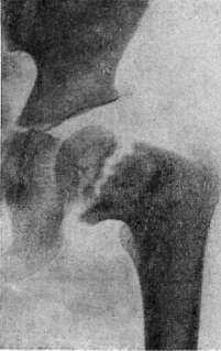
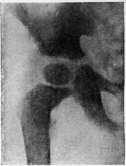
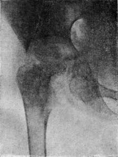
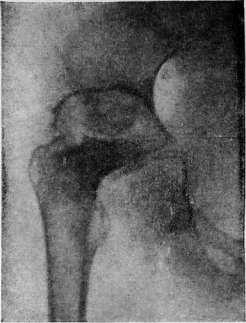
Figure 1. Central neck focus; joint not yet affected. Figure 2. Evolution of tuberculous coxitis-I stage; parietal neck focus with sequestrum; joint not involved. Figure 3. Evolution of tuberculous coxitis (same case)-II stage; joint involved; destruction and rarefaction of the head; expanded acetabulum; height of the disease. Figure 4. Evolution of tuberculous coxitis (same case)-III stage; subsidence of the process, reparative phenomena.
Figure 5. Epimetaphyseal tuberculous osteitis of the lower end of the femur; latent course. Fig. 6. Spina ventosa; proliferative periostitis of the metacarpal bone. Figure 7. Spina ventosa diaphysaria. Figure 8. Spina ventosa with fistulas.

Figure 1. Circular plaster cast: a, b-improperly applied; c-properly applied.
Local conservative treatment aims primarily to create maximum rest for easily damaged, unstable tuberculous tissues. For most patients, elementary rest is achieved by lying on a flat, non-sagging bed. The problem of rest is more completely solved by orthopedic measures-immobilization and unloading. Immobilization (see) is best achieved by plaster casts-circular (figure 1), embracing two adjacent joints (in spondylitis-large corsets with collars for upper thoracic lesions), or splints, to which also belong plaster beds applied in spondylitis and coxitis. Unloading is achieved by the horizontal (lying) position of the patient, by applying continuous traction, relaxing his muscular contracture. During the entire active period of the disease, patients must lie (on average 2-3 years), and then, when the process subsides, determined clinically, radiographically and laboratory-wise, they can be very gradually accustomed to get up and sit, but necessarily in orthopedic apparatuses, first plaster, then removable, gelatin-formalin or celluloid, and on crutches, without putting weight on the diseased limb. Such frequent in T. of joints muscular contractures are at first successfully corrected by gradual traction with relatively small weights, later they easily pass into persistent vicious positions, already associated with changes in the joints themselves (arthrogenic contractures), which cannot always be corrected without forcible straightening, and the latter is dangerous in T. Therefore, it is easier to prevent contractures than to treat them conservatively. Even more difficult to correct is the so frequent displacement of joint ends-subluxations and dislocations, often requiring operative interventions. Abscesses threatening to rupture require, on the one hand, especially careful treatment of the primary process by immobilization, and on the other hand, timely evacuation of the liquid content by means of punctures (see also Spondylitis). The latter are indicated mainly for progressive and large abscesses with tense walls and should be performed with thick long needles aseptically from the overlying healthy tissues by oblique punctures with skin displacement (fig. 2). To liquefy the thick and crumbly content, a 10% glycerin emulsion of iodoform can be introduced. The frequency of punctures is determined by the filling of the abscess and the tension of its walls. Opening of closed abscesses and their open drainage are inadmissible. In particularly stubborn cases, one can attempt to enucleate the entire abscess together with the capsule. Forming fistulas should be protected from secondary infection by carefully performed aseptic dressings without any irrigations. In case of infection of the fistula and formation of purulent infiltrates, wide incisions are indicated. In protracted cases with scanty content, sometimes
Figure 2. Rules for puncturing a cold abscess: A and E - incorrect technique of direct puncture - straight channel; B and G - correct technique; puncture from the side - zigzag channel; D and E - incorrect technique; F - correct technique.
For the treatment of fistulas, forced treatment is used by injecting 1-3cm3 of a 10% solution of CuSO4+ZnSO4, (Franke). In general, fistulas are easier to prevent than to treat.
* Surgical interventions that fall into the category of local measures should only complement the main sanatorium-orthopedic conservative treatment and be combined with it into one general system. Part of the surgeons (Leningrad school) has recently resorted to surgical removal of these delimited foci for the final elimination of the process: in bones - by necrectomy and curettage followed by iodoform-vaseline plugging, in joints - by so-called economical resections. Timely removal of para-articular nest-like lesions prevents the development of secondary arthritis (radical-prophylactic operation), and timely amputation in progressive purulent lesions of the joints prevents the development of amyloidosis and saves from death. A significant part of surgeons (Krasnobayev et al.) considers this radicalism not justified. Among the therapeutic-adjuvant interventions that improve the results of conservative treatment, osteoplastic fixations of the spine (see Spondylitis) and extra-articular arthrodesis in coxitis are particularly well-known. Various interventions proposed to change the course of the process itself - blocking devices (R. Lavall, Vreden), osteotomies (Kozlovsky), sympathectomies (Leriche), neurectomies (Molotkov), etc. have not yet gone beyond the development stage and therefore cannot be recommended for mass application. Finally, to correct persistent deformities, so-called corrective operations are used, primarily osteotomies, which are indicated mainly in complete bony ankylosis. The indications for the listed interventions are determined by age, location, stage, and complications. The old formula of Sorrel that conservative treatment is indicated for children, resections for adults, and amputations for the elderly, while generally retaining its significance, is being reconsidered at the present time, since according to clinical observations by Korneyev and others, resections can be successfully applied in children, especially older ones, if only operated on in the stage of subsidence. Such interventions are indicated mainly on the knee joint, where the operation, so-called extra-articular economical resection according to Korneyev (Fig. 3), applied as the final act of treatment to remove the delimited focus during subsidence of the process, leads to healing of the process with ankylosis in the correct position without disturbing bone growth (Geliknova). This experience should be transferred with great caution to the hip joint, where intra-focal interventions are best combined with additional extra-articular arthrodesis to obtain a strong ankylosis. The shoulder joint more often requires surgical intervention than the elbow and ankle joints. On the latter, it is recommended to remove the frequently affected talus bone - to perform so-called astragalectomy (de la Haye, Sorrel). Finally, mention should be made of operations for fistulous tuberculosis, if the source of the latter is an infected bone cavity, usually with a sequestrum. If there is an isolated lesion in the bone, curettage of the cavity followed by iodoform-vaseline tamponade according to Korneyev (filling with a paste of composition 30:100) is indicated. This method has particularly justified itself in tuberculosis of the calcaneus and in ;spina ventosa.
Figure 3. Extra-articular economical resection of the knee joint. (According to Korneyev.)
Organization of the fight against bone and joint tuberculosis. The organization of the fight against bone and joint tuberculosis should be based on the social significance of these diseases as a factor of mass disability of the younger generation. Preventive measures require primarily the timeliness of providing medical assistance, for which it is necessary to have a special network of specialized institutions and to carry out a special plan. The basis of such a plan should be: 1) wide coverage of the entire population with qualified assistance; 2) identification of all patients in the given area and especially identification of early forms; 3) conducting dispensarization of these patients and providing them with all types of assistance - medical and social; 4) timely treatment of fresh, non-neglected cases in order to prevent complications; 5) demarcation of the tasks of treatment from the tasks of caring for cripples and fistulous chronic patients; 6) unification of stationary, outpatient, and home (patronage) assistance into one general system having an 'staged' character. In addition, in each region there should be created its own support point - a clinical institution, which should become a scientific-methodological center for the entire periphery.
p- Cornet. XI. Tuberculosis in children. Statistical data. The ratio between infection, morbidity, case fatality, and mortality from T. in different periods of childhood reflects the significance and influence of the age factor on its course. The large amount of accumulated data on the state of tuberculin reactions in children of different age groups indicates a regular increase with age in the number of children infected with tuberculosis. The significant variation in the percentage of positive tuberculin reactions in children of the same age according to different authors is mainly due to the different nature of the material examined. The numerical data primarily from older authors, based on examinations of selected material, sick children, dispensaries, and consultations, naturally exceed the data on the number of positive tuberculin reactions in children's groups, in nursery, kindergarten, and school children. For example, when examining 5,000 nursery children in Moscow, a positive Pirke reaction was noted at age 0-1 year - 5%, 1-2 years - 13%, 2-3 years - 24% (Vasich, 1925-26) - a significantly lower percentage than what various authors usually indicate for children of this age; when examining nursery children of the Zamoskvoretsky district of Moscow and nursery children of the city of Orekhovo-Zuevo in 1932, this percentage was even lower. - In full accordance with the data on tuberculin reactions are also the data from autopsy material, confirming the increase in infection of children with their age. Older data from classical authors and more recent data note an increase with age in the number of tub. findings at autopsy, reaching, according to Negeli, at age 18 years up to 96%. This increase is due to the finding of limited tub. lesions, often a hidden focus in children who died not from T., but from accidental disease. Thus, in this autopsy material, with age, the quantitative ratio between cases with severe tub. lesions and accidental findings of small tub. changes in children who died from other diseases changes. In the first year of life, the percentage of cases with severe tub. changes in relation to all cases with findings of tub. changes at autopsy approaches 100, but with age it decreases, reaching according to Negeli at age 18 years 29%. This regular increase with age in children of the number of tub. lesions discovered accidentally at autopsy is noted by all statistical data. In children of the first years of life, a hidden primary focus or limited lesion of the bronchial glands is much less frequently noted as an accidental finding at autopsy. Infection at this age leads to expressed disease and the more pronounced, the younger the child. To the same extent as the fact of increasing infection with T. with age, it is considered established that there is a decrease with age in case fatality among infected children. The highest case fatality is noted among children of the first years of life, especially the first year. In the 'pre-tuberculin period', when the entire circle of infected T. children was not taken into account and only severe cases of the disease were recognized, case fatality of infected T. children of the first year of life according to these old data was 100%. With the introduction of tuberculin tests into practice and improved recognition of T. in children, the circle of accounted for infected and sick T. children significantly expanded, and thus the indicated case fatality figures also changed. Thanks to good accounting and careful observation of infected children, especially from bacillary foci, it became clear that even among children who became infected in the first months of life, there is a significant percentage of survival. 'The average case fatality figure' for children infected in the first year of life, according to the combined material of various authors, approaches 60%. This 'average case fatality figure' has a very relative meaning, as it is derived from the most diverse material, with case fatality ranging from 20% and lower among a wider circle of infected children from dispensary material and 80-100% case fatality from hospital, clinic material, i.e., selected severe material covering only a part of infected T. children. Despite the changed in recent years, more favorable assessment of the prognosis of T. in a child infected even in the first year of life, the position that case fatality in the first months and years of life significantly exceeds case fatality in subsequent periods of childhood remains absolutely unshakable. This higher case fatality in early childhood, especially in the first year of life, causes higher mortality from T., in this period significantly exceeding mortality from T. in older children. Mortality from T. in infancy and early childhood is the most significant, many times exceeding mortality in other age groups of childhood; decreasing in subsequent years, it sharply decreases after the age of 4, giving again a significant increase during the period of puberty (see Statistics of T., table 3 - mortality in the city of Moscow). With this high mortality from T. in children of the first year of life, it is relatively small compared to the high overall mortality from other diseases in this period of life. Tuberculosis as a cause of death in children of this age occupies one of the last places, far behind mortality from 'congenital weakness', digestive and nutritional disorders, pneumonias, and acute infectious diseases. Thus, the ratio between infection, morbidity, case fatality, and mortality in different periods of childhood is such that we have in infancy and early childhood, with a relatively small number of infected T. children, a large number of sick children, with severe forms of the disease, giving high case fatality and mortality. With age, infection with T. increases, but the percentage of severely ill children decreases, the tendency to generalization of the process becomes less pronounced, thus case fatality and mortality decrease. The ratio between the number of infected and sick T. children in early age sharply differs from this ratio in subsequent periods of childhood. Among nursery-age children infected with T., the percentage of sick children with expressed signs of the disease and clinical identification of lesion localization is significantly higher than in preschool and school children. All this gives T. particularly great social significance in infancy and early childhood. The severe course and high mortality from T. in the pubertal period also force special attention to be paid to the fight against T. in adolescence. Methods of infection and routes of penetration of the tubercle bacillus into the child's body - see above, the causative agent of T. Fundamentals of pathogenesis. The pathogenetic interpretation of tuberculosis in children in recent years has been under significant influence of Ranke's teaching on the cyclic course of tuberculosis in humans (see above - pathological anatomy and pathogenesis). From this very point of view, it is very important to note that the Ranke's pathomorphological characterization of its second stage is essentially a characterization of childhood T. The tendency to generalization of the process, to its spread by all ways - per continuitatem, lymphogenously, hematogenously, and intracanalicularly (bronchogenously), expressed localization in lymph glands, first of all intrathoracic, tendency to perifocal inflammation around foci, characterizing the second stage of Ranke, find their expression in the picture of T. in children and the more sharply, the younger the child. The regular decrease with age in children of the number of severe forms of T. and decrease in mortality vividly testify to the great significance of the age factor and reflect the peculiarities of the age-related reaction of the body to tuberculous infection. Age, however, is not the only factor determining the reactivity of the body and the form of tuberculous disease. We also encounter in adults with late infection the picture and forms of childhood T. (see Tuberculosis of the lungs, clinical forms of pulmonary T.). In infancy and early age we encounter, along with severe forms of the disease with all the expressed characteristic features of 'the second stage of Ranke', also mild forms of the disease without significant anatomical spread of the lesion. Within the framework of the peculiarities of age-related reaction characteristic of the child, we have a very polymorphic course of T. in him, conditioned by a whole complex of constitutional and conditional factors, genotypic and paratypic influences. A whole series of exogenous factors - mode and character of infection, massiveness of infection, virulence of the causative agent, superinfection - are given great importance in relation to their influence on the course and outcome of tuberculous disease in the child. The significance and influence of the entire social environment in which the child is located, in particular the state of care and feeding, as well as the influence of intercurrent diseases, on the form and course of T. in the child are very clear. Acute infectious diseases (measles, influenza, etc.) are particularly important activators of T. in the child, often contributing to the development of miliary T. and meningitis. It is not always easy to take into account the exogenous and endogenous reasons leading to exacerbation of the process in the child and causing the characteristic wavy course of tuberculous disease.
Periods of exacerbation, which are expressed pathomorphologically in the appearance of perifocal infiltrations around old foci, in the appearance of new foci, metastases or the eruption of miliary tubercles in different organs, are replaced by a period of remission. Along with taking into account individual influences of the complex paratypic complex, one must also strive to take into account genotypic influences, individual resistance or lability to tuberculous infection. This influence of genotypic factors on the course of the tuberculous process is still a little-developed problem. In the picture and course of the tuberculous disease in a child, which represents an equation with many unknowns, the influence of elements of the social environment is very significant and clear. Clinical forms and course of T. in children. The main characteristic features of childhood T. find their most vivid expression in infants and young children. It is precisely at this age that significant development of tuberculous lymphoma in the bronchial and mesenteric glands, marked perifocal inflammatory lesions around foci and primarily the primary focus in the lung, and a tendency for the process to spread to different organs and its generalization are noted in tuberculous children. But despite this pronounced tendency of the process at this age to a more severe course, even in the first months of life, localized forms with insignificant spread of the disease, running relatively favorably, are encountered. As the child grows older, the percentage of children infected with tuberculosis without any signs or with mild signs of the disease increases, the number of children with limited and insignificant lesions of the bronchial glands, not extending beyond the formation of the primary complex, increases, the tendency to multiple spread of the process in different organs decreases, and the percentage of miliary T. and tuberculous meningitis decreases. The onset of tuberculous disease even in children in the first years of life does not always manifest noticeably. The described symptoms—digestive disturbances, cessation of weight gain, poor appetite and mood, etc.—of course cannot be considered pathognomonic for T. and rarely draw attention to T. Initial fever, noted by some at the time of appearance of the tuberculin reaction as a sign of the beginning of tuberculous disease, is rarely observed. Tuberculous invasion and the pre-allergic period in the vast majority of cases proceed covertly. The pre-allergic period, i.e., the period from infection to the appearance of the first tuberculin reaction, lasts on average from 6 to 8 weeks. By the time the tuberculin reaction appears, intrathoracic changes in the form of lesions in the lung, associated with the primary focus, are noted in some cases. In a significant percentage of cases, and the greater the older the child, tuberculous invasion throughout the subsequent observation period manifests itself only by the presence of a positive tuberculin reaction. These T.-infected children, who give a positive tuberculin reaction on examination but show no clinical signs of the disease or general disturbances, nor local changes in the form of specific localization, according to the classification adopted at the IV Congress, belong to the group 'status allergicus'. T.-infected children who do not show pronounced and clear clinical signs of intrathoracic localization but show signs of general disturbance, such as malnutrition, anemia, subfebrile temperature in the presence of peripheral polyadenopathy, are according to the same classification considered to have 'chronic tuberculous intoxication'. The difficulty in determining the quantitative and qualitative nature of the lesions of the bronchial glands by physical examination methods and X-ray (see Bronchadenitis) forces one to especially carefully consider general disturbances in the body, especially their complex: nutritional status, temperature reaction, morphological changes in the blood, ESR, etc. Thus, symptoms of general disturbance, characterizing the degree of tuberculous toxemia, are a very important addition to the data of physical examination and X-ray, allowing together with them to form a judgment about the nature of the process. Nevertheless, one cannot but emphasize that the assessment of many symptoms, such as disturbed nutritional status, anemia, as signs of tuberculous intoxication, must be done with great caution due to the tendency of children, especially infants and young children, to develop dystrophy and anemia under the influence of the most diverse alimentary and infectious factors. To the same extent this applies to older children, especially in assessing unexplained fever and subfebrile temperature, which occurs in inflammatory lesions of the nasopharynx, hidden pyelitis, vegetative instability of thermoregulation, etc. The picture of general disturbances in bronchadenitis is very varied and polymorphic, and in many cases with pronounced bronchadenitis there is neither noticeable weight loss nor anemia nor temperature reaction. All this has recently caused a skeptical attitude towards the symptom complex 'chronic tuberculous intoxication'. In any case, the existing opposition in the proposed classification of the IV Congress between forms of tuberculous disease without clearly expressed clinical localization and so-called local forms must be considered unsuccessful, just as it is impermissible to strive to oppose the group 'chronic tuberculous intoxication' to 'bronchadenitis'. This opposition is to a large extent based on an underestimation of X-ray examination in the diagnosis of T. in children and an overestimation of the diagnostic value of individual symptoms and their combination in T. in children. Tuberculosis of the bronchial glands in the form of extensive and massive lesions, creating conditions for characteristic clinical symptoms and X-ray data, is encountered the more frequently the younger the child (see Bronchadenitis). In the presence of a mandatory reaction from the bronchial glands in the form of greater or lesser spread of changes in different groups, sometimes exceeding in size the lesions in the lung, the most polymorphic picture of pulmonary changes is noted (see Tuberculosis of the lungs, T. of the lungs in children). The younger the child, the more sharply expressed is the tendency to generalization of the process, and each severe case of T. in a young child we are justified in considering as a generalized form, in which a significant part of the localization remains hidden. Localizations in the bronchial and mesenteric glands, solitary tubercles of the brain, parenchymatous organs, T. of the intestine, etc., often remain hidden. In children in the first year of life, such forms with hidden generalization often proceed under the guise of severe nutritional disturbance, severe atrophy. The classical expression of hematogenous generalization is miliary T. According to the course and nature of the eruption, acute, subacute and chronic forms of miliary T. are distinguished. Acute miliary T. proceeds as an acute infectious disease in cases with fresh infection or in cases where it joined a slightly expressed lesion in the lung and bronchial glands. In some cases it proceeds with pronounced pulmonary phenomena (pulmonary form of acute miliary T.): dyspnea, cyanosis, with scanty percussion and auscultatory findings and significant temperature reaction. X-ray examination in most cases reveals the characteristic picture of scattered small-focal lesions of the lung in miliary T.—'marbled lung', 'starlit sky', etc. (see Tuberculosis of the lungs, T. of the lungs in children). Where acute miliary T. proceeds as an indefinite febrile disease (typhoid form, septic form), the X-ray of the chest also often unexpectedly reveals the picture of scattered T. of the lungs. T. meningitis often in these forms quickly cuts short the child's life. In other cases of miliary T., meningeal phenomena early come to the forefront in the clinical picture, and these forms rightly bear the name of meningeal forms of acute miliary T. (see Meningitis, tuberculous meningitis). In cases with subacute course, along with clinically often hidden tubercular eruption, expressed localizations in organs appear: tuberculides of the skin, solitary tubercles in the brain and parenchymatous organs, T. otitis and mastoiditis, etc. In the clinical picture, there is as it were a wavy course of the disease in the form of periodic exacerbations leading to new eruptions and the appearance of new metastases. Chronic miliary T. is a rare form with a hidden course. Extrapulmonary localization of the tuberculous process with a tendency to spread in different organs is often encountered in young children. The younger the child, the more sharply expressed is the multiplicity of external localization: skin T. (scrofuloderma), tuberculous lymphadenitis, bone T. (spina ventosa) and their combination with intrathoracic lesions—bronchadenitis and pulmonary lesion, predominantly of an infiltrative nature. External T. as an independent clinical form is noted more frequently from 2-3 years of age. Tuberculosis of the skin. Among skin lesions in children are encountered: a) papular, or papulonecrotic tuberculides, b) scrofuloderma, c) lichen scrofulosorum, d) more rarely—lupus vulgaris (see.
Tuberculous skin diseases). Tuberculides appear on different parts of the skin, preferably on the buttocks, back, on the extensor surfaces of the limbs, on the face, and on the fingers of the limbs. According to Zimbler's observations, especially in infants, tuberculides often accompany severe forms of T. and thus have not only valuable diagnostic significance but also to a certain degree prognostic significance. Scrofuloderma, on the other hand, often combines with other extrapulmonary localizations and with forms of intrathoracic T. with a favorable course. Tuberculous lymphadenitis. T. peripheral lymphadenitis in the form of a pronounced limited lesion of a separate group of lymph glands as an independent clinical form occurs mainly in preschool and school-age children. At the same time, anatomic lesions of peripheral lymph glands in severe cases of T. in children, especially infants and young children, occur, according to autopsy data, very frequently. These T.-affected peripheral lymph glands are not always clinically detectable, especially the deeper ones, difficult to palpate. Palpable significantly enlarged and markedly dense lymph glands in tuberculous children ('gland-stones,' in Kisel's expression) may arouse suspicion regarding their tuberculous nature, but this does not give the right to consider that the nature of the palpable glands, their size and density are pathognomonic symptoms of T. The uniform polyadenia described in T. with equally expressed density of all or the majority of groups of peripheral glands and the picture of micropolyadenia cannot be considered pathognomonic symptoms of T. Thoracic glands are often palpable in tuberculous children, but they also occur in children with other chronic diseases of the lungs and pleura. Other extrapulmonary localizations of T. (see Gonitis, Spondylitis, Coxitis). In early childhood, lesions of the small bones of the hands and feet are often noted (see Hand, Foot), often lesions of the flat bones of the skull and face, in many cases widespread lesions of many bones, both tubular and flat-forms with multiple bone localization.-T. otitis-mastoiditis, sluggishly and insidiously proceeding, are frequent localizations in tuberculous children.-Flickering kerato-conjunctivitis is an extremely frequent tuberculous manifestation. Keratitis in T. in children, differing in great persistence, often lead to a decrease and loss of vision (see Keratitis). Scrofulotuberculosis. Scrofula. Since Cherny gave a classic description of the exudative diathesis, we began to distinguish from the mass of 'scrofulous children,' on the one hand, tuberculous diseases with predominantly external localization, on the other-non-tuberculous cases with lesions of the skin and mucous membranes, which we attribute to manifestations of the exudative diathesis. However, this group of tuberculous children for the most part has such a peculiar clinical picture and character of the course of the process that it is quite appropriate to retain the name scrofula, or better, scrofulotuberculosis, in relation to this group of children. In these children, along with extrapulmonary localization of the process-tuberculosis of the lymph glands, skin, small bones, flictenae-non-specific inflammatory changes of the skin and mucous membranes are noted: impetiginous-exzemal lesions of the skin, runny nose, thickening of the upper lip, giving the child a special habitus-facies scrofulosa. According to the accepted interpretation, these forms represent a combination of T. with an exudative-lymphatic diathesis. T. in an exudative, lymphatic individual gives such a peculiar picture of scrofula. Not denying the significance of constitutional peculiarities in the manifestation of this peculiar reaction of the child's organism to tuberculous infection, one must also consider that the nature of nutrition-carbohydrate food (bread, potatoes)-and the environmental conditions in which the child lives (pauperism) have a great influence on the manifestation of this peculiar reactivity of the organism and play an important role in the genesis of these forms of tuberculosis. Diagnosis. 1. An important basis for diagnosis is first of all the history, which often gives indications of contact with tuberculous parents, who often do not know about their T., or with other family members, often with tuberculous old people, 'elderly coughing,' with tuberculous neighbors in the same apartment (apartment contact). A history collected with due fullness, reflecting the mortality of other children in this family, gives not only material for the correct recognition of the disease in the child but often reveals the previously hidden source of infection. Anamnestic data about past illnesses, in particular about acute infectious diseases, diseases of the respiratory tract, pneumonias, about the nature and development of the present disease, are very important for diagnosis and differential diagnosis. 2. Clinical symptoms. The diagnostic value of clinical symptoms is based not on the evaluation of the pathognomonicity of each of them individually, but, as with any disease, on their comparison with each other, with the data of the history, with the data of X-rays, with the data of tuberculin reactions, and with the data of other studies. In such a comparison, individual clinical symptoms acquire much greater diagnostic significance than in their isolated evaluation. A pronounced fever is by no means mandatory even in severe, far-advanced cases of tuberculous disease. Generalized forms, miliary T., disseminated T. of the lungs, caseous pneumonia often proceed, especially in infants and young children, without a pronounced fever, apyretically (see Tuberculosis of the lungs, T. of the lungs in children). The widespread opinion that a subfebrile temperature is a frequent, if not constant, sign of latent T. found its critical evaluation among pediatricians much earlier than among therapists. Cough, its duration and character have great diagnostic significance, often allowing one to judge the localization of the lesion: a resonant bitonal cough-in bronchial adenitis, with abundant sputum-in bronchiectasis, with abundant purulent discharge-in lung abscesses and suppurating cavities (see) etc. (see Bronchial adenitis, Tuberculosis of the lungs).-Dyspnea-as both an increase in the frequency of respiration and a change in its character-attracts attention and in many cases characterizes the form of the lesion: expiratory stridor in tumorous bronchial adenitis, increased respiration along with scanty physical changes in miliary T. of the lungs etc. (see Tuberculosis of the lungs). Disorders of digestion and nutrition, so often accompanying T. in a child, especially in the first year of life, by no means should always be regarded as a manifestation of tuberculous disease, but are often caused in tuberculous children by other etiological factors: alimentary, enteral and parenteral infections, intercurrent diseases etc. Not always a pronounced and widespread tuberculous disease even in a young child leads to a sharp violation of the child's nutrition, to a pronounced dystrophy. In the presence of a sharply expressed picture of digestive and nutritional disorders-acute or chronic-in a tuberculous child, especially with little spread of the process, one must always think of another, besides T., etiology of these disorders. Along with this, widespread tuberculous disease often proceeds in a child under the guise of a severe nutritional disorder, and in some cases of such severe atrophy, a significant part of the localization of the tuberculous process remains hidden: intestinal localization of the process, lesions of the mesenteric glands (tabes mesaraica of old authors), hidden universal tuberculous lymphadenitis (see Tabes mesaraica). Percussive and auscultatory data in their comparison with each other and in subsequent comparison with the data of fluoroscopy and X-ray have great diagnostic significance (see Tuberculosis of the lungs, T. of the lungs in children). 3. Fluoroscopy and X-ray (see Tuberculosis of the lungs, X-ray diagnosis, T. of the lungs in children). 4. Tuberculin diagnosis. A positive tuberculin reaction means that this child is infected with T., but it does not testify that this child is sick with T., it does not give grounds to judge the degree of spread of the tuberculous process in the organism, its 'activity,' it also does not give grounds for prognosis. However, the fact that the number of infected and sick with T. sharply diverges only in older children, and converges and approaches in children of the first years of life, gives precisely in early childhood the tuberculin reaction great diagnostic significance. The younger the child, the more likely this or that lung lesion, bone lesion or other unexplained disease turns out to be tuberculous with a positive tuberculin reaction. The older the child, the more often we in difficult diagnostic cases encounter phenomena of non-tuberculous disease in a child infected with T. A negative tuberculin reaction does not always mean that the child is free from T.
The reaction can be negative: a) in a child infected with T. in the pre-allergic period (see above); b) in a tuberculous child with an advanced case of caseous pneumonia, generalized T.—'negative anergy' (as opposed to 'positive anergy', when the body, being in good condition, stops reacting to tuberculin, which is observed, for example, after tuberculin therapy); c) in a tuberculous child during an acute infectious disease, especially measles, but sometimes also with whooping cough, diphtheria, etc. Occasionally, temporary anergy has to be noted in tuberculous children without being able to find any explanation for it. Often it is possible to note a positive Pirke reaction only after its repetition in a week. In some cases, with pronounced tuberculous disease, the positive reaction persistently fails to appear only for the intracutaneous Mantoux reaction, while the Pirke reaction remains negative for a very long time. Explanations for all these facts have to be sought in the state of the autonomic nervous system, which plays a major role in the mechanism of allergy. Of the proposed methods of using tuberculin for diagnostic purposes, Cimbler uses only the skin (see Pirke reaction) and intracutaneous (see Mantoux reaction) reactions; the subcutaneous reaction (Hamburger) is unnecessary and not harmless. The intracutaneous reaction (Mantoux) is recommended to be performed only after it has been established that the skin reaction is negative; otherwise, without knowing the degree of allergy, starting immediately with the intracutaneous reaction, one can obtain a sharply expressed general (fever, poor condition), local (large redness, swelling, lymphangitis, regional lymphadenitis), and sometimes also a focal reaction (exacerbation in the lung). The Mantoux reaction is performed, starting with a dilution of 1:10,000 (No. 4), increasing with a negative reaction at intervals of 5-7 days the concentration of tuberculin in the following order: 1:1,000 (No. 3), 1:100 (No. 2), 1:10 (No. 1). The greater sensitivity of the intracutaneous reaction compared to the skin reaction is a significant advantage, but at the same time it is necessary to evaluate it with great caution and to consider only a clearly expressed papule and redness significantly exceeding the control reaction as a positive Mantoux reaction; the size of the redness and papule should be at least 10 mm. As a control liquid, it is best to use condensed broth diluted in physiological salt solution according to the dilution of tuberculin, or a 0.25% solution of Acid carbolic in physiological solution. The age of the child and its condition cannot be a contraindication for performing the Pirke reaction; severe condition and high fever with a strong suspicion of a tuberculous nature of the disease can be a contraindication for performing the Mantoux reaction. 5. Examination of sputum for Koch's bacilli is an exceptionally important and valuable method, the data of which often allow a final decision in favor of tuberculosis diagnosis. 6. The morphological composition of the blood and the erythrocyte sedimentation rate (ESR) serve as an aid in characterizing the process, its activity, degree of intoxication, predetermine1 the approach to therapy and provide material for prognosis, but the direct significance of these data for diagnosis should not be overestimated. T. in childhood does not belong to anemia-causing diseases; moderate anemia develops slowly with it, and a severe degree of anemia, especially with unexpressed tuberculous disease, is always associated with another etiology besides T. and often finds its explanation in the presence of secondary infection. 7. Serological reactions with deviation of complement have not found practical application in the diagnosis of T. in children. Therapy. The basis for treating T. in a child is a rational hygienic-dietary regimen. Rational feeding and nutrition, which constitute the main prerequisite for the correct growth and development of the child, occupy an important place in this regimen. Diet therapy, based on the principle of preferring some food ingredient to the detriment of another, has not found application in T. in children. The important physiological significance of carbohydrates for a growing organism does not give the right to limit their intake at the expense of increasing the amount of protein and fat. At the same time, taking into account the arguments recently put forward in favor of the specifically beneficial effect of food protein in T. (increasing the buffer capacity of the body), one should strive to ensure that the child's food contains no less than 20% protein. Along with complete proteins, it is necessary to provide the food with fat containing additional factors A and B. Therefore, the administration of cod liver oil is mandatory for a tuberculous child. The food should be rich in other vitamins (C), and should be tasty in view of the persistent anorexia in most tuberculous children and contain a certain amount of fiber (fruits, vegetables). Proper care and regimen, hygiene in the child's environment, maximum use of fresh air are also the basis for treating a tuberculous child. There are no contraindications for treating a tuberculous child with fresh air, depending on the child's condition, just as there should be no contraindications depending on the weather. Tuberculous children use fresh air regardless of the air temperature and other meteorological factors, provided protection from overheating or excessive cooling in a place protected from strong wind, snow, and rain. The number of hours spent outdoors varies depending on climatic conditions, time of year, and meteorological conditions from 3-6-8 hours to round-the-clock stay outdoors. The beneficial effect of aerotherapy consists in affecting the child's metabolism, increasing oxidative processes in the body, improving the child's appetite, sleep, and mood. The effect of aeration is more noticeable and intense with simultaneous direct exposure of the child's skin to a significant amount of reflected sun rays, during air baths. Heliotherapy, i.e., the use of direct sun rays, should be regarded as a significantly more drastic physical agent, for which there are its own indications and contraindications. Caution in sun treatment of young children is dictated primarily by the well-known danger of overheating with all its consequences for children of this age. Sun treatment is contraindicated in cases of a tendency for the process to spread, with hemoptysis, etc. Heliotherapy is especially indicated for extrapulmonary T.: bone and joint, T. of the skin, lymphadenitis, etc. For these same forms, the use of the mercury-quartz lamp is also indicated, while for pulmonary forms caution is necessary. The indications and contraindications for any other irritation therapy (Reiz-therapie), to which various modifications of protein therapy can be attributed, including treatment with sera, hemotherapy, etc., are based on the same approach. Specific irritation therapy, which includes tuberculin therapy, has few supporters in T. in children. Drug treatment for T. is limited to the use of a limited arsenal of symptomatic agents: narcotic—belladonna, codeine, luminal—for severe cough and pronounced compression symptoms in tuberculous bronchial adenitis; iron preparations, liver preparations—for accompanying anemias. The popular use of calcium preparations is not based on a sufficiently solid theoretical basis, if only because it is difficult to imagine how demineralization and decalcification in T., based on a deep disturbance of intermediate metabolism, can be corrected by simply increasing the amount of calcium received by the child. This favorable effect on mineral metabolism and increased calcium retention is rather to be expected from the use of ultraviolet rays, preparations of irradiated ergosterol or parathormone. According to Cimbler's observations, a beneficial effect is achieved by the use of vigantol and parathyreocrin with simultaneous administration of calcium preparations in certain forms of tuberculosis, especially in bone and joint T. Methods of collapse therapy—see Artificial pneumothorax, Phrenicectomy, Thoracoplasty. Prevention of T. in children is expressed: 1) in measures aimed at protecting the child from infection, and 2) in measures aimed at increasing the child's resistance in the fight against the infection that has penetrated his body. The severe course and high mortality among T.-infected children in the first years of life, especially children in the first year of life, place first the protection of children of this age from infection, which in the vast majority of cases occurs from contact with a sick family member or a sick apartment dweller (intra-family, intra-apartment contact). This places first before the women's consultation and the tuberculosis dispensary the task of careful accounting and detection of pregnant women suffering from T. and in contact with bacillary and active tuberculous patients. Newborns from bacillary, severely ill mothers must be separated from them immediately after birth and placed in a baby home.
A significant percentage of infection in infants does not occur from sick mothers, but from sick fathers and other family members, which forces the anti-tuberculosis organization to raise the question of the widespread hospitalization of tuberculosis patients with severe forms of tuberculosis. Isolating an infant from the source of infection remains the most important link in the prevention of T. in children of this age. This separation, even if brief (6 weeks-2 months), from a bacillary patient is necessary also for newborns vaccinated by Calmette, since a certain period is required immediately after vaccination for immunity to develop. The danger of infection not only from family members but also outside the family, from chance encounters and unidentified sources of infection, which increases with age, forces concentration on the hygienic environment surrounding the child, sharpens attention during home visits, and requires attention to this issue in the educational work of the consultation. Another aspect of preventive work, along with the fight against infection, is the struggle for the proper growth and development of the child, ensuring the necessary resistance of the body to infection. This aspect of prevention is fundamental and decisive in all periods of childhood. All measures to create the most favorable environment for the child, conditions of care, and rational nutrition become the most important link in the prevention of T. in children. Creating a healthy children's collective, developing a wide network of high-quality infant care centers, developing differentiated infant beds, the work of consultations, health work in kindergartens and schools, creating differentiated groups in the latter, and networks of health schools thus constitute the foundation of all anti-tuberculosis work. The presence of a large number of hidden bacilli excretors among early childhood children with various forms of T., proven by experiments on guinea pigs and cultures on special media, forces the separation of tuberculous early childhood children with mild symptoms of the disease from uninfected children in children's collectives and infant care centers by organizing separate sanatorium groups for such children. Anti-tuberculosis institutions for children of different age periods, see above - Organization of the fight against tuberculosis. Vaccination by Calmette (BCG). The high mortality rate among T. infected children of the earliest period of childhood has long forced the search for preventive methods through active immunization using one vaccine or another. Of the attempts in this direction, the most successful has been vaccination by Calmette (see above - Causative agent of tuberculosis and Bacteria, Calmette-Guérin bacilli). In our country, vaccination is carried out according to a resolution of the Council of People's Commissars in 14 cities of the RSFSR (Moscow, Leningrad, Ivanovo, Saratov, Voronezh, Gorky, Rostov-on-Don, Smolensk, Kuibyshev, Sverdlovsk, Kazan, Ufa, Yalta, Alma-Ata), as well as in some major industrial centers of the Moscow region (Kalinin, Tula, etc.) and in major cities of the Ukrainian SSR.
Related articles
Cite this page
“Tuberculosis.” Soviet Medical Encyclopedia. English translation of Bolshaya Meditsinskaya Entsiklopediya, 1st ed. (Moscow, 1928–1936), ed. N. A. Semashko. https://sovietmedicalencyclopedia.pages.dev/article/tuberculosis/