Keratitis
Historical document, translated for reference. It reflects medical knowledge of the 1920s–30s and is not medical advice.
Summary
Keratitis is inflammation of the cornea, one of the most common lesions of the anterior eye segment. The article discusses its diverse etiology, pathological anatomy, clinical classification, and the cellular processes involved in corneal inflammation.
Encyclopedia article (1928–1936)
Keratitis. Contents: Etiology.....................573 Pathological anatomy of K...........574 General clinical manifestations and course of K......57G Individual forms of K...............57J Keratitis, keratitis (from Greek keras-horn), inflammation of the cornea, one of the most common lesions of the anterior eye segment. In clinical terms, the concept of K. includes not only inflammatory conditions of the cornea but also various degenerative changes, trophic disorders, etc.; however, such clinical forms that do not correspond to the essence of changes occurring in inflammation should not be included in the description of keratitis. The etiology of corneal inflammations is extremely diverse, and in this regard, it is first necessary to distinguish primary K., i.e., those occurring without connection to lesions of other parts of the eye, and secondary K., when corneal inflammation is a complication of inflammations of other membranes, most often the conjunctiva of the eye, or the result of the spread of the process from adjacent parts of the eye. The main causes of K. in the vast majority of cases are infections, and these infections occur either by the exogenous route, i.e., by the entry of some pathogen from outside, or by the endogenous route in the presence of general infections such as lues, tbc, etc. Among the causative agents of exogenous K., very diverse microbial forms are encountered, but these are predominantly pyogenic species: pneumococcus, streptococcus, staphylococcus, Pseudomonas aeruginosa, Escherichia coli, Koch-Weeks bacillus (Bac. Koch-Weeks, Bacillus influenzae), etc. In addition to infections, K. sometimes develop on the basis of various constitutional anomalies, resp. diatheses, such as exudative diathesis, arthrism, gout, etc. Furthermore, some forms of K. are due to trophic disorders resulting from lesions of trophic centers and nerves of the cornea. Finally, causes of K. can also be traumatic injuries and various physical-chemical agents causing wounds to the cornea, its burns, etc. The classification of K. presents great difficulties, since it is not possible to base it on any single principle, and it is not possible to divide K. by etiology. Not to mention that not all forms of K. have an established cause, far from all K. known by etiology can be distinguished into defined forms, since the same cause very often causes diverse clinical pictures. The classification most satisfying practical needs is one based on division of K. by clinical picture and by basic pathological-anatomical changes. On the basis of such principles, K. are divided into two main groups: according to the nature of pathological-anatomical changes—into purulent and non-purulent K., and according to the degree of spread—into limited and diffuse, superficial and deep. Such division, providing a satisfactory scheme, still cannot encompass all forms of K., and there is a whole series of transitional forms and types that do not fit into this classification. Pathological anatomy of K. Pathological-anatomical studies of the cornea during inflammation have played the greatest role not only for understanding the picture of K., but also for studying many aspects of inflammation in general, since the cornea by its structure represents a very accessible and interesting object for observing inflammatory phenomena. But despite numerous and long-standing observations, far from all aspects of corneal inflammation appear to be clarified; for this reason, works devoted to the study of the problem of corneal inflammation continue to appear to the present day (Marchand, Lohlein, Schonemann, etc.). Each inflammation of the cornea is accompanied by an increase in cellular elements in one or another part of the cornea, which in clinical examinations is expressed in the form of varying degrees of cloudiness of the cornea. As experiment and spontaneously occurring K. teach, this cloudiness, due to infiltration, can sometimes arise within a few hours and especially rapidly in corneal infections. The question of the origin of the cellular elements that make up the inflammatory infiltrate in various forms of K. still causes many debates. In this regard, two main opinions are opposed: some (Grawitz and his school) attribute the origin of the cells of the inflammatory infiltrate only to the permanent elements of the cornea, while others since the classical works of Leber on inflammation consider that the infiltrate is predominantly formed by white blood cells migrating into the cornea (Cohnheim, Marchand, Orth, Lubarsch, etc.). Hippel (Hippel; 1928), summarizing modern research in this field, states that the emigration of leukocytes from the vessels of the marginal network as the basis for infiltration in purulent K. no longer raises any doubts and that the processes of proliferation of permanent corneal cells occur only in the sense of participation in regeneration, but even here they are secondary in importance. The participation of so-called histiocytes for early stages of K. is not proven. In later stages of purulent inflammations of the cornea, as well as in non-purulent K., lymphocytes are predominantly observed in the infiltrate. Infiltration of the cornea, varying

Figure 1.
Figure 2.
Figure 1. Corneal infiltrate: 1- epithelium; 2- Bowman's membrane; 3- Descemet's membrane. Figure 2. Corneal ulcer: 7- epithelium; 2- Bowman's membrane; 3- Descemet's membrane. In terms of its cellular composition, it is accompanied by various changes in the permanent elements of the cornea. In addition to the phenomena of proliferation of corneal corpuscles, they undergo degenerative changes - they swell, disintegrate, the lamellae of the cornea also undergo fine-grained clouding, swelling and may disintegrate into a granular mass. In cases of virulent infections, when an infiltrate forms, necrosis of corneal tissue occurs, which is expressed as softening of the stroma; this leads to the formation of either a limited corneal abscess with subsequent development of an ulcer or widespread purulent infiltration, causing destruction of corneal tissue throughout its entire extent and thickness. In cases of non-purulent K., the infiltration of the cornea is in some cases predominantly lymphocytic, in others - plasmacellular, and in mild cases it is not accompanied by sharp changes in the corneal stroma itself; usually, however, in such processes, as for example in typical parenchymatous K., the permanent corpuscles of the cornea, its lamellae undergo both proliferative and degenerative and disintegrative changes (fig. 1 and 2).- Along with the phenomena of corneal infiltration in all kinds of K., vascularization of it is observed at one or another stage of the process due to the new formation of vessels from the marginal vascular network; the newly formed vessels are either in the subepithelial, superficial layers of the cornea or between the lamellae of the cornea in the deep layers of its stroma. The vessels are usually accompanied by strands of newly formed connective tissue. Furthermore, in corneal inflammation, a number of inflammatory and degenerative changes are found in the corneal epithelium. In addition to the phenomena of inflammation in the corneal membrane itself, the harmful agents causing it also exert remote influences on the vessels of the iris and ciliary body, which is expressed by the exit of white blood cells into the anterior chamber, clinically giving a picture of hypopyon, and patho-anatomically manifests as an accumulation of leukocytes with fibrin in the anterior chamber, both in its angle and often on the posterior surface of the cornea, especially in the area corresponding to the purulent infiltration of the cornea, in the form of extensive accumulations of cellular elements; sometimes, however, on the posterior surface of the cornea, so-called precipitates are found, i.e., limited conglomerates of cells and fibrin. General symptomatology and course of K. Despite the diversity of individual forms of K., a number of objective and subjective symptoms are more or less constant and common. Among the objective signs, clouding of the cornea comes first, and the color of these cloudings, their saturation depend on the nature of the infiltrate, its density, and the depth of its position in the cornea. Small non-purulent infiltrates have a bluish, gray color, dense purulent infiltrates give a yellowish, white color, and the denser the infiltration and the more the corneal parenchyma is involved in the process, the more saturated the clouding. Characteristic for inflammatory infiltration in its progressive period is the indistinct demarcation of the clouding from the surrounding transparent parts of the cornea. The formed corneal infiltrate is usually accompanied by a change in the epithelial covering over the focus of inflammation - it appears uneven, as if pockmarked; in a number of cases, the infiltrate protrudes above the surface of the cornea. Non-purulent infiltrates, having reached one or another size, in their regressive period gradually decrease and in a number of cases may not leave behind a persistent clouding, completely resorbing; in other cases, cloudings of the cornea remain, due to destructive changes in the corneal stroma and replacement by connective tissue (corneal spots, leukomas--see Cornea). Purulent infiltrates usually quickly lead to disintegration of corneal tissue, rejection of the epithelial covering and formation of a tissue defect - corneal ulcers (ulcus corneae). The different localization of ulcers both on the surface of the cornea and in depth, the different etiology, determine their division into separate clinical forms, but common to them at the height of the process is the deepening in the cornea at the site of the defect, loss of the mirror reflex, clouding to one degree or another. In their course, all corneal ulcers have progressive and regressive stages. The symptoms of the progressive period come down to the fact that the ulcer has a dirty floor, a densely cloudy edge or edges of yellowish-gray or white color, the ulcer over a large extent is surrounded by a gray border and often from it in various directions go thin strips into the still transparent parts of the cornea; this is infiltration spreading from the site of the ulcerative process into the surrounding healthy tissue, thanks to which the spread of the ulcer, its progression, takes place. In certain forms of ulcers (the so-called creeping corneal ulcer--fig. 3), one edge of the ulcer has a marked progressive character, is strongly infiltrated and creeps along the surface of the cornea and gradually destroys it. In other cases, the ulcer has a tendency to progress in depth, which is characterized by an increase in the saturation of the color of the ulcer floor: it becomes intensely yellow, white. Sometimes during the course of the ulcerative process both phenomena are observed - the spread of the ulcer over the surface of the cornea and in depth, as a result of which the entire cornea can be destroyed by purulent

Figure 3. Creeping corneal ulcer: /- epithelium; г- Bowman's membrane; 3- Descemet's membrane.
an ulcerative process. The ulcerative process as a rule ends in the formation of scars of the cornea, either superficial or deep (macula corneae, leucoma). An ulcer spreading in depth can lead to perforation of the cornea, which entails a series of complications depending on the size of the perforation and its localization. With small sizes of perforation and with its location in the center of the cornea, the consequence of this will be the outflow of fluid from the anterior chamber and subsequently the approximation of the lens to the posterior surface of the cornea, and if this contact of the lens at the site of perforation is prolonged, then a clouding of the anterior capsule will develop (anterior capsular cataract); in favorable cases, the perforation hole can close quickly, the anterior chamber will be restored, and the lens will return to its original position. A severe consequence of limited destruction of the cornea is the formation of a corneal fistula, i.e., a long-unhealing opening in the cornea, which sometimes, if measures are not taken, leads to the death of the eye. If the perforation is extensive, and especially if it occurs in the periphery of the cornea, then the iris inevitably falls into the resulting opening (prolapsus iridis), which during subsequent ulcer healing grows into the scar of the cornea, and forms the so-called leucoma adhaerens—an opacity adherent to the iris; with small sizes, this adhesion is more often called an anterior synechia (synechia anterior). Extensive prolapses of the iris with widespread destruction of the cornea throughout its thickness are in a number of cases accompanied during subsequent healing by the bulging of the corneal and iris scar in the form of the so-called staphyloma (staphyloma corneae), which can be partial or complete depending on whether the entire cornea or part of it is bulged (see Cornea).-The regressive period of the ulcer is characterized above all by the so-called cleansing of the ulcer, i.e., the bottom of the ulcer, its edges become more transparent due to the rejection and resorption of the infiltrate, the bottom of the ulcer becomes smoother, its edges from being undermined, sometimes eroded, become rounded, the surface of the cleansing ulcer begins to give a mirror reflex, which indicates the covering of the defect with epithelium, the onset of the healing process, repair of the ulcer. This process of the regressive period of the ulcer is not always observed in relation to the entire ulcer on the surface, but in some cases it is limited only to its part, while in other parts of it the ulcer progresses. In some cases, the ulcerative defect is not completely restored for a long time, and a depression remains in the cornea, covered with a shiny epithelium; this condition of the regressive period of the ulcer is called a 'facet'. Many forms of K. are accompanied by vascularization of the cornea, and the presence or absence of newly formed vessels in the cornea at the height of the process allows one to divide K. into vascular or avascular K. (k. vasculosa, k. avasculosa). The formation of new vessels varies depending on whether it accompanies deep or superficial K. For superficial K., a characteristic feature is the direct transition of vessels from the vessels of the conjunctiva, well visible in the area of the limbus and further spreading tree-like over the surface of the corneal membrane, and in some cases the formation of new vessels is limited, in others it spreads over the entire surface of the cornea, increasing its cloudiness and giving a reddish tint to the affected cornea. In some forms of deep K., the vascularization of the cornea is due to the formation of new vessels from the deep network of episcleral vessels, and the vessels run in the cornea in straight trunks, closely adjacent to each other and in places indenting into the cornea in the form of a broom or brush. Due to the fact that these vessels are formed deeply, tracing their origin from the episcleral vessels is not always possible. Each K. is accompanied to one degree or another by phenomena of hyperemia in the anterior part of the eyeball: injection of vessels in the area around the cornea, phenomena of the so-called pericorneal or ciliary injection, characteristic not only of K. but also of inflammation of the iris, ciliary body, sclera. Pericorneal hyperemia, due to the dilation of vessels of the anterior ciliary arteries and veins, is distinguished by its violet color; but along with pericorneal injection in K., we often see the so-called conjunctival hyperemia due to the dilation of the vessels of the eyeball's conjunctiva. In addition to hyperemia, in a number of cases, especially in severe purulent K., from the side of the conjunctiva and sclera, edema of it (chemosis conjunctivae) is observed, then edema of the eyelids, redness of their covering. The usual symptoms of K. are also photophobia, lacrimation, and often blepharospasm and pain as a result of reflex irritation of the sensory nerves of the cornea or involvement of the iris or ciliary body in the inflammatory process. The addition of iritis or iridocyclitis to the inflammation of the cornea is a very frequent phenomenon and not only in purulent K. or deep non-purulent, but sometimes also in superficial and limited ones (see separate table, fig. 1), and moreover, a parallelism between the severity of K. and the phenomena of irritation is by no means always observed, and sometimes superficial and limited processes are accompanied by exclusively sharply expressed phenomena of irritation and vice versa. Iritis, especially in purulent K., is often accompanied by the formation of hypopyon, giving a picture of hypopyon-keratitis. A constant symptom of K. should also be considered the disturbance of vision. The degree of visual impairment depends on the extent and localization of the inflammatory foci, and it is understandable that K. located in the central areas of the cornea especially reduce vision, and foci of inflammation lying in the periphery have less effect on it; but it should be noted that the accompanying K. phenomena of irritation sometimes even with weak changes in the cornea cause extraordinary disturbances of vision. Individual forms of K. Into the first group of K., according to the classification indicated above, one should include superficial non-purulent limited K.; of these, a typical representative is vesicular K., k. vesiculosa, s. herpes corneae. This form of K. is characterized by the appearance on the surface of the cornea of small, pinhead-sized vesicles, often arranged in groups; the process arises acutely with phenomena of severe irritation of the eye, and the vesicles, having existed for a very short time, burst, leaving a superficial loss of substance of the cornea—erosion of it (erosio corneae); only in exceptionally severe cases do deep ulcers form. A variety of herpes corneae is k. dendritica—dendritic K. (fig. 4), developing after the appearance of a group of vesicles in the form of a superficial ulceration having the appearance of tree-like branching strips, often ending in thickenings. The vesicles in herpes corneae are analogous to herpetic vesicles on the skin (herpes labialis, faciei), and they arise, like the latter, in various febrile diseases (hence the name herpes corneae febrilis). Research first by Gruter, and then by Dorr, Lowenstein and many others, proved that herpes has an infectious origin, is due to a special filterable virus, and can be transferred to the cornea of a rabbit (see Herpes). Herpes corneae is furthermore part of a more serious lesion due to a neurotropic virus, the so-called herpes zoster ophthalmicus. The diagnosis of herpes corneae is facilitated by examination of the cornea when a solution of fluorescein is instilled, which stains the slightest defects in the cornea. Treatment—general, directed against the general infections in which herpes is observed (aspirin, quinine, etc.); locally—bandage, disinfecting ointments (xeroform, irol, etc.), dionin; with irritation of the iris—atropin, in the absence of irritation—pilocarpine. Sometimes chemical cauterization (T-ra Jodi), thermal cauterization (galvanocautery) is useful. [*. } A frequent form of superficial K. is pannus (pannus); it is observed first of all as an accompanying inflammation of the cornea in trachoma, and then as one of the forms of the so-called scrofulous K. (see below). Superficial K. are the so-called recurrent traumatic erosions (erosiones traumat.), which arise after superficial injuries causing desquamation of the epithelium and the formation of superficial defects of the cornea (erosions). After healing, such an erosion recurs after a certain time with phenomena of severe irritation and pain in the eye; at the site of the former erosion, a vesicle develops, similar to a herpetic one which quickly bursts, and due to this an erosion forms again. Such recurrences can occur many times, usually arising in the morning upon opening the eyes. In the interval (calm state of the eye), an abnormal condition of the epithelium at the site of injury is noted with a slit lamp, which explains its easy vulnerability.-In treatment, sometimes scraping of the diseased epithelium is necessary to obtain regeneration of the normal. Parenchymatous K.
(k. parenchy-raatosa, s. interstitialis)—the next very important form of non-suppurative K., but only of the deep type. Parenchymatous K. occupies a special place among inflammations of the cornea as a typical manifestation of congenital syphilis. Since the time of Hutchinson, it has been recognized as one of the most important symptoms of late congenital syphilis, and since then, especially since the introduction of serological reactions into the diagnosis of syphilis, it has been conclusively proven that syphilis indeed has the predominant etiological role in this process. Even if one considers the assertion of some researchers, led by Igersheimer, that syphilis is the sole cause of parenchymatous K. to be insufficiently substantiated, the majority of ophthalmologists still attribute 90-95% of all cases of this K. to syphilis. Among other causes of parenchymatous K., until recently, especially after the observations of E. Hippel (1914), tuberculosis was given considerable importance, and there are researchers who attribute a large percentage of cases to tbc. But with the refinement of the diagnosis of tbc and syphilis, it had to be recognized that a typical primary parenchymatous K. of tuberculous origin is a very rare phenomenon. Parenchymatous K. is observed extremely rarely in connection with various acute infections—pneumonia, influenza, malaria, etc., but the evidence of these cases is questioned by many. Despite the predominant role of congenital syphilis in the etiology of parenchymatous K., there is still a small number of cases of this process on the basis of acquired syphilis (2-1%, according to Heine) in its secondary or tertiary period. Pathological anatomy and experimental study of parenchymatous K. Regarding the pathological-anatomical changes in parenchymatous K., it should be said that in the doctrine of the histology and pathogenesis of parenchymatous K., the study of experimentally obtained parenchymatous K. in animals when infected with syphilitic products or cultures of the pale spirochete is of great importance, especially when obtaining parenchymatous keratitis by the metastatic route. The most important works in this field in relation to the eye belong to Igersheimer. He succeeded in obtaining metastatic parenchymatous K. in a rabbit through infection via the carotid artery after 2 months, and when infecting via the testis in the same animal, parenchymatous K. was obtained after 4-9 months. In experimental K., it became possible to study the pathological-anatomical and early manifestations of parenchymatous K., and it was established that in the very earliest period of parenchymatous K., in addition to lymphocytic and eosinophilic infiltration of the deep layers of the cornea and neovascularization, there is marked involvement of the corneal parenchyma. Pathological-anatomical studies of parenchymatous K. have been performed in humans in very rare cases, which is quite understandable, since parenchymatous K. in itself cannot provide a reason for enucleation. The main studies of early parenchymatous K. in humans are the studies of Elschnig (1905), who managed to examine the eyes 3 weeks after the onset of the disease in one eye and 7 weeks in the other; other authors in pathological-anatomical studies dealt with old cases. Elschnig emphasizes the widespread degenerative changes in the lamellae of the cornea and in the permanent cells of the cornea, which are observed even in the early period of parenchymatous K., along with the phenomena of infiltration and expansion of the juice canals filled with a fine-grained mass from the decay products of cellular elements. Along with these phenomena, there is neovascularization, proliferation of permanent corneal cells, and subsequently, resorption of infiltrates and replacement of foci of necrosis with newly formed connective tissue. The epithelium of the cornea above the foci of inflammation undergoes various changes, degeneration, edema. The endothelium of Descemet's membrane rarely remains normal; even in fresh cases, its vacuolization and disappearance in places are visible. On the posterior surface of the cornea, the so-called precipitates are encountered (Fig. 5). The pathogenesis of parenchymatous K., while recognizing the predominant role of syphilis in its etiology, remains to this day insufficiently clear. From Igersheimer's point of view, the origin of parenchymatous K. needs to be explained as a true spirochetal disease of the cornea. That spirochetes can penetrate the cornea is evidenced by findings in the corneas of fetuses with congenital syphilis; spirochetes are sometimes found in enormous numbers in experimental parenchymatous K.; they were also found by Igersheimer in one case of parenchymatous K. in late hereditary syphilis in humans. According to Igersheimer, the penetration of the pale spirochete occurs at the very earliest age and most likely during the intrauterine period; once in the cornea, the spirochete either directly causes an inflammatory reaction in the form of parenchymatous K. (such is the parenchym. K. in congenital syphilitics of newborn or infant age) or can remain in the cornea for a long time without causing a reaction, but by its presence it causes a restructuring of the tissue ("Umstimmung der Gewebe") and creates an increased sensitivity of it. Under this assumption, Igersheimer considers that parenchymatous K. arises as an anaphylactic reaction, with the spirochetes in the cornea playing the role of antigen and the circulating toxins in the blood of the syphilitic playing the role of antibody. A number of other authors (Wessely, Schieck, etc.) also accept the anaphylactic origin of parenchymatous K. Some (Elschnig and others) consider parenchymatous K. as a predominantly degenerative process and its cause as the circulating toxins in the blood. Some authors express a connection between parenchymatous keratitis and disorders of the functions of the endocrine glands. Course. Parenchymatous K. is manifested in typical cases by the appearance near the edge of the cornea of a deeply located clouding of a light gray color, which when examined with a magnifying glass or especially with a slit lamp consists of separate infiltrates in the form of dots, strips, etc. The process usually begins with moderate phenomena of hyperemia of the eye, photophobia, and lacrimation. Subsequently, the clouding begins to spread over the cornea, and the epithelial covering of the cornea becomes matte, pitted, and as if covered with fat. Soon after the clouding, newly formed vessels also appear, extending into the deep layers of the cornea and arranged in the form of a tuft or broom. Increasing in intensity, the process can involve the entire cornea, and in some cases it becomes diffusely cloudy, sometimes intensely white. At the height of the process, in addition to deep vessels, superficial

Figure 5. Parenchymatous keratitis in the stage of progression: 1-epithelium; 2-Bowman's membrane; 3-infiltrate; 4-Descemet's membrane; 5-conjunctiva; 6-sclera.
products or cultures of the pale spirochete, especially when obtaining parenchymatous keratitis by the metastatic route. The most important works in this field in relation to the eye belong to Igersheimer. He succeeded in obtaining metastatic parenchymatous K. in a rabbit through infection via the carotid artery after 2 months, and when infecting via the testis in the same animal, parenchymatous K. was obtained after 4-9 months. In experimental K., it became possible to study the pathological-anatomical and early manifestations of parenchymatous K., and it was established that in the very earliest period of parenchymatous K., in addition to lymphocytic and eosinophilic infiltration of the deep layers of the cornea and neovascularization, there is marked involvement of the corneal parenchyma. Pathological-anatomical studies of parenchymatous K. have been performed in humans in very rare cases, which is quite understandable, since parenchymatous K. in itself cannot provide a reason for enucleation. The main studies of early parenchymatous K. in humans are the studies of Elschnig (1905), who managed to examine the eyes 3 weeks after the onset of the disease in one eye and 7 weeks in the other; other authors in pathological-anatomical studies dealt with old cases. Elschnig emphasizes the widespread degenerative changes in the lamellae of the cornea and in the permanent cells of the cornea, which are observed even in the early period of parenchymatous K., along with the phenomena of infiltration and expansion of the juice canals filled with a fine-grained mass from the decay products of cellular elements. Along with these phenomena, there is neovascularization, proliferation of permanent corneal cells, and subsequently, resorption of infiltrates and replacement of foci of necrosis with newly formed connective tissue. The epithelium of the cornea above the foci of inflammation undergoes various changes, degeneration, edema. The endothelium of Descemet's membrane rarely remains normal; even in fresh cases, its vacuolization and disappearance in places are visible. On the posterior surface of the cornea, the so-called precipitates are encountered (Fig. 5). The pathogenesis of parenchymatous K., while recognizing the predominant role of syphilis in its etiology, remains to this day insufficiently clear. From Igersheimer's point of view, the origin of parenchymatous K. needs to be explained as a true spirochetal disease of the cornea. That spirochetes can penetrate the cornea is evidenced by findings in the corneas of fetuses with congenital syphilis; spirochetes are sometimes found in enormous numbers in experimental parenchymatous K.; they were also found by Igersheimer in one case of parenchymatous K. in late hereditary syphilis in humans. According to Igersheimer, the penetration of the pale spirochete occurs at the very earliest age and most likely during the intrauterine period; once in the cornea, the spirochete either directly causes an inflammatory reaction in the form of parenchymatous K. (such is the parenchym. K. in congenital syphilitics of newborn or infant age) or can remain in the cornea for a long time without causing a reaction, but by its presence it causes a restructuring of the tissue ("Umstimmung der Gewebe") and creates an increased sensitivity of it. Under this assumption, Igersheimer considers that parenchymatous K. arises as an anaphylactic reaction, with the spirochetes in the cornea playing the role of antigen and the circulating toxins in the blood of the syphilitic playing the role of antibody. A number of other authors (Wessely, Schieck, etc.) also accept the anaphylactic origin of parenchymatous K. Some (Elschnig and others) consider parenchymatous K. as a predominantly degenerative process and its cause as the circulating toxins in the blood. Some authors express a connection between parenchymatous keratitis and disorders of the functions of the endocrine glands. Course. Parenchymatous K. is manifested in typical cases by the appearance near the edge of the cornea of a deeply located clouding of a light gray color, which when examined with a magnifying glass or especially with a slit lamp consists of separate infiltrates in the form of dots, strips, etc. The process usually begins with moderate phenomena of hyperemia of the eye, photophobia, and lacrimation. Subsequently, the clouding begins to spread over the cornea, and the epithelial covering of the cornea becomes matte, pitted, and as if covered with fat. Soon after the clouding, newly formed vessels also appear, extending into the deep layers of the cornea and arranged in the form of a tuft or broom. Increasing in intensity, the process can involve the entire cornea, and in some cases it becomes diffusely cloudy, sometimes intensely white. At the height of the process, in addition to deep vessels, superficial
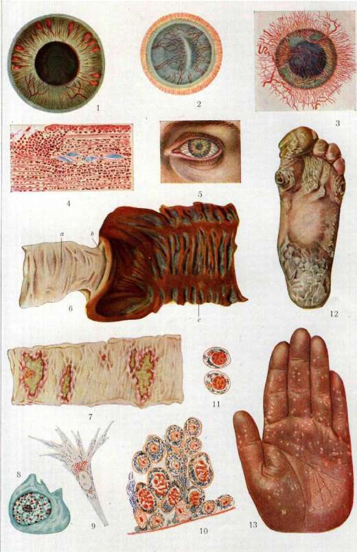
Ei стч fic/rufftiffti, h'rpnmttjfit,. КишечшШ, /{.ичпм,
In the regressive period, the opacities begin to resolve, first at the periphery; the central parts of the cornea clear last. Deviation from the typical forms of parenchymatous keratitis consists in the fact that infiltration appears not at the periphery but in the center of the cornea, and from there the process spreads to the periphery, in severe cases involving the entire cornea. In rare cases, the opacity is limited to only the central parts of the cornea (k. parenchymatosa centralis). Furthermore, parenchymatous keratitis may sometimes manifest in the form of separate, deeply situated point-like infiltrates arranged in a ring-shaped opacity in the center of the cornea (k. annularis Vnssius'a). A rare exception are forms of parenchymatous keratitis in which vessels do not develop in the cornea (k. parenchymatosa avasculosa). Such atypical forms are more often encountered in patients of older age (over 20 years) and mostly in connection with acquired syphilis, and they run with mild signs of irritation. In tuberculous parenchymatous keratitis, there is a tendency for the process to produce mainly nodular infiltrates in the form of separate nodes among diffusely clouded cornea, and the process usually does not involve the entire cornea, but it must still be said that in a number of described cases of tuberculous parenchymatous keratitis, their picture was identical to the typical syphilitic parenchymatous keratitis. All forms of parenchymatous keratitis in a large percentage of cases are accompanied by inflammation of the iris (according to Igersheimer in 50% of cases). Anterior chorioretinitis (chorio-retinitis anterior) is also often encountered in parenchymatous keratitis; these lesions of the vascular tract are considered as two processes independent of each other but arising from the same cause - syphilis. The course of parenchymatous keratitis is prolonged, and in it two stages must be distinguished: the progressive stage, lasting several weeks (4-8), and the regressive stage, usually very prolonged (several months and even up to a year). The favorite age for the appearance of parenchymatous keratitis is 6-20 years (most often 7-14); but keratitis can manifest itself in the first years of life and even in the intrauterine period, as well as in later years, and exclusively rarely after 35-40 years. In later age periods, parenchymatous keratitis is usually caused by acquired syphilis. Parenchymatous keratitis affects both eyes, but usually not simultaneously. Recurrences of parenchymatous keratitis are sometimes observed (14-17%). For tuberculous parenchymatous keratitis, unilateral involvement is noted. Diagnosis of parenchymatous keratitis. The clinical picture of parenchymatous keratitis in typical cases is so characteristic that the diagnosis is not difficult; sometimes the etiological diagnosis is difficult, although in the vast majority of cases with the usual form of parenchymatous keratitis, there are signs of congenital syphilis, characteristic changes of the skull, etc., and especially valuable is the RW reaction, which is observed with great constancy in parenchymatous keratitis (up to 90%, according to Igersheimer and others). A negative RW, positive to tuberculin, and especially with a positive focal reaction to tuberculin, gives the right to establish a diagnosis of tuberculous parenchymatous keratitis. But the most difficult and sometimes insoluble cases are those of parenchymatous keratitis in the presence of both syphilis and tbc; here the clinical picture as well as the results of combined therapy should be taken into particular consideration. The prognosis in parenchymatous keratitis depends on the intensity of the process and in general should be serious, since parenchymatous keratitis, despite treatment, leaves behind scar opacity, reducing vision in almost 40% to significant limits (below 0.2) and in rare cases even to blindness. In prognosis, it must be taken into account that final conclusions about the state of vision can be made only after a long time (not less than a year) after the inflammation has subsided. Prevention of syphilitic parenchymatous keratitis can be achieved by applying early specific treatment to children with congenital syphilis. Treatment of parenchymatous keratitis must be both general and local. General, depending on etiology, and therefore most often antisyphilitic, is indicated in the usual forms; in tuberculous parenchymatous keratitis, tuberculin therapy has been used with success. In mixed infection (lues + tbc), combined therapy is recommended. Local therapy is symptomatic - mydriatic agents (atropine, scopolamine, etc.); in view of the involvement of the iris - heat in various forms; in the regressive period - resorbent agents (dionin, yellow mercury ointment), vibrational massage. Close to parenchymatous keratitis and also arising on the basis of syphilis, but acquired, is syphilitic point deep keratitis of Mauthner (k. punctata syphilitica Mauthner's). It is characterized by the formation in the deep layers of the cornea of grayish plaques, which usually develop rapidly but can also disappear just as rapidly, especially under the influence of specific treatment. Sclerosing keratitis (k. scleroticans, s. sclerosificans) also belongs to deep non-purulent keratitis, but unlike typical parenchymatous keratitis, this keratitis is almost always a secondary process due to the spread of inflammation to the cornea from the sclera, sometimes from the anterior chamber, iris, or ciliary body. The cause of this keratitis is predominantly tbc, much more rarely - syphilis, gout, rheumatism. The process usually begins with existing scleritis or the presence of tuberculous nodules in the corneo-scleral area or in the iris, with the appearance near the edge of the cornea of deep opacities in the form of tongues, which soon take on a saturated white color, similar to the color of the sclera (hence the name). The keratitis spreads slowly, sometimes in separate small foci, sometimes, with the fusion of the latter, in the form of large infiltrates, but still the opacity as a rule does not involve the entire cornea. The epithelium over the area of infiltration changes just as in parenchymatous keratitis; just as in the latter, vascularization of the cornea is observed, but less pronounced. The formed infiltrates in their reverse development do not resolve but are replaced by persistent connective tissue, giving permanent scar opacities. The diagnosis is based on the clinical picture, etiology is established by examination for tbc. The prognosis is unfavorable. Treatment is the same as for parenchymatous keratitis. To deep keratitis belongs the rarely occurring disciform keratitis (k. disciformis), which owes its name to the opacity in the form of a disk, arising in the deep layers of the cornea, in its center, and this opacity is particularly dense in the center of the disk. A characteristic feature is the reduced sensitivity of the cornea in the area of infiltration. The causes are unknown; some connect them with trauma. The next group of keratitis are transitional forms from non-purulent to purulent keratitis. These include the important in practical respect inflammations of the cornea in the so-called scrofula in children. Clinically, scrofulous keratitis manifests in 3 forms: 1) phlyctena of the cornea (phlyctaena corneae); 2) fascicular keratitis (k. fascicularis) and 3) scrofulous pannus (pannus scrophulosus). All forms of scrofulous keratitis are characterized by the fact that they are observed along with other scrofulous lesions of the eyes. Phlyctenae of the cornea represent separate superficial infiltrates in the form of small nodules, usually located near the periphery of the cornea, often at the limbus itself, in their character and origin analogous to phlyctenae of the conjunctiva, with which they appear simultaneously. Phlyctenae sometimes later resolve, but more often they break down into ulcers, leaving scar opacities of the cornea (macula corneae). In the process of regression, the formation of new vessels approaching the infiltrate is often noted. Fascicular keratitis is expressed by the appearance (also near the periphery) of an infiltrate similar to a phlyctena, breaking down into an ulcer, to which vessels approach in the form of a bundle, and the ulcer, having an edge toward the center infiltrated and toward the periphery cleared, can move toward the center; the outcome of this process is an opacity of the cornea in the form of a triangle - traces of the former bundle of vessels and infiltrate. Scrofulous pannus is characterized by the development in the cornea of superficial vessels approaching from the limbus to multiple infiltrates of the cornea, scattered mainly at its periphery; in severe cases, the entire cornea can be occupied by vessels and many infiltrates-phlyctenae (see separate table, fig. 5). Scrofulous keratitis as a rule are accompanied by severe signs of irritation (photophobia, lacrimation, blepharospasm). For all of them, a prolonged course and a pronounced tendency to recurrences are characteristic. The diagnosis of scrofulous keratitis, in addition to the usually typical local symptoms, is confirmed by the presence of general signs of scrofula and other eye diseases on the basis of scrofula. The prognosis is in general favorable; scrofulous keratitis, despite the duration of the course, leave superficial opacities of the corneal membrane, usually only at the periphery, not causing significant disturbance of vision. Treatment first of all against scrofula (general strengthening, dietary, climatic, light therapy, etc.). Locally - yellow mercury ointment (1-2%), calomel, with hyperemia of the iris - atropine, in its absence - pilocarpin. Purulent keratitis.
Among the individual forms of purulent keratitis, corneal ulcers should be distinguished, and among these—the most important in practical terms—the creeping ulcer (ulcus corneae serpens). The cause of this ulcer is in most cases the pneumococcus, less frequently the streptococcus, and the diplobacillus of Morax-Axenfeld (Diplobac. Morax-Axenfeld). The onset of the ulcer is very often preceded by trauma, sometimes insignificant, not always easily established in the medical history. The traumatic origin of the ulcer explains why it occurs in persons of various occupational categories as a professional eye disease. A contributing factor to the development of pneumococcal infection is dacryocystitis, observed in 50-60% of cases of ulcus corneae serpens. In childhood, the creeping corneal ulcer occurs rarely. The pathological anatomy of the creeping ulcer has been described above; it should be noted that the first detailed studies of the creeping ulcer were given as early as 1896 by Uhthoff and Axenfeld. Arising from a small infiltration consisting of pus cells and rapidly breaking down, the ulcer from the very beginning is characterized by the following features: one of its edges appears strongly infiltrated in the form of a ridge, and the infiltration extends into the healthy corneal tissue, while on the opposite edge of the ulcer early signs of regression are noticeable: the epithelium, multiplying, covers the edge of the ulcer, descends onto its bottom, which is freed from infiltration (Fig. 3). The advancing edge subsequently appears undermined due to necrosis and softening of the tissue. On microscopic examination, a mass of pneumococci or other causative agent is found in the sections of the infiltration of the advancing edge and between the layers of the still unchanged cornea (see separate table, Fig. 4). In the anterior chamber, a collection of purulent elements is found in its angle (hypopyon); also on the posterior surface of the cornea in the area of the ulcer, a collection of pus cells is observed. The endothelium of Descemet's membrane undergoes changes—it becomes necrotic and falls off. The course and symptomatology of the creeping ulcer. At the very beginning, a grayish-yellow infiltration appears, usually in the center of the cornea—the area most susceptible to external influences; it rapidly breaks down into a superficial ulcer, taking on a characteristic appearance: one edge (the advancing one) is of a saturated yellowish-white color, while the bottom and the other edge are translucent. Early, a frequent companion of the creeping ulcer appears—the hypopyon, due to which the process is called keratitis-hypopyon (Fig. 6). The hypopyon is associated with the developing iritis from toxic influences—it is sterile. Rarely does the ulcer remain limited to the involvement of the central parts of the cornea; in most cases, it creeps along the surface with its advancing edge, sometimes destroying the cornea throughout its entire length; in a number of cases, the ulcer progresses into the depth of the cornea, causing destruction of the cornea through its entire thickness with all the consequences. It is necessary to note the clinically established fact that after the perforation of the cornea
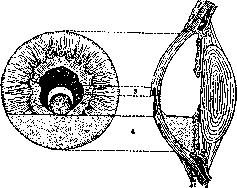
Figure 6. Creeping corneal ulcer: 1—conjunctiva; 2—sclera; 3—advancing edge; 4—hypopyon.
the infectious process in the membrane often stops and scarring occurs. Diagnosis is etiologically established based on bacteriological examination of the infiltrate from the progressive edge of the ulcer; here the causative agent is found, as in a pure culture. The prognosis is always serious: the ulcer often leads to complete blindness. Prevention of this ulcer as a professional disease consists of protecting the eyes from damage; a more specific prevention is the timely treatment of dacryocystitis in persons who, due to their professional activities, are at risk of eye damage. Recently, after any injury, no matter how small, optochin (Optochinum hydrochloricum) has been recommended for prevention. Treatment is directed toward destroying the infectious focus, and here thermocauterization is successfully applied, especially with small ulcers. Chemical cauterization (Tinctura Iodi) and cauterization with zinc sulfate (according to Fregger) have less importance. Further, various bactericidal agents (collargol, xeroform, etc.) are used, and against pneumococcus, optochin is widely used in the form of drops (1-2% solution), ointment, or in the form of applying a tampon to the ulcer for 2 minutes; some introduce optochin by iontophoresis. In diplobacillary ulcers, treatment with zinc sulfate may be effective. Serum treatment has not yet found wide application, as there are not yet sufficiently effective antipneumococcal sera. Among surgical methods, paracentesis of the cornea is used, indicated in hypopyon, especially of significant size; in addition to draining the pus, paracentesis is beneficial because it enhances the eye's fight against the infection. In advanced cases, when the ulcer threatens to perforate, Saemisch's operation is used, consisting of an incision of the cornea over the entire length of the ulcer, as splitting the ulcer often leads to a halt in the process of corneal destruction. Recently, Sondermann proposed instead of Saemisch's operation to perform trepanation of the ulcer, considering that such operation is subsequently accompanied by fewer complications (Weinshock). As an emergency operation in ulceration, the lacrimal sac should be removed if it has purulent inflammation. In addition to these measures, favorable results are obtained in the treatment of creeping ulcers with light; for this purpose, the lamp of Birch-Hirschfeld is particularly used. Among other ulcers, typical from a clinical standpoint are catarrhal ulcers (ulcus catarrhale). They form from the breakdown of marginal infiltrates observed in chronic and acute catarrhs of the conjunctiva. These ulcers appear as arcuate defects in the periphery of the cornea with indistinctly infiltrated bases, usually separated from the edge of the cornea by a strip of normal tissue. Their cause is in some cases infection with Morax-Axenfeld's diplococcus, sometimes with pneumococcus and others, sometimes bacteriological research yields no result. Catarrhal ulcers are more common in elderly people; recurrences occur, sometimes associated with the time of year (autumn, spring). The prognosis is favorable; under the influence of symptomatic treatment, the process resolves in a short time. Infinitely more dangerous purulent keratitis arise on the basis of acute infectious conjunctivitis as secondary purulent keratitis, most often in the form of diffuse keratitis (k. purulenta diffusa); such are the keratitis in gonorrhea of newborns and adults, in diphtheria of the conjunctiva. Among rare forms of purulent keratitis, mycotic mold keratitis (k. mycotica aspergillina) should be noted, observed due to infection with mold fungi and primarily from infection with Aspergillus fumigatus. The process usually arises after injury in the form of formation of an infiltrate in the center of the cornea with subsequent breakdown into an ulcer; the focus appears dry, granular, yellowish-white in color. It is separated at the height of development from surrounding parts by a demarcation groove, sometimes hypopyon is present. The diagnosis is clarified by bacterioscopic examination: in a smear - the mycelium of the fungus. Treatment is thermocauterization or curettage of the focus. Annular keratitis, or abscess (k. annularis; Ringabscess - according to German authors), belongs to rare infections of the cornea, usually also after trauma or after operations. The causative agents are the bacillus pyocyaneus (Bac. pyocyaneus), pneumococcus, streptococcus, etc. A purulent infiltrate arises in the deep layers of the cornea in the form of a yellowish ring lying in the periphery of the cornea. The process quickly spreads to the middle parts, and the entire cornea is destroyed; sometimes panophthalmitis arises. Among ulcerative forms of keratitis, keratitis in acne rosacea - k. rosacea, observed either simultaneously with the skin disease acne or sometimes long after this lesion, should be further noted. The cause of k. rosacea is not well known, just as the cause of the skin affliction. The process is expressed by the appearance of marginal infiltrates, sometimes superficial, sometimes deep, breaking down into an ulcer with undermined edges; the ulcer is accompanied by vascularization, heals poorly; recurrences often occur; sometimes the ulcer spreads toward the center. K. is often accompanied by the formation in the conjunctiva of peculiar vascular ectasias in the form of plaques. The characteristic feature of the process is its bilateral nature. Treatment is symptomatic; recently, radiotherapy has been successfully applied. Rare and at the same time very characteristic is ulcus corneae rodens, first described in 1867 by Mooren (Mooren). This ulcer has a progressive course, usually arising with sharp phenomena of irritation from an infiltrate in the periphery of the cornea. The ulcer takes a sickle shape and then slowly spreads in spurts along the periphery and toward the center, sometimes involving the entire cornea, while the ulcer has a progressive, more infiltrated, somewhat undermined edge; other parts of the ulcer are vascularized and gradually scar over. There is never perforation of the cornea, nor hypopyon. Sometimes anesthesia or hypesthesia of the cornea is noted. The causes of ulcus corneae rodens are interpreted differently. Bacteriological investigations are unsuccessful; Junius attributes to it a neuropathic origin, Triebenstein disputes the very independence of the process, asserting that this ulcer can develop from keratitis of various etiology under unknown conditions, possibly in trophic disorders. Treatment is symptomatic. Apart from ordinary keratitis, the following forms also stand out: 1. Neuroparalytic keratitis (k. neuroparalytica) develops due to a lesion of the first branch of the trigeminal nerve, its paralysis - spontaneous or after removal of Gasser's ganglion or the introduction of alcohol into it, etc. The pathogenesis is not entirely clear. Since the time of Magendie, this keratitis has been associated with trophic disorders of the cornea in lesions of the trigeminal nerve. Some consider that the cause of keratitis is the traumatization of the cornea due to its anesthesia. However, most consider trauma merely a favorable factor, and in the basis of keratitis they see trophic disorders. Clinically, the process is expressed by the appearance of clouding in the center of the cornea with complete anesthesia of it, desquamation of the epithelium, rapidly spreading over the entire surface of the cornea except for a narrow band along the periphery. Subsequently, the clouding increases, ulcers appear, especially in the center of the cornea, sometimes destroying the cornea throughout its entire thickness; but usually it does not come to extensive ulcers. The prognosis is unfavorable, as therapy is ineffective: persistent cloudings remain. 2. Keratitis from non-closure of the eyelids (k. elaeophthalmos) most often develops in paralysis of the facial nerve, but also in cicatricial ectropion of the eyelids, strong protrusion of the eye, etc. The process begins with the parts of the eye, constantly exposed to the action of air (conjunctiva, cornea), drying out; the secretion of the conjunctiva dries on its surface, and sometimes on the cornea; the epithelium, drying out, breaks down, falls off, erosions appear, most often at the bottom of the cornea, exposed even during sleep. Secondary infections sometimes cause severe purulent keratitis as well. Prevention of such keratitis consists in timely closure of the palpebral fissure by plastic operations in ectropion, suturing of the eyelids in paralysis of the n. facialis, application of ointment dressings, etc. When keratitis develops, the same care for closure of the palpebral fissure is required.
Related articles
Mentioned in
Cite this page
“Keratitis.” Soviet Medical Encyclopedia. English translation of Bolshaya Meditsinskaya Entsiklopediya, 1st ed. (Moscow, 1928–1936), ed. N. A. Semashko. https://sovietmedicalencyclopedia.pages.dev/article/keratitis/