Pertussis
Historical document, translated for reference. It reflects medical knowledge of the 1920s–30s and is not medical advice.
Summary
This article from the 1928–1936 Soviet Great Medical Encyclopedia details the history, etiology, and epidemiology of pertussis (whooping cough). It traces the disease's historical descriptions, the scientific consensus on its bacterial cause (Bordet-Gengou bacillus), and patterns of epidemic spread.
Encyclopedia article (1928–1936)
PERTUSSIS (pertussis, tussis convulsiva), an acute infectious disease characterized by a peculiar spasmodic cough and a cyclical course. The first mention of pertussis dates back to 1578, when an epidemic of pertussis with enormous mortality occurred in Paris and was described by Guillaume de Baillou, dean of the University of Paris. In 1678, pertussis raged in London; a description of this epidemic was made by Willis. Subsequently, there were more frequent descriptions of pertussis epidemics in many countries. Sydenham analyzed the pertussis epidemics of 1670 and 1679 in England. Further, in 1695, pertussis was the cause of increased infant mortality in Paris and Rome. In 1724, pertussis epidemics were observed in England, Austria, and Alsace. In the same year, the very name of the disease "pertussis" appeared for the first time. During the first 30 years of the 18th century (1703–1733), pertussis covered almost all of Europe, having assumed, according to the description by Sticker, the character of a pandemic. From Europe, pertussis also spread to America. Pertussis gained even greater distribution and malignancy in Europe in the second half of the 18th century and the first half of the 19th century. The same picture was observed in America. In the 18th and 19th centuries, pertussis penetrated almost all parts of the world, including tropical countries (the Antilles, Brazil, Argentina, etc.). From the second half of the 19th century, pertussis epidemics in Europe became milder. At the same time, according to data provided by Kölsch, a milder course occurred in places where pertussis had firmly settled; upon its appearance in new places, the severe character of the disease was noted again. At the present time, in many cities, and especially in large ones, constant endemics of pertussis are observed, turning from time to time into epidemics. In villages, on the contrary, as a rule, only epidemics occur. At the same time, individual sporadic cases can generate entire epidemics. Etiology. Since the appearance of the first description of pertussis, i.e., since 1578, various kinds of views regarding its etiology have existed and exist to the present time. According to one view, pertussis is a pure neurosis, independent of infection. Another view admits that various pathogens can cause the picture of typical pertussis if the child's increased nervous excitability contributes to this. Thus, Szontagh still asserts that the bacterial flora in pertussis is inconstant and of secondary importance, and that catarrh of the upper respiratory tract with various bacterial flora in children with a nervous-arthritic diathesis usually proceeds under the guise of pertussis. Both of these views have few supporters at the moment. The prevailing and generally accepted view now is that of pertussis as a specific infectious disease caused by a specific characteristic microbe, discovered in 1900–1909 by the Belgian scientists Bordet and Gengou and named after the authors. It is interesting to note that the view of pertussis as a specific contagious disease existed even in the period of the first historically established appearance of pertussis. In times closer to us, Trousseau especially ardently supported the idea of specificity, proceeding from the epidemic nature of pertussis, its contagiousness, the cyclical nature of its course, the characteristic symptomatology, and in particular the character of the spasmodic coughing fits. Intensive and persistent work to find the pathogen of pertussis was conducted even before the discovery by Bordet and Gengou. Thus, in 1887, Afanasyev isolated a streptobacillus from the bronchial secretion in 20 out of 29 cases of pertussis, which had a spindle-shaped or rod-shaped form and grew on sugar agar at 30°. In young dogs and cats, the introduction of this microbe into the trachea caused pertussis-like attacks, and sometimes pulmonary complications. Leaving aside descriptions of cocci and Protozoa as the pathogen of pertussis, one can note the findings of small rods similar to the influenza bacillus. All these studies helped Bordet and Gengou, who used a more complete methodology and immunity reactions and in 1906–1909, after 6 years of work, achieved the ability to cultivate the pertussis microbe and study it in pure culture experimentally. A whole series of authors confirmed these conclusions. If an examination is performed in the catarrhal period of pertussis and at the beginning of the spasmodic period, it is possible to obtain corresponding positive results in the majority of those examined. In the sputum of children suffering from other diseases, pertussis bacilli are absent. According to Inada's statistics, the pertussis bacillus was found in 78 out of 81 examined pertussis patients, and, conversely, in 18 patients suffering from other diseases, a negative result was obtained. Approximately the same results are obtained in analogous mass examinations. Positive results are also obtained in relation to the immune reaction: the serum of convalescents specifically agglutinated the pertussis bacillus, and when using the complement fixation method, the presence of a specific amboceptor was revealed. Experimental verification gave the same positive results. This verification was widely conducted by V. Klimenko on puppies, kittens, and monkeys. By introducing a culture of the pertussis bacillus into the trachea with a laryngeal syringe, he managed to induce a pertussis-like disease in the animals. Two control monkeys that were in contact with the infected ones also fell ill with an analogous form in Klimenko's experiments. Fraenkel obtained similar results in two monkeys. Arnheim does not consider these experiments entirely conclusive, given that the pertussis bacillus was not isolated from the sick animals. Inada himself performed impeccable experiments on monkeys and, after a 13-day incubation, obtained a full picture of pertussis, which lasted 45 days and had a correctly cyclical character with a 23–25-day spasmodic period. The pertussis bacillus was isolated from the sick animals. Inada's experiments must be recognized as sufficiently convincing. The pertussis bacillus, which is located intracellularly and extracellularly, has rounded edges and stains well with ordinary aniline dyes. With carbol methylene blue, the ends of the rod stain more strongly than the middle for 1/2–2 minutes. The rod does not stain by Gram, does not form spores, and does not have flagella. Under aerobic conditions, it is cultivated on blood agar, better on Bordet-Gengou glycerin-potato blood agar, prepared with the blood of a rabbit, dog, or human; agar with pigeon blood is of little use (Klimenko). On this nutrient medium, after two days at 37°, very small, barely visible, strongly protruding, round, shiny colonies are formed. Upon further cultivation of the pertussis bacillus under laboratory conditions, cultures begin to grow on ascitic and serum media or even on simple agar, which the influenza bacillus does not do. From a culture of the pertussis bacillus, Bordet and Gengou managed to isolate a necrotizing endotoxin, which is easily destroyed under the influence of light and air. Guinea pigs that received 1 1/2–2 mg of a 2–3-day agar culture intraperitoneally died after 1–2 days with the presence of numerous hemorrhages, hyperemia, and strong effusions into the pleural and pericardial cavities. Almost analogous changes are observed with the introduction of killed cultures. The pertussis bacillus is pathogenic for many other animals: mice, puppies, kittens, pigeons, piglets, horses, etc. The virulence of the pertussis bacillus isolated from a pertussis patient varies. A differential diagnosis must be made with the influenza bacillus, which is frequently encountered in the sputum of pertussis patients. In this case, one is guided by the greater monomorphism of the pertussis bacillus, the slowness of growth, the presence of hemolysis, the ability to grow in laboratory conditions on simple agar, and finally, serodiagnosis. The resistance of the pertussis bacillus is extremely low. It dies at 56°. Epidemiology. The pertussis epidemic in cities proceeds in the form of waves, more often of a regular character. At the same time, the state of lull separating two adjacent epidemic waves lasts 1–4 years. This periodicity takes place in all countries (Katanyants, Vladimirov, Baginsky, Dopter, Debre, etc.). The epidemic, after a rapid start, subsides, only to rise again, more often in the form of irregular jumps, which little by little lead to an epidemic state. The spread itself is irregular. The wave engulfs a house, a street, a block, affects neighboring streets and blocks, moves from village to village, and captures entire regions and countries. Individual waves produce an unequal number of diseases, mainly in connection with population density. Statistics from all countries indicate that 80% of pertussis cases fall on the age under 5 years and only 20% fall on older age groups. According to data by Unruch, covering 1,952 cases of pertussis in Dresden, the age curve of pertussis morbidity rises to the 7-year age group and then begins to decline, and at the age of 8, it still remains higher compared to the curve of infancy. Deviations from these provisions depend on the speed of the onset of new epidemics. With a rapid alternation of epidemics, younger ages are affected; with a rarer one, on the contrary, older groups are also involved to a greater extent (Runner, etc.).
As a rule, the greatest susceptibility falls on the age from 2 to 5 years. On the other hand, pertussis has been described in 80-year-old elderly people. Although pertussis is found everywhere, it is encountered less frequently in tropical countries and runs a milder course due to a smaller number of complications from the respiratory tract. Mode of infection. The pertussis bacillus is found on the mucous membrane of the upper respiratory tract, which presents a picture of a local inflammatory process in the form of superficial necrotic changes. These changes, as shown experimentally, are caused by the endotoxin of the pertussis bacillus. Due to the lack of generalization, the pertussis bacillus is found only in the sputum of the patient. The bacillus enters the air from the respiratory tract. In view of the extremely low resistance of the pertussis bacillus, as a rule, a direct method of transmission of pertussis from a patient to a healthy person takes place. The role of transmission by a third party, as well as through items of care and daily use, if it occurs at all, is practically extremely negligible, and the descriptions of old authors about such transmission of pertussis are explained, perhaps, by infection with the help of unclear and therefore overlooked forms of pertussis (formes frustes). Theoretically, of course, it is possible to admit that in exceptionally rare cases, in the presence of a non-dried virus, such an infection can occur. There are also no sufficient data on the transmission of pertussis through animals, despite positive experiments on infecting the latter. Even if such a possibility were confirmed, then even then, no practical significance could be attributed to this method. Thus, the direct method of transmission of pertussis through patients remains. This method of transmission has also been confirmed bacteriologically by examining sputum after a child's cough over a Petri dish with a corresponding medium. At the same time, it was possible to clarify that the radius of virus dispersion and the phase of the active state of the latter are small. This is also in agreement with old epidemiological observations. Filatov also noted that pertussis infection rarely spreads in hospitals through a corridor and from ward to ward. Old disputes about the periods of the contagious state of a pertussis patient have also received clarification in modern times from the bacteriological method. It is considered established that a pertussis patient is contagious in the catarrhal period and in the initial stage of the spasmodic period. According to Danish data, the pertussis bacillus can be isolated in the catarrhal period in all patients, at the beginning of the spasmodic period also in almost all, during the first two weeks of the spasmodic period in 2/3, in the 3rd week in 1/3, and later only as a rare exception; after five weeks, the pertussis bacillus is not detected at all. At the present time, a number of authors (Weil and others) consider a child from the 3rd week of spasmodic cough to be non-contagious. Dopter considers a child non-contagious after 25 days. In view of the detection of the pertussis bacillus in patients in the 3rd and even in rare cases in the 4th week, it is more correct to consider a child non-contagious no earlier than after 4 weeks from the beginning of the spasmodic attacks. Weil's assertion that the pertussis bacilli found in the 3rd–4th week are non-virulent requires confirmation. In addition to patients with the typical form of pertussis, cases that run atypically and are therefore very often not diagnosed (formes frustes) play an extremely large role in its spread. With the discovery of the pathogen, these forms received their explanation. Delcourt in Brussels, during school epidemics, found a positive complement fixation reaction in many schoolchildren without the presence of a clinical picture of pertussis. Madsen and Barbier found the pertussis bacillus in children who were not sick with pertussis and were together with pertussis patients. The significance of formes frustes in the epidemiology of pertussis has been definitively established by bacteriological means. Thus, Delcourt managed to establish the dispersion of the pertussis bacillus through a number of schools by a singing teacher who showed a positive complement fixation reaction upon examination. The question of the transmission of pertussis through healthy bacillus carriers should for now be considered open. Individual literary references to carriers of the pertussis bacillus appear even now. Attempts to use the skin reaction for sensitivity to pertussis may, in case of success, play a large role in epidemiology and prevention. In those who have had pertussis, immunity is obtained for life. Cases of repeated pertussis infections are casuistic in nature and, moreover, not absolutely reliable. M. Danilevich. Statistics and geographical distribution. 1. Mortality. Pertussis is one of the most common and most severe childhood infections, claiming many tens of thousands of victims annually. During the first decade of the 20th century, no less than 662,000 children died in Europe, not counting Russia and the Balkans. Table 1 provides the absolute numbers of those who died from pertussis in some European countries for pre-war and recent years. Table 1. Number of those who died from pertussis in some European countries for 1911-13 and 1925-27. Countries 1911 1912 1913 1925 1926 1927 England 7,844 8,407 5,458 6,058 5,884 3,681 Belgium 2,085 1,923 1,867 1,300 1,271 863 Germany 11,990 13,669 11,632 6,299 4,066 4,202 Spain 2,549 2,492 2,932 1,766 1,410 940 Italy 6,833 6,721 4,882 262 218 201 France 2,915 2,363 3,112 1,737 506 850 Switzerland 394 446 550 827 Scotland 2,549 2,492 2,932. Table 2 provides mortality rates from pertussis (on average per year per 100,000 population) for some European countries, as well as for the USA. Table 2. Mortality from pertussis (on average per year per 100,000 population). Countries 1901-05 1906-10 1911-15 1916-20 1921-25 1924-27 England 51.6 38 32.9 20.0 14.2 11.6** Belgium 47.6 52 24.6 26.3 11.5 8.5** Germany 29.6 38 18.8 19.3 10.4 Holland 70.2 29 16.8 16.3 8.5 Italy 45.9 24 19.3 17.6 12.0 Spain 65.5 18 11.6 13.3 8.8 7.1 Switzerland 35.3 18 17.3 10.2 10.0 7.3** USA 24.3 52 14.6 12.0 9.0 7.8 Sweden 18.8 25.3 39.4 12.0 5.1 9.4 Scotland 59.8 34.0 21.2 14.4 16.4 12.7 11.2 11.1 11.1 12.6 35.8 6.4***. In the 20th century, mortality from pertussis is lower than in the last decades of the 19th century. In all countries, a decrease is noted after the World War. Mortality from pertussis in individual countries in recent years is provided in Table 3. Table 3. Mortality from pertussis in some countries for 1923-28 (per 100,000 population). Countries 1923 1924 1925 1926 1927 1928 Austria 5.8 6.7 6.5 10.5 10.5 5.9 England 15.6 4.8 9.0 10.0 5.7 4.0 Belgium 17.0 20.2 5.5 10.4 6.0 10.0 Bulgaria 10.7 9.4 10.9 5.5 4.8 7.0 Hungary 8.2 10.0 8.9 4.2 6.1 11.3 Germany 10.0 5.1 17.3 6.3 7.4 8.2 Holland 7.3 7.5 7.9 10.3 5.4 Denmark 6.2 21.9 Spain 12.0 Romania 6.5 Czechoslovakia 7.3 Switzerland 12.5 Sweden 6.7 Scotland 5.0 USA 35.5. Unlike measles (see), there are no sharp fluctuations in the level of mortality between individual countries. The highest mortality rates are noted for all years in Scotland. Table 4 gives an idea of the level of mortality from pertussis in Russian cities. The figures in this table speak of a tendency towards a decrease in mortality from pertussis. Table 4. Mortality from pertussis in Moscow, Leningrad, and Odessa for 1881-1928 (on average per year per 100,000 population). Years Moscow Leningrad Odessa 1881-85 26.7 21.0 1886-90 19.7 20.8 1891-95 17.3 25.2 1896-1900 15.0 20.0 1901-05 19.0 21.0 17.6 1906-10 17.9 24.0 9.3 1911-13 19.1 11.6 10.2 1914-18 13.3 14.8 9.9 1919-23 10.8 13.4 10.5 1924-28 9.8 13.2 10.7. Table 5. Mortality from pertussis in Moscow per 100,000 children under 14 years of age. Years Mortality Years Mortality 1892-1901 82.9 1917 17.9 1902-1905 92.5 1918 21.3 1906-1909 85.3 1920 36.1 1910-1913 79.6 1923 47.1 1926 34.5. To compare mortality from pertussis in Russian cities with that in large cities of Western Europe, Table 6 provides mortality rates from pertussis over a long period of years in Hamburg, London, and Paris. Figure 1. Mortality from pertussis in England from 1861 to 1927 (on average per year per 100,000 population). Table 6. Mortality from pertussis in Hamburg, London, and Paris (on average per year per 100,000 population). Years Hamburg London Paris 1831-40 34.1 41.6 1851-60 35.2 45.6 1871-80 46.4 1881-90 39.3 1906-10 17.0 1910-13 15.5 22.2 9.3 1925-28 13.9 11.1 6.9.
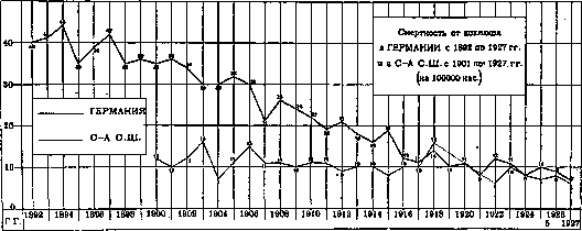
Figure. When calculating mortality from pertussis in relation to the child population (aged up to 14 years), we obtain the following indicators (Table 5). Consequently, in recent years, we have had an actual decrease in mortality in relation to the child population. Mortality from pertussis in Moscow and Leningrad over the last 20 years of the 19th century was recorded as lower than in Hamburg and London; in the 20th century, it has remained at approximately the same level as in those cities (a sharp decline in recent years is noted in Hamburg); mortality in Odessa remains at approximately the same level as in Paris. Over the 70 years of the 19th century (for London, over 60 years), mortality from pertussis in these cities changed little; the decline began only in the 20th century. Figures 1-3 show the trend of mortality from pertussis in England from 1861-1927, in Germany from 1892 to 1927, and in the USA from 1901. By city, it shows very large fluctuations; mortality from pertussis in most cities remains at approximately the same height. The highest mortality from it in recent years is noted in Belfast, Birmingham, Glasgow, and Liverpool, i.e., in the cities of Great Britain. It is also high in Le
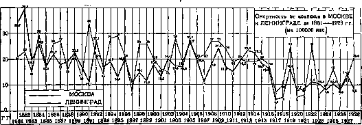
Figure 3. 1927, and for Moscow and Leningrad from 1881-1928. The curves provide an idea of both the decrease in mortality from pertussis and the periodic fluctuations of pertussis epidemics. The periodicity of epidemics is more pronounced in large cities (Moscow, Leningrad) than in entire countries. They usually repeat every 2-3 years. Epidemic waves are not as pronounced in Leningrad, and are significantly lower in Moscow and Odessa. As for non-European cities, high mortality rates from pertussis are noted in almost all cities of Japan, in some cities of North America (Mexico), and South America (Guayaquil, Quito, etc.). Cities in Australia show low mortality rates from pertussis.
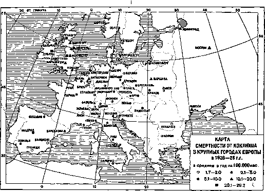
Figure 4.
- Johannesburg .... I
- Rio de Janeiro . . . I
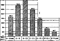
Table 8.
Age 0-1 yr. . . . 1-4 » . 5 years and older Total 65.4 32.6 2.0 100.0 51.2 47.0 1.8 57.6 40.7 1.7 63.7 35.0 1.3 75.5 22.9 1.4 100.0 Figure 5. Table 10. Mortality from pertussis by age groups in Paris in 1904-13 (per 100,000 children of corresponding age). Age Died per 100,000 children Age Died per 100,000 children 0-3 months . . . 166.9 4-5 years . . . . 39.7 3-6 » . . . 293.6 0-1 yr . . . . . . 283.5 6-12 » . . . 352.3 0-3 » . . . . . 226.4 1-2 yr . . . . 244.9 3-5 years . . . . 53.3 2-3 » . . . 139.0 0-5 » . . . . 158.2 3-4 » . . . . 66.7 Died per 100,000 children of corresponding age in 1926 in Moscow and Leningrad (Table 9): Table 11. Mortality from childhood infections in various countries in 1925-26 (per 1,000 born in these years). Countries Pertussis Measles Scarlet Fever Diphtheria 1925 1926 1925 1926 1925 1926 1925 1926 England . . . 3.8 2.8 1.6 1.2 0.04 0.03 0.17 0.18 Belgium . . . 5.8 - 3.3 - 0.6 - 0.7 - Hungary . . . - 1.1 - 0.3 - 0.1 - 0.3 Germany . . . 3.5 3.4 2.0 1.3 0.06 0.04 0.4 0.3 Holland . . 1.6 2.9 0.9 - 0.03 0.02 0.08 0.07 Denmark . . 4.0 5.5 0.5 0.7 - 0.01 0.08 0.2 Irish Free State . . . 3.3 1.8 1.6 0.5 0.03 0.03 0.13 0.25 Northern Ireland . . . 4.8 2.0 1.6 2.6 0.1 0.1 0.1 0.1 France . . . 1.6 1.2 1.0 1.2 0.04 0.05 0.27 0.29 Sweden* . . . 3.9 2.5 0.3 1.4 0.09 0.04 0.07 0.15 Scotland . . 7.6 2.4 1.4 2.4 0.1 0.2 0.5 0.5 Canada . . . - 3.3 - 1.1 - 0.1 - 0.2 USA . . . . 1.8 - 0.3 0.1 0.1 0.3 0.3 New Zealand . . . 0.3 2.4 0.1 0.07 - 0.07 0.25 * 1923-24
Pertussis claims a large number of victims even among children of the earliest age in the first months of their life. The highest mortality of children from pertussis is observed in the second half of their first year of life. In this, pertussis differs from measles, in which mortality in the first year is relatively low. Of all infectious diseases of early childhood, pertussis claims the largest number of victims among infants. Table 11 shows mortality rates in the 1st year of life from 4 infections: pertussis, measles, scarlet fever, and diphtheria. Mortality from each infection is calculated per 1,000 live births in the same year. Characteristic of pertussis is unequal mortality by sex: more girls die than boys. Table 12 shows the corresponding mortality rates for Prussia in 1896-1900 and Germany in 1926-27. Pertussis Table 12. Mortality from pertussis by age and sex (per 100,000 children of the corresponding group). Table 16. Age Prussia 1896-1900 Germany 1926 Boys Girls Boys Girls Boys Girls 0-1 yr. 1-2 » 2-3 » 3-5 yrs. 923 375 117 45 27.4 32 For every 100 boys who died from pertussis, there are girls: Table 13. Cities and Countries Years Number of deaths Budapest Vienna . . . Paris . . New York Germany Australia 1886-1905 1886-1899 1886-1905 1886-1904 1925-1927 1901-1905
3. Lethality. It is difficult to calculate lethality for pertussis, because far from all patients are registered. If we proceed from the figures of registered patients and deaths, then lethality (per 100 cases) in some countries will give the following figures (Table 14).
Table 14. Countries 1923 1924 1925 1926 1927 Denmark Norway Switzerland 2.6 1.8 13.3 2.5 1.4 7.3 1.9 1.2 8.7 1.8 1.0 9.9 1.8 10.4
Countries 1925 1926 1927 1928 USSR . . . . . . . . 25.1 29.5 29.6 28.9 Bulgaria . 15.3 10.0 10.6 13.8 Hungary . 6.2 6.3 5.3 11.6 Denmark . . 60.4 88.2 52.3 55.9 Norway . 43.3 46.3 - 46.0 Poland . . 3.8 2.3 3.3 4.0 Romania 10.2 6.6 8.7 16.9 Switzerland 7.6 5.6 4.9 6.9

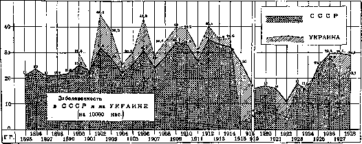
The highest morbidity rates for pertussis are noted in Denmark and Norway, which is possibly explained to a certain extent by more complete registration of patients in these countries. Table 17 presents morbidity rates for pertussis in the USSR (Russia) from 1896 and for Ukraine from 1901. Table 17. Number of pertussis patients registered in the USSR and Ukraine (average per year per 10,000 population). Years Ukraine 1896-1900 . . . 21.9 1901-05 . . . . 23.0 32.1 1906-10 . . . . 30.4 37.1 1911-13 . . . . 31.5 35.3 1914-16 . . . . 25.3 14.3 1919-23 . . . . 15.6 8.5 1924-28 . . . . 25.4 22.9
Figure 6 shows the morbidity of pertussis in the USSR and Ukraine by individual years. The curves provide an idea of the periodic fluctuations in pertussis morbidity. The low morbidity figures during the war years are likely explained both by the incompleteness of registration and by the decrease in the number of the child population. Morbidity by individual republics of the USSR is given in Table 18 (per 10,000 population).
Table 15. Republics of 1927. These figures, of course, provide only the most approximate idea of the mortality from pertussis. Hospital statistics do not provide a clear idea of it either, since only the most severe cases end up in hospitals. In German hospitals, the case fatality rate for 1889–1897 was about 12%. In the Kiel Polyclinic (Germany) for 1884–1895, the following number of pertussis patients were treated: 1,080 boys and 1,219 girls; per 100 patients, 5.9 boys, 5.0 girls, and 5.4 of both sexes died. 4. Morbidity. The number of pertussis patients registered in individual countries is shown in Table 15. Table 15. Number of pertussis patients registered in individual countries in 1920–1928 (absolute figures). [Table data omitted]. Morbidity (number of registered pertussis patients) per 10,000 population for some countries for 1925–1928 is shown in Table 16.
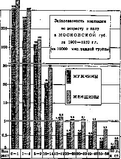
5. The morbidity of pertussis by age and sex in the Moscow Governorate and the city of Moscow is shown in Table 19. Table 19. Morbidity of pertussis by age and sex in the Moscow Governorate and the city of Moscow (per 10,000 population of the corresponding group). [Table data omitted]. The morbidity of women is higher than that of men in all age groups. Children aged up to 1 year and from 1 to 4 years get sick with pertussis most often. After 15 years of age, pertussis is observed in isolated cases (Figure 7). In individual cases, pertussis is encountered even in the first days of a child's life. Feer (Basel) had a patient who contracted pertussis at the age of 3 days; he died at 6 weeks of age.
6. Pertussis and social conditions. Mortality from pertussis is significantly higher among the poor segments of the population than among the affluent. A lack of air and light in living quarters, dampness, and nutritional deficiencies—all of this increases the mortality rate from pertussis. In large cities, in poor neighborhoods with overcrowded apartments, mortality from pertussis is higher than in affluent neighborhoods. For corresponding figures, see Measles.
7. Seasonality of pertussis morbidity. Figure 8 shows the monthly distribution of pertussis in Denmark and Hungary for 1925–1927 and in the USSR and Ukraine for 1926–1928. In the USSR and Ukraine, maximum morbidity is observed from June to September. In Denmark, the June maximum was observed in 1926; in 1925, the maximum fell in March; in 1927, in January. In Hungary, two maximums are noted—in January and in the summer months. I. Dobreytser.
Pathological anatomy. Anatomically, in pertussis, there is a widespread catarrh of the mucous membrane of the respiratory tract from the nose and pharynx down to the bronchi, with the posterior part of the glottis and the region of the tracheal bifurcation being the most intensely affected, i.e., the places which, according to Kohts, represent sections of the mucous membrane that trigger the cough reflex. In 2/3 and more of cases, catarrhal inflammation of the bronchi and bronchopneumonic foci of various sizes are found during autopsy, in individual cases accompanied by pleurisy (fibrinous and exudative). Very characteristic of pertussis lesions is the presence of peribronchitis, which in its picture almost completely resembles that of measles. Dilatations of the small bronchi and bronchioles should be classified as not infrequent changes. In prolonged cases, obliterating bronchiolitis has sometimes been observed. Emphysema of the mediastinum with subcutaneous emphysema is rarely observed. Findings of various tuberculous changes are quite frequent. On the part of the heart, dilatation of the right ventricle is found, sometimes together with hypertrophy of its muscle. In the pathologico-anatomical picture of pertussis, air embolism, arising on the basis of ruptures of the pulmonary alveoli during coughing fits, occupies a prominent place. This embolism can apparently be considered the primary cause of so-called pertussis eclampsia (Neuburger). In some cases, cerebral edema and phenomena taken for simple meningitis occur.
Clinical picture. The duration of the incubation period is 4–11 days. The course of pertussis.
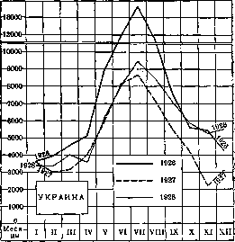
Figure 7. Figure 6. Figure 8. Monthly distribution of pertussis morbidity.
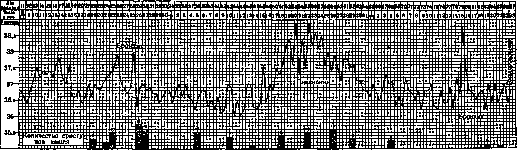
Hemorrhages in internal organs, and especially in the brain, occupy a significant place. These hemorrhages may be related to venous congestion in the systemic circulation, which in turn is associated with coughing fits. In the brain substance itself, the hemorrhages are multiple and small in nature; in the meninges, they are more extensive. Hemorrhages are most often localized in the white matter of the brain. There are indications that in the pathogenesis of cerebral phenomena observed in Pertussis (degeneration of nerve cells, proliferation of glia), the greatest significance can be divided into three periods: the initial-catarrhal, the period of spasmodic (convulsive) cough, and the period of resolution. The first stage, catarrhal, differs little from an ordinary catarrh of the upper respiratory tract. There is a dry cough, sometimes a runny nose, often accompanied by sneezing. In some cases, phenomena of acute laryngitis are noted, which may take on the character of false croup. In this first stage of the disease, the child is capricious, loses appetite, and has a fever. The temperature has an irregular character but can also be normal. Upon examination, there is a slight catarrhal process in the upper respiratory tract; dry rales are heard in the lungs. The cough is distinguished by its persistence, does not yield to remedies usually used in such cases, continues to intensify, and, especially at night, begins to change, gradually acquiring the character of distinct fits, ending with the expulsion (even in small children) of viscous sputum, and sometimes also vomiting. Soon the cough takes on a definitely convulsive character, and the patient enters the second, spasmodic period of the disease. By this time, the temperature stabilizes at normal figures. The first stage lasts on average about two weeks; in infants, it is less prolonged. In the spasmodic period, the fits become more frequent and take on a typical character. The fits are often preceded by various phenomena in the form of tickling in the throat, sneezing, a painful sensation in the chest, and general malaise. The child often anticipates the approach of a fit—stops playing, hides in the mother's dress, runs into a corner of the room, etc. The fit consists of rapidly following cough impulses, which from time to time are interrupted by a characteristic whistling, labored inspiration. Such alternation is repeated 3-6 times within 1-3 minutes, after which the fit ends with the expulsion of thick mucus, clear or mixed with blood, and often with vomiting of food masses. Sometimes, after a short break, a second, shorter fit is observed, which may be followed by a third. After this, the child quickly calms down and, at night, soon falls asleep. Some children, especially small ones, on the contrary, lie in a state of exhaustion after the fit. During the paroxysm, the child makes a heavy impression. Due to the narrowing of the glottis during the fit, air penetrates the chest with difficulty, as a result of which an asphyctic state is created. In some cases, it comes to incontinence of urine and feces, and prolapse of the rectum. The puffy face of the child turns red, blue, the cutaneous veins swell, the visible mucous membranes and, especially, the protruded tongue become cyanotic; abundant secretion of tears appears, bleeding from the nose, and in rare cases from the ear canal due to rupture of the eardrum, ecchymoses in the conjunctiva and subcutaneous tissue. In the intervals between fits, there may be no special pathological phenomena. The patient does not cough and breathes calmly. Only the puffiness of the face remains, and especially the swelling of the eyelids, dilation of veins around the eyes, ecchymoses, and a small ulcer on the frenulum of the tongue, formed as a result of the scratching of the lower part of the tongue, protruded during the fit, against the lower incisors. Naturally, such an ulcer does not occur in children who do not have teeth. In the lungs, phenomena of emphysema and a small number of rales may be noted. The pulse during the fit is significantly accelerated; in the intervals between fits, it is slightly accelerated. A predominance of the tone of the parasympathetic nervous system is noted [increase in the Aschner phenomenon, lively reaction to pilocarpine and atropine (Koltypin)]. Regarding the blood, general leukocytosis is observed with a simultaneous relative lymphocytosis, with a shift to myelocytes or to young forms; in many cases, Türk's irritation cells also appear in the blood. The urine is reduced in quantity, has an acid reaction, and contains a large amount of uric acid. In some cases, sugar is found in the urine, and the test with alimentary glycosuria is positive. The duration of the spasmodic period is from 2 weeks to 2 months or more, on average 4-6 weeks. At the end of the spasmodic period, the strength and frequency of the fits decrease, they become less agonizing, and the whistling inspirations shorten and weaken. The sputum becomes thick, green, and then ceases to be excreted at the end of the fit. The usual cough is mixed into the coughing fits themselves. Eventually, the disease imperceptibly passes into the stage of resolution, lasting 2-4 weeks or more. It is difficult to establish the clinically exact moment of the end of Pertussis. Fits can be observed every day; they can appear again after disappearing under the influence of physical, mental, or pathological causes after a long period (runny nose, bronchitis, etc.). The cause of such 'tails' is the tendency to cough in fits left to the child by Pertussis. The entire disease lasts on average about 1 1/2 - 2 months or more. The degree of severity of the disease can vary depending on the strength of the virus, and mainly on the age, the state of the organism, and the conditions in which the patient is located. By severity, Pertussis can be divided into severe, moderate, and mild forms. The first form is characterized by the severity of individual symptoms, the strength, and the number of fits. The severe character in such cases manifests itself already in the initial catarrhal state of the disease. The temperature reaches high figures, the pulse is accelerated, and general malaise is expressed to a strong degree. The cough is of a continuous, agonizing nature. In the presence of a generally severe condition, typical spasmodic fits develop only after 15-20 days. The fits are long (1/4 hour), violent, repeat every 1/4-1/2 hour, and are accompanied by phenomena of circulatory disturbance and vomiting. This form is especially severe for small children. In the latter, the fit itself can proceed with only cough impulses without whistling sounds, i.e., without inspirations, in the presence of, consequently, prolonged apnea. The child may die during the fit from asphyxia or from convulsions, which may occur not only during the fit but also shortly after its end. More often, in these forms, children die from added complications and especially from complications of the respiratory tract. Light forms of Pertussis proceed in the form of a weakened but typical disease (Fig. 9) or in the form of atypical, erased forms. The weakened typical form passes with a normal temperature and is characterized by the weakness of the fits, their insignificant frequency, and also the small influence of the disease on the general condition of the child. The atypical erased form (formes frustes) is characterized either by the absence of convulsive cough or its weak expression. Such a form proceeds under the guise of a simple runny nose, vulgar bronchitis, and sometimes it is possible to catch a hint of a fit during an ordinary cough. On the other hand, the appearance of the patient, and especially the puffiness of the eyelids, can also take place in such cases. Formes frustes are more common in adults but can also occur in infants. In general, these include complications in the form of severe vomiting, incontinence of urine, prolapse of the rectum, and ulcers on the frenulum of the tongue. Of the complications of the second group, the main, fundamental place must be occupied by lesions of the respiratory tract, starting with rhinitis, laryngitis, bronchitis, and ending with capillary bronchitis and pneumonias, complicated in turn by pleurisy; all these complications, especially bronchopneumonia, not only in the vast majority of cases occur in small children.
Figure 9. Case of pertussis in a 3-year-6-month-old child (mild form). In adults, the spasmodic character of the cough and the characteristic wheezing are expressed more weakly and less distinctly. The quantity and quality of complications are also closely related to the age of the patient and their environment. All complications in pertussis can be divided into 3 main groups: pathological conditions associated with the increased severity of the symptoms of pertussis itself, complications actually caused by extraneous secondary microbes, and finally, consequences resulting from having had pertussis (sequelae). The first group includes, first of all, pulmonary emphysema with its subspecies in the form of interlobular emphysema, accompanied by subcutaneous emphysema. In second place are various types of hemorrhages, associated mainly with venous congestion due to the attacks. This may include bleeding from the nose, the retropharyngeal space, and in rare cases from the bronchi and the external auditory canal; furthermore, frequent hemorrhages into the conjunctiva are observed, and rarer ones into the eyelid tissue, and in exceptionally severe forms, hemorrhages into the skin tissue. To this same group of complications must be attributed convulsions and spasms of the larynx, which in some cases are the direct cause of sudden death, as well as paralyses in the form of hemiplegia, more rarely monoplegia, and very rarely in the form of peripheral neuritis. The cause of central paralyses is also more often hemorrhages and less often inflammatory processes. To this same first group of complications (up to 3 years), but they are also the main cause of death in the latter (Figs. 10-12). Thus, according to Katanyants, out of 1,305 cases that had complications and died, 85% showed bronchopneumonia, with 62% falling in the first year of life. Predisposing causes, besides age, here, just as with measles, are the condition of the child before the disease, housing and hygienic conditions, the lack of proper care, and the presence in the patient's environment of a large amount of infectious material (secondary infection). The etiology of these bronchopneumonias is mixed; more often they, like in measles, are caused by pneumococci, but can also be caused by other flora, including streptococci. As a rule, the pneumonias have the character of bronchopneumonia; lobar, fibrinous pneumonias belong to rare complications. Usually, pneumonias develop not at the beginning of pertussis, but in the spasmodic period. Besides pleurisy, one of the rare complications of bronchopneumonia is bronchiectasis.
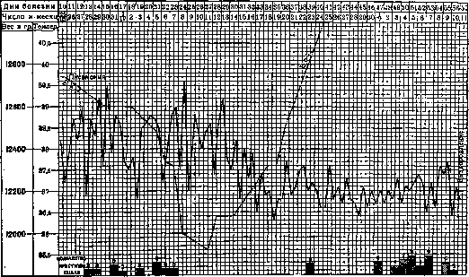
Figure 10. Cases of pertussis complicated by pneumonia in a 4-year-old child (recovery). Complications of the second group from other organs in the form of otitis, lesions of the digestive tract, endocarditis, pericarditis, and nephritis are encountered rarely. Among the consequences of pertussis, it is necessary to note chronic emphysema and bronchiectasis in very rare cases. Complications from the nervous system also show almost complete resolution. Only as an exception do persistent paralyses, loss of hearing, and vision remain. Rare cases have been described where pertussis was the cause of mental disorders in the form of hypochondria, melancholia, and even dementia. Lesions of the spinal cord in the form of sclerosis en plaques and Friedreich's disease must also be attributed to exceptional rarities. The combination of pertussis with other infections most often worsens the course of both combined infections. Such worsening is especially strongly felt when combining pertussis with measles. Broncho-
Figure 11. Case of pertussis complicated by bronchitis and pneumonia in a 3-year-8-month-old child (recovery). pneumonia here is a constant complication. Ecchymoses often appear. According to Roger's data, such patients died in 2/3 of cases. Significantly less dangerous is the combination of pertussis with scarlet fever and diphtheria. When combined with scarlet fever, the number of attacks may even temporarily decrease. The connection of pertussis with tuberculosis is of extremely great importance; here the same picture occurs as in the combination of measles and influenza with tuberculosis. Pertussis activates pre-existing tuberculous foci and itself in turn, in relation to diagnosis, this stage of pertussis does not present special difficulties. True, pertussis-like attacks can sometimes be observed with foreign bodies in the bronchi, with bronchial asthma, with influenza, with tuberculosis, and especially with tuberculosis of the bronchial glands. But in all these conditions, besides the presence or absence of signs typical for each of them, the diagnosis is aided by the increase in the strength of the attacks characteristic of pertussis, their intensification, the expectoration of viscous sputum, and ulcers on the frenulum.
with measles and especially with influenza. Characteristic for pertussis is the absence or weak expression of catarrh of the mucous membranes, the intrusiveness and persistence of the cough, which do not correspond to the physical findings. The picture of the convulsive period is characteristic, and therefore
It develops luxuriantly on a tuberculous soil. The flourishing of tuberculosis under the influence of pertussis occurs in the form of pleuropneumonia, peritonitis, meningitis, and broncho- and tracheoadenitis. Clinical diagnosis of pertussis in the catarrhal period is difficult. Here, a mix-up is possible. In recent times, in addition to X-rays, immunoreactions and bacteriological findings of the pertussis bacillus have come to the aid of diagnosis. Prognosis in pertussis follows from the above. Treatment. The basis of pertussis treatment should be general hygienic rules regarding regimen, care, and nutrition. In relation to pertussis, abundant ventilation of the lungs with clean, fresh air must be applied in the widest possible form. The very method of providing the patient with clean air depends on the circumstances and is not uniform. Many Russian pediatricians (Filatov, Kisel) are particularly strong supporters of treating pertussis with clean air, even cold air. According to Kisel, complications are not observed in pertussis-affected children who spend a lot of time outdoors. On the other hand, in a pertussis patient, especially one who is weakened, cold, damp air can cause a worsening of the disease symptoms. In poor housing conditions, stale air must, of course, be replaced with fresh air, even if it is cold. Bed rest is applied only during a febrile state. Bathing and wet rubdowns, if they were used before the illness, can be continued during the disease. Exceptionally great attention should be paid to the cleanliness of the environment surrounding the patient. The most severe complication, which is the main cause of high mortality in pertussis, is bronchopneumonia, which is precisely caused not by the pertussis bacillus, but by extraneous microbes. These extraneous microbes are often picked up by the patient from their surrounding environment. In this regard, protecting the pertussis patient from influenza infection is also important. Great attention should also be paid to nutrition, especially in children suffering from frequent vomiting. Such children should be fed often in small portions and given food after a coughing fit. The food should be high in calories and nutritionally complete. A corresponding diet can also be used for therapeutic purposes in children with constitutional disorders (spasmophilia). Improvement of these disorders in connection with appropriate nutrition is also reflected in the general course of the pertussis process itself. An extremely large number of drugs have been proposed for the treatment of pertussis. There are no specific medicinal agents. In all periods, alkalis can be used in the form of alkaline mineral waters or simply a 2% soda solution in hot milk. In the presence of catarrhal symptoms (rales), an expectorant mixture (ipecacuanha, senega) can be prescribed. Among narcotic agents, bromine preparations, chloral hydrate, ether, etc., are used. In severe attacks, morphine preparations (pantopon, codeine, etc.) can be prescribed for short periods. Most authors also use atropine preparations. Thus, to this day, the Henoch-Heubner mixture with calcium bromide, antipyrine, and belladonna is prescribed quite widely. Attempts are being made to use physiotherapy for treatment in the form of X-ray therapy and quartz light irradiation. Complications in pertussis are treated on general grounds. Therapy with vaccines and sera has so far yielded little. The use of anti-pertussis anti-endotoxic sera has not yielded positive results (Bordet and Gengou, Klimenko). In recent times, specific vaccine therapy has also found quite wide application in the treatment of pertussis. In this case, as a rule, the same vaccines are used as for prophylaxis. Among non-specific vaccines, the Korshun combined scarlet fever vaccine has found relatively wide application in the USSR. Some authors are negative about vaccine therapy, while others note its favorable effect. But the magnitude, and above all the nature of these results, are not definite and demonstrative enough to unconditionally attribute the therapeutic effect to vaccine therapy. In view of the peculiarities of the course of pertussis, the weakening of symptoms and significant rapid improvements can also occur spontaneously. The serum of pertussis convalescents (Debré, Davydov, and others) has also been used in the treatment of pertussis with positive results. Prophylaxis of pertussis is based on data from etiology and epidemiology. Sanitary measures in the fight against the pertussis danger are reduced to the registration of cases of the disease, isolation, and, in a relatively limited form, to disinfection. Registration of cases of the disease with accurate early diagnosis, especially of atypical cases, using not only clinical but also bacteriological methods, will facilitate the implementation of isolation. Isolation in relation to a pertussis patient should be applied in children's institutions, and if the surrounding environment allows, then in the family as well. Pregnant women in the family who have never had pertussis should be separated from pertussis patients. Such separation is especially required when the due date is near. If the mother is ill with pertussis during childbirth, the newborn must be separated immediately after birth. It is necessary to isolate the patient until the end of the contagious period. We are currently in a transitional state regarding the use of new data on the duration of the contagious period. So far, only a part of the countries have taken these data into account and reorganized sanitary measures accordingly. Denmark is leading here, where (according to an order of the Ministry of Public Education) children can attend schools, nurseries, and other children's institutions one month after the onset of convulsive coughing. In the USSR, according to the rules approved on January 1, 1925, by the Scientific Medical Council, the duration of the contagious period in pertussis is calculated as 40 days from the onset of coughing. In France, children are allowed to go to school only after 4 weeks from the onset of the disease, provided that the coughing fits have ceased. Brothers and sisters of the patient are prohibited from attending school for 21 days. In the RSFSR, this period is 11 days. Dopter suggests either shortening the isolation period to 1 month after the 1st attack or limiting it to obtaining negative results from a twice-performed bacteriological examination. Regarding children who have been in contact with a pertussis patient, he suggests establishing a quarantine for a period equal to the duration of the incubation period and calculated from the moment the possibility of infection is excluded. In this case, the child is released from quarantine only in the absence of coughing and suspicions of the disease. Here, too, the method of bacteriological examination must gain citizenship. The method of isolation for the purpose of protecting children from a possible encounter with a pertussis patient should be widely and systematically applied in children's polyclinics, hospitals, mother and child homes, and other children's institutions. Children's hospitals should have small wards, and for newly admitted patients, boxes. In extreme cases, it is desirable to separate children from each other with partitions or screens, observing the appropriate principles of care based on the observance of thorough cleanliness. The amount of air per patient should be sufficient (50 m3). In closed, and partly in semi-closed children's institutions, arriving children must undergo quarantine in special rooms for a period of 21 days. Closing schools during pertussis epidemics is inexpedient. The implementation of the principle of isolation in children's outpatient clinics, which are currently a frequent source of the spread of the disease, is of particularly great importance. In addition to filters and examination upon admission, during the entire stay of the child in the polyclinic, they should have as little contact as possible with other children. All children with acute catarrhal symptoms and with a cough should be received separately, in special isolated boxes. Disinfection plays a relatively small role in the fight against pertussis. Vomit, sputum, and the patient's surrounding household items should be disinfected. Disinfection here is carried out not so much for the purpose of destroying the unstable pertussis virus, but to free the patient's surrounding environment from so-called secondary microbes. In view of the difficulty of fighting pertussis with sanitary measures, specific prophylaxis of pertussis has attracted particularly great attention from medical thought. At the present time, immunization is carried out in the form of active—with various types of vaccines—and passive—with sera of pertussis convalescents and sera of adults. In recent years, active immunization against pertussis has been used on a fairly large scale.
The vaccines used are different in their composition and methods of preparation. Most authors used a vaccine made from the pertussis bacillus; some used a mixed vaccine made from the pertussis bacillus, influenza bacillus, pneumococci, and streptococci; an autogroup vaccine was also used. The results, in general, with relatively few exceptions, were favorable. Nevertheless, to turn the idea into a method, many more studies and much more factual data are required. Polyvalent vaccines were used for the purpose of obtaining immunity against the pathogens that are most often the etiological cause of complications. The method of passive immunization with the serum of pertussis convalescents was developed particularly fully by Debré. According to the Degkwitz method (see Degkwitz vaccinations), the serum is taken in the 4th week from the onset of the spasmodic cough. It is at this time that the blood of patients contains the greatest amount of immune bodies. According to Debré, the injection of serum is performed in the following doses: 3 cm3 for children under 3 years old, 3-6 cm3 for children 3-10 years old, and 3-8 cm3 for older children. When injected in the first days of the incubation period, the disease in most cases does not occur; when used at the end of the incubation period, it is more often possible to obtain not the prevention of the disease, but only a mitigation of the course of the disease (Debré).
M. Danilevich.
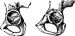
Figure 1. Initial tubercular foci in the region of the acetabulum in children.
its functions. A predisposing factor in the development of the disease is any weakening of the organism, in particular infections—measles, scarlet fever, pertussis; as for trauma, its significance is disputed by many. The role of social factors in the development of tubercular coxitis—see Tuberculosis, Joints. The pathological-anatomical process of the development of infection in coxitis proceeds in the same way as in other joints (see). The disease may be limited to the lesion predominantly of the joint capsule (synovial form), proceed with more or less pronounced clinical phenomena, and may end without significant anatomical and functional defects. The process may begin and develop predominantly in the bone tissue (osseous form) and, under favorable conditions, stop early without causing deep deformations of the joint. Much more often, in the synovial form of coxitis, and even more so in the osseous form, destructive changes pass from one tissue to another, spread to the cartilaginous articular covering and bone, and lead to more severe anatomical and functional lesions.
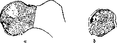
Figure 2. Initial forms of tubercular foci in the epiphysis of the femur: a—wedge-shaped infarcts of the femoral head; b—tubercular focus of the head, penetrating through the epiphyseal cartilage into the region of the metaphysis.
Nevertheless, it is rarely possible to establish in which cases the synovial form has passed into the osseous form and vice versa; therefore, some deny the existence of the synovial form altogether, recognizing only the osseous one. Local features in the development of the pathological-anatomical picture of coxitis reduce to the following. Depending on the localization of the primary focus in the bone, the initial lesion is concentrated in the acetabulum (Fig. 1), in the region of its epiphyseal cartilage in children, coxitis, or in the epiphysis of the head and neck of the femur, sometimes taking here the typical form of wedge-shaped infarcts (Fig. 2). Deeper destruction in both the osseous and synovial forms can be accompanied by extensive destruction of both the articular cavity and
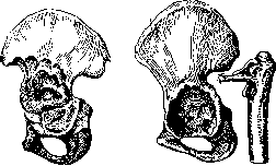
Figure 3.
Figure
4. Figure 3. Diffuse lesion of the pelvic bone. Figure 4. Coxitis with the greatest lesion of the head and neck of the femur. os ilii (Fig. 3), as well as the articular end of the femur (Fig. 4) and even its greater trochanter. Gradually deepening, the process can cause perforation of the acetabulum and involve the tissues of the small pelvis (Fig. 5) or, developing in the bone of the head and neck, be directed predominantly towards the joint capsule and the femur, causing perforation of the bone focus into the joint cavity (Fig. 6) or, in rarer cases, being accompanied by its extra-articular opening (see below, Fig. 12). In parallel with these phenomena, there occurs the development of pathological postures—contractures in the region of the hip joint, mainly in the form of flexion contracture, in the position of abduction (abductio) of the femur in the initial stage and adduction (adductio) with internal rotation in the final stage of coxitis. Destructive changes can reach an extreme degree, leading to pathological dislocations (Fig. 7), sequestra, muscle atrophy, and gross deformations with significant shortening of the diseased limb. The sequestra that form are encountered in coxitis, as in other joints, either in the form of so-called sand or more voluminous pieces of bone, lying freely among the fungous masses of the focus (Figure 6) or often for a long time retaining a connection with the main bone tissue. In some cases, with the loss of connection with the cartilage, the entire epiphyseal part of the femoral head can be sequestered. Dislocations in coxitis can occur with more or less sharply altered forms of the acetabulum or the articular end of the femur, whereby the displaced end of the femur is sometimes held within the limits of the acetabulum (subluxation) with the help of a negligible remnant of the neck
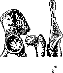
Figure 5. a—perforation of the acetabulum in coxitis; b—perforation that caused the formation of an intrapelvic abscess.
of the femur (Fig. 4 and 8a); with a destroyed acetabulum, it completely exits its limits and is fixed at the outer surface of the ilium (Fig. 8b). In children, with a central focus of the head in the region of the fovea, cases of complete dislocation of the unchanged head from an acetabulum that has preserved its shape can occur. This type of dislocation is caused by the early destruction of the lig. teres in the presence of a large effusion in the joint (Fig. 9). Abscesses (Fig. 10) accompany no less than 50% of all cases of coxitis and in 25-30% are complicated by fistulas, often with secondary infection; these forms differ sharply in the severity and duration of their course in comparison with closed ones. In closed forms, the process sometimes proceeds according to the type of caries
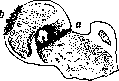
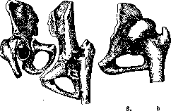
Figure 7.
a
Fig.
Figure 7. Dislocation of the femur in coxitis. Figure 8. Subluxation of the femur in destruction: a—of the head and neck of the femur, b—of the acetabulum. caries sicca and is characterized by the destruction of the head (without sharply pronounced infiltration, without the accumulation of pus). Along with articular forms, there can be cases of the development of extra-articular bone foci, manifested by clinical signs of articular coxitis. Such foci, proceeding "dry" or with the formation of abscesses, arise either in close proximity to the articular surface (Fig. 11 and 12) or in more distant regions of the femur or pelvic bone (Fig. 13). In their further course, these para- (juxta-) articular foci in severe cases of the course can eventually involve the joint cavity in the process, while in favorable cases they complete the entire cycle of their development without the participation of the joint and end with the preservation of full mobility of the joint. Course of the process. In the overwhelming majority of cases, the process develops slowly, gradually. Initially, there is noted less desire for movement, loss of appetite, lethargy, emaciation, slight increases in temperature; pain is often observed

Figure 9. Distended
joint capsule with exudate. coxitis in the knee or sacrum. Over time, fatigue and diffuse pain in the leg are added, intensifying after functional strain
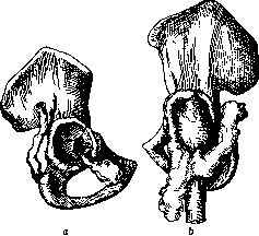
Figure 10. a—perforation of the acetabulum; cold abscesses and fistulas in the region of the adductor muscles and the buttock; b—simpler spread of the abscess along the anterolateral surface of the thigh. (Diagram.)
lesions, and only subsequently do complaints of pain in the joint itself appear. Often children cry out at night and, upon waking, are in a position usually of flexion contracture, adduction, and ankylosis (Fig. 14). Simultaneously with the pathological positioning of the hip (flexio and adductio), the position of the pelvis changes, and following this, the spine as well. The pelvis gradually assumes an oblique position: the side of the pelvis corresponding to the diseased hip is raised; at the same time, the entire pelvis tilts forward, and in parallel with this, a more or less pronounced lordosis and scoliosis develop. Over time, these changes become persistent and are accompanied by deformation of the pelvis; it becomes asymmetrical. Radiologically, one can detect gross changes in both the shape and the internal structure of the bones. The entire half of the pelvis on the side of the diseased joint appears atrophied, and all the bones are thinner. These deformations of the pelvis can be so strongly pronounced that extremely unfavorable conditions for childbirth are created (see Pelvis). Changes in the position of the pelvis initially have a functional character, but then serious deformations of the skeleton occur. The duration of the course is determined by the form of the process, the resistance of the organism, age, complicating factors, and the nature of the treatment. As a rule, it can be said that closed forms in childhood, with rational treatment, result in
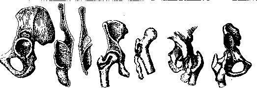
Figure 11.
Figure 12.
Figure 13.
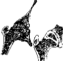
Figure 11. A focus located in the vicinity of the ilium, not communicating with the joint. Figure 12. Extra-articular (osseous) lesion of the femur in the region of the hip joint. Figure 13. Para-articular foci in the pubic and iliac bones with the formation of cold abscesses. complain of pain in the joint. These cries at night are generally characteristic of tuberculous patients. Limping, which initially manifests only upon fatigue, then becomes constant. Over time, in connection with the extreme diversity of the localization of the tuberculous focus (in the head, neck, femur, acetabulum, in the bursa), a varied picture emerges. As for the pains, they are different—from barely expressed to extremely severe, such that the slightest touch is accompanied by extreme tenderness. A sharp exacerbation of pain is usually associated with the perforation of a focal lesion into the joint cavity. As the process develops, the limitation of mobility and the tendency toward contractures increase; subsequently, in connection with destructive changes, mobility decreases more and more, and finally, the limb becomes fixed, with the process subsiding on average in 2-3 years, while complete recovery drags on for much longer: up to 6-8 years. Recognition of a pronounced form of coxitis presents no difficulties. However, the initial stages of the disease are often diagnosed with great difficulty, and prolonged observation is required so as not to make an erroneous diagnosis. A thorough assessment of the anamnesis, subjective and objective data, with blood examination, temperature, and repeated X-ray examination, usually makes it possible to [see Fig. 14: Coxitis with fixation of the deformed head in the acetabulum (fibrous ankylosis)].
Old cases
to clarify the nature of the process; as for tuberculin tests, their positive nature cannot have decisive significance; negative tests have somewhat greater significance. For a detailed examination of the patient, it is necessary to undress them completely and make them walk in order to catch defects in their gait. The examination must be conducted the entire time, necessarily comparing both sides; then it is easier to catch those small changes which, in the aggregate, can give definite suspicions of a developing process. By tapping on the trochanter, one checks the sensitivity on both sides; tenderness upon percussion is one of the important signs. Among the objective signs of incipient Pertussis, the smoothing of the inguinal and gluteal folds, swelling of the glands on the affected side, muscle atrophy, and thickening of the skin fold (Alexandrov's symptom) are significant; more precise data are obtained by examining mobility. It is important to check the completeness of all types of movements of the hip joint: flexion, extension, rotation in both directions, adduction, and abduction; even small defects in them are of great significance, which is reinforced by the presence of other factors. One of the most valuable methods of examination, especially in the initial stages of Pertussis, is radiography. In all doubtful cases, it is necessary to have images of both sides in a strictly symmetrical setup. To obtain a radiograph in a different setup, one can take an image by bending the thigh at a 90° angle and abducting it (Lauenstein's position). Comparison of clear radiographs of both hip joints often provides decisive data. Moderate atrophy, a difference in the size of the joint space, and the contours of the head provide material for judging the nature of the process. In doubtful cases, only repeated control examinations decide the matter. In later stages of the process's development, when there is already lameness, atrophy, significant limitation of mobility, and pathological positioning, the diagnosis of Pertussis does not cause difficulties. Doubts may arise regarding the nature of the pathological-anatomical process; here one must remember infectious arthritis, osteomyelitis of the neck, Perthes disease, consequences of trauma, etc. In that stage of the process where there are gross destructive changes, an X-ray picture is necessary not for diagnostic purposes, but for determining the nature and degree of destruction and regenerative phenomena. In tuberculous lesions, both these processes proceed in parallel, but at first, destruction prevails, and phenomena of regeneration are difficult to catch, and only with the passage of time are signs of regeneration expressed more or less well on clear radiographs. On radiographs, one can trace the restoration of the pattern of the cancellous substance: the structure of the trabeculae becomes increasingly distinct, the corticalis thickens; along with this, one can often observe phenomena of transformation, adaptation to new conditions of statics and function: the destroyed remnant of the head and the altered acetabulum adapt to each other in such a way that one can speak of a new joint [see separate table (art. Bone), fig. 15]. Much more often we see in the final stage of Pertussis, especially where secondary infection joins, the formation of ankylosis, usually in a position of flexion and adduction. Such positioning as a rule leads to sharply pronounced lordosis; simultaneously, on the radiograph, one can establish significant changes on the part of the corresponding half of the pelvis; it lags in its development, and the entire bone appears atrophic. Cases of pathological dislocations in Pertussis are caused mainly by gross destruction of the acetabulum and its expansion upward; the head of the femur, also more or less deformed, is displaced and can slip out of the acetabular cavity entirely. [See separate table (art. Bone), fig. 16 and 17.] In a number of cases, destruction can be accompanied by perforation of the floor of the acetabulum, and then by palpating the region of the iliac fossa and examination per rectum, it is possible to determine the presence of an infiltrate or abscess. At the same time, it is extremely important to establish this phenomenon, as it significantly worsens the course of the process, and it must be kept in mind when choosing therapeutic measures. Radiographic examination can help clarify the existence of perforation of the acetabulum. On a radiograph, perforation is usually diagnosed when a part of the deformed head protrudes into the pelvic cavity, or in those rare cases where intrapelvic luxation takes place. The treatment of Pertussis is divided into general and local. As with any tuberculous patient, the general condition of the coxitis patient must be raised, and their resistance strengthened. The longest possible stay in the fresh air, observance of strict cleanliness of the body, and rational nutrition—all these factors are of paramount importance in the general plan of treatment. The best is sanatorium treatment. Besides these measures of a general nature, the planned distribution of the patient's time is also of great importance; this type of patient requires bed rest for sometimes 1, 2, 3, or more years. It is clear that this time, besides treatment procedures, must be filled with reasonable entertainment, a correspondingly developed system of study, and even labor processes; all this reflects beautifully on the well-being of the patients; not lagging behind their healthy comrades, they calmly reconcile themselves to the burdensome stay in bed. As for the significance of climate, the advantages of staying in the mountains or on the seashore, it must be said that in some respects their advantages are beyond doubt (in the question of conducting sun baths, in the favorable influence on the patient's psyche), but still, the center of gravity is not in where the treatment is conducted, but in how it is conducted. Along with general treatment, it is necessary to conduct systematic orthopedic treatment. In this regard, one of the most important factors is the prevention of pathological positioning—contractures (see). This is achieved by fixation of the affected limb in a certain position with the help of traction or plaster casts, circular or open—by the type of bed splints. Both traction and fixation with splints or closed circular casts, while preventing the formation of contractures, at the same time immobilize the affected joint, and immobilization (see) in bone tuberculosis is an indispensable condition for successful treatment. Bearing in mind the constant tendency in Pertussis to form contractures in the position of adduction, it is necessary to give the thigh a position
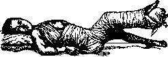
Figure 15. Fixation in pathological positioning, according to Finck.
abduction, and if adduction is already present, it is necessary to persistently combat it, but without resorting to any harsh measures. Traction for coxitis has until now usually been carried out with the help of significant weights, and there was a tendency to gradually increase these weights. Now some authors think that significant weights can be the cause of an exacerbation of the process and that, in general, the optimum of their action corresponds not to the maximum weight, but to some minimum, which fluctuates within significant limits. In any case, the criterion should be the relaxation of contracted muscles without unpleasant subjective sensations. Usually, in children, one can limit oneself to a weight of 200–800 g; at the same time, it must be distributed over the thigh and lower leg. As soon as the desired correction is achieved, it is considered rational to gradually reduce the weight to 100–50 g and carry out further fixation with a circular bandage or an open splint-bed, covering the torso from behind up to the angles of the shoulder blades and even higher. It is also necessary to combat the tendency of the entire limb to rotate outward, which is always present with hip abduction, which is especially noticeable by the position of the foot and the patella, as well as the positioning of the foot in the position of pes equinus. If the latter can be corrected quite easily, then outward rotation is corrected with significant difficulties; the commonly used Ménard supports and plaster splints with a plate at the base do not achieve the goal: while holding the foot in the correct position, they cannot rotate the thigh. Correction of contractures in coxitis represents one of the most difficult tasks. Some major orthopedists (Ludloff, J. v. Finck) even suggest not correcting them at all, but fixing them in the pathological position in which they arrive for treatment, achieving ankylosis, and subsequently correcting them by osteotomy (Fig. 15). Likewise, for some authors, the question of which treatment result is preferable is debatable: ankylosis in a good position or a mobile joint; it would seem that the latter outcome is better; however, observations have shown that joints with mobility preserved after coxitis are inferior, insufficiently stable, extremely painful, and very easily give rise to exacerbations (relapses) of the disease. Only coxitis in children at an early age without gross destruction comparatively more often ends in functionally more stable joints. It should be borne in mind that bony ankylosis is obtained relatively rarely; as a rule, it is fibrous ankylosis (Fig. 14). Thus, in this controversial issue, the answer is determined to a certain extent by the age of the patient. In general, however, ankylosis in a good position, i.e., in a position of slight flexio et abductio, provides a stable limb that is convenient for function. At the present time, the correction of pathological positions in coxitis by applying more or less gross force, usually under anesthesia, has been completely abandoned. This inevitably entailed significant trauma, ruptures, and hemorrhage in loco morbi, often pathological fractures, and resulted not only in a sharp focal exacerbation but also in cases of generalization of the process (meningitis, miliary tuberculosis). Only a few orthopedists attempt, and even then very cautiously, to correct the pathological position under anesthesia only insofar as it can be done without violence. A very important, responsible question in the treatment of coxitis is the question of the patient getting up. The determination of this period depends on the sum of general and local phenomena: temperature, general condition, the presence of complications (abscesses), secondary infection, contractures, the degree of destruction, its nature, the position of the head, etc. A good radiograph is an indispensable condition and is of decisive importance in determining these moments. It provides an answer to the question about the degree and nature of the destructive phenomena, about the position of the bone head,
as well as the degree
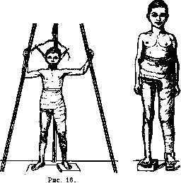
Figure 17.
of repair, and if there is a series of radiographs taken under uniform conditions, then it is possible to obtain extremely valuable material for judging the increase in reparative phenomena, and consequently, the moment when one can begin to load the joint. This question is of essential importance; we have two schools of thought regarding it. Some, led by Lorenz, believe that loading the joint, provided it is fixed [at the same time, it is sufficient to fix it with a short bandage up to the knee (Gipshose) (Fig. 16)], leads sooner to the formation of ankylosis. Others hold the opposite point of view and protect the joint from loading for as long as possible. Accordingly, patients are mobilized in a long plaster splint, where the ankle joint is also captured; they are allowed to walk on crutches, and in order not to touch the ground with the diseased leg, the healthy one is placed on a higher heel. There is another method of unloading the diseased joint—this is a bandage with a stirrup (Fig. 17), where
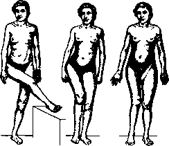
Figure 18. Contracture in the position of flexion and adduction before and after osteotomy.
the fulcrum is transferred to the tuber ischii. Instead of non-removable, more or less bulky plaster bandages, one can subsequently use removable celluloid or splint-sleeve apparatuses constructed on the same principle, i.e., with a stirrup and with supports on the tuber ischii. The healthy leg is also raised here either with a high heel or with an orthopedic shoe. Such apparatuses must be worn for a long time, often for years, keeping in mind that the subsidence of the process proceeds slowly; one should part with them gradually; at night—a splint-bed. There is also a middle approach; it boils down to the following: at the moment when all phenomena subside, and on radiographs, there is a clear prevalence of regenerative processes over destruction, it is permitted to lightly load the joint. It is believed that in this way, one can accelerate the development of reparative phenomena. Patients with a bandage fixing up to the middle of the lower leg walk on crutches, slightly loading the diseased leg, and gradually increase the load. Along with these purely conservative methods of treating coxitis, there are others: the introduction of various kinds of medicinal substances directly into the diseased joint, as well as into the surrounding tissues (muscles). The most common substances are preparations of iodine, creosote, naphthol, iodoform, etc. Proponents of these injections are Calot, Gotz, Grekov, Finikov. It must be said, however, that after a period of enthusiasm, these methods did not find wide distribution and meet with objections from many orthopedists. Abscesses in coxitis. About 50% of all cases of coxitis are accompanied by abscesses and about 25-30% by fistulas. The fate of these abscesses is different: some are resorbed, some calcify, some open, and then fistulas are formed; in the latter case, a secondary infection easily joins. It must be borne in mind that even closed abscesses in some cases can be infected by a secondary infection. This infection can penetrate hematogenously; but more often it is introduced during punctures. (Treatment of abscesses in coxitis—see Joints.)—In addition to the orthopedic techniques mentioned above, conservative methods of treatment also include heliotherapy, treatment with artificial light (quartz lamp, arc lamp), radiotherapy, and mud therapy. The basic principle of heliotherapy—careful dosage along with individualization—must be especially strictly carried out in severe forms of coxitis. One should not strive for a significant lengthening of the irradiation sessions; one must avoid overheating in every possible way. The best system will be one where the actual sunbath is alternated with an air bath, and the latter with rest. In the correct alternation of sessions lies the key to the success of heliotherapy. At the beginning of treatment, it is recommended not to subject the hip joint area to irradiation and to expose it only gradually, guided by the reaction. As for artificial light sources, the most popular are the quartz lamp and the arc lamp. Regarding the latter, in a number of northern countries (Sweden, Norway, Denmark, Finland), a solid body of material has been accumulated in its favor
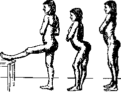
Figure 19. Contracture in the position of flexion and adduction before and after osteotomy.
Application. X-ray therapy, which finds supporters in some clinics for the treatment of bone tuberculosis, is almost never used for coxitis. The objections raised in this regard mainly boil down to the difficulties of dosage and the fear of adversely affecting the epiphyseal lines and thus disrupting the normal growth of the limb. In the field of specific therapy, tuberculin therapy for coxitis, there are no extensive observations and little literary data; it should be considered established that cases of moderate severity are suitable for tuberculin therapy, and that treatment must be carried out extremely carefully, starting with the smallest doses. Mud therapy for coxitis, see Joints. A certain percentage of coxitis cases in the final stage are fixed by fibrous or bony ankylosis, and rarely in a good position. The most frequent position is flexion and adduction. Even mild degrees of adduction cause many inconveniences and therefore require correction; it is carried out by an operation known as osteotomy (Figs. 18 and 19). After placing the hip in a position of slight abduction, a plaster cast is applied for a period of about 8-10 weeks, but usually, it is possible to move around in the cast on crutches after just 6 weeks. In bilateral coxitis, it is better to perform arthroplasty on only one joint. The arthroplasty operation for ankylosis after tuberculous coxitis was until recently performed only as an exception. Such restraint is explained on the one hand by the fear of causing an exacerbation of the process, and on the other by the fact that this operation is technically difficult and accompanied by great trauma. Hence, it is understandable that the operation can be started only when a significant period has passed after the complete subsidence of the process. Recently, there has been a tendency to somewhat expand the indications in this area. Prophylaxis of tuberculous coxitis—see Joints. Operative treatment of coxitis was until recently the dominant method; extensive operations were performed—resections (Bardenheuer), during which not only the head, neck, and trochanter were removed, but also part of the femoral diaphysis; this led to extreme deformity; even when the wounds closed, the resulting limb was so unsuitable that it was necessary to move with the help of crutches. These operations were often accompanied by secondary infection, which frequently led patients to death; it is not surprising, therefore, that the results obtained by Rollier turned out to be better, and for a number of years the guiding position on the issue of treating bone tuberculosis was: 'Any tuberculosis is cured if one has the patience not to operate on it' (Rollier). But this did not last long, and voices began to be raised more and more often in favor of the necessity to operate in certain cases. Thus, Ludloff, Fritz König, and others pointed out the necessity of removing isolated foci; Waldenström considered it necessary to widely resect all severe, protracted cases of coxitis. König, expanding these indications, believed that in cases of mixed infection, it is generally necessary to resect the joint. A number of orthopedists approached the operative treatment of coxitis from another side, namely: they strive for fixation of the joint by forming an extra-arti-
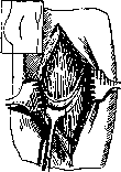
Figure 20. Hass's operation for extra-articular fixation in coxitis (first step).
cular ankylosis, some (Hibbs, Hass) with the help of the trochanter, others with the help of plates taken from the tibia (Albee). In technical terms, the operation boils down to the following: a depression is formed above the acetabulum, a part of the trochanter is chipped off and shifted so that it
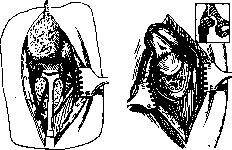
Figure 21. Hass's operation—second and third steps.
does not lose its connection with its base, and its end is driven into the formed depression (Figs. 20 and 21) (Hass), or the knocked-off part of the trochanter is turned 180° around its axis and then driven in with its lower end (Hibbs) (Fig. 22). After this, the pelvis and thigh are fixed in a plaster cast until a strong ankylosis is formed. Albee's operation is clearly visible in the accompanying figure (Figure 23). Robertson Lavalle proposes changing the type of blood circulation in the area of tuberculous foci. Around the tuberculous nest, in his opinion, a zone of dilated
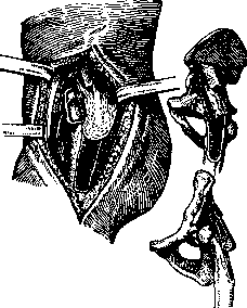
Figure 22. Hibbs's operation
for extra-articular fixation in coxitis. vessels with extremely slowed blood circulation is formed; in order to facilitate the outflow of venous blood and thereby create conditions for the rapid healing of the process, Lavalle proposes introducing bone plates into the area of the affected focus by operative means. The plate must be inserted exactly to a specific section of this zone. In individual cases, another plate is inserted, which
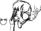
is connected to the first and is located under the skin—this should increase the outflow of venous blood from the affected area. Lavalle aroused interest in his proposal especially by his assertion about the exceptional speed of the process's liquidation (from 1 to 6 months), and he recommends starting early movement in the diseased joints and allows patients to get up early. R. Lavalle's proposal caused lively debate and very sharp criticism. This method, nevertheless, finds followers.
Figure 23. Diagram of Albee's operation—extra-articular arthrodesis.
In a somewhat modified form, the method was tested by Prof. Vreden, who at the last surgical congress gave a report on this issue, which also provoked lively criticism. This issue is still in the stage of clinical verification. Infectious coxitis, in comparison with tuberculous coxitis, is encountered much less frequently. Statistical data exist only in relation to a few infections: thus, among gonorrheal lesions of other joints, the hip joint accounts for 5%; in polyarthritis infectiosa (according to Stoll's statistics) 4.1%. Infectious coxitis can be the result of direct infection of the joint, for example, during trauma or upon the spread of infection from neighboring foci, in cancer, phlegmons of neighboring tissue areas, or it is a complication of general infectious diseases and purulent foci. Such a source of infection can be any focus in the body (furuncle, phlegmon, erysipelas, angina, osteomyelitis) or a general infectious disease—scarlet fever, measles, influenza, pneumonia, diphtheria, typhus, postpartum sepsis, etc. In infectious coxitis, the synovial form is more often encountered in the form of serous, fibrinous, or purulent arthritis. Serous exudate has the appearance of a yellowish liquid with a greater or lesser significant amount of pus corpuscles; it also differs from normal synovial fluid by a higher specific gravity. Fibrinous exudate is characterized by the presence of a greater or lesser significant amount of fibers, flakes, and whitish shreds of tissue. In purulent infectious coxitis, along with the accumulation of purulent fluid, there are also gross changes in the capsule; it is inflamed and unevenly thickened, loosened. Much less frequently, the infection enters some part of the bone close to the joint, develops there as osteomyelitis, and then secondarily involves the joint in the process. Infectious coxitis usually develops violently, as a complication of a general infectious process, and is accompanied by high temperature, a severe general condition, intense pain, a feeling of tension, and swelling in the area of the affected joint; very soon, a contracture in the position of flexion and adduction is formed. Pain in individual infections can be expressed extremely strongly; this is especially characteristic of gonorrheal lesions. Less often we encounter the so-called subacute form, where all these phenomena develop more gradually. As a rule, the process ends with a more or less strongly expressed limitation of mobility in a pathological position. The articular capsule can be so stretched that the head of the bone easily slips out of the joint, and cases of pathological dislocations are a frequent complication of infectious coxitis. In purulent forms, pathological dislocations can develop as a result of destruction of the head or the acetabulum. Cases of pathological fractures of the neck during its osteomyelitis are not rare; it usually proceeds like coxitis. The entire picture of acute infectious coxitis can develop within a few weeks; following this, the temperature begins to fall, acute phenomena subside, and complications emerge—abscesses, pathological dislocations, etc. Diagnosis of infectious coxitis in fresh cases usually does not present difficulties; diagnosis is somewhat more difficult in subacute forms. The violent development of the process, the connection with infection, and the X-ray picture usually easily resolve difficulties. One has to differentiate it from osteomyelitis of the femoral neck and tuberculous coxitis. Treatment varies depending on which form of coxitis is present and in what stage. In the initial stages, strict rest and traction are necessary to prevent pathological positions. Accumulation of fluid in the joint requires puncture and aspiration of the accumulated fluid and repetition of this procedure upon new accumulation. In case of accumulation of purulent fluid, besides strict rest, punctures, and washing of the joint with various solutions (Rivanol, etc.), it is often necessary to resort to wide opening of the joint. The choice of one or another method of treatment is determined by the form and stage of the disease. Often, carefully adjusted traction with a small weight leads in a short time to complete relief of pain, a drop in temperature, the contracture is smoothed out, and the entire picture of the disease changes sharply for the better. Here, just as with tuberculous coxitis, it is necessary to give the thigh a position of slight abduction. Depending on the nature of the infectious coxitis, as well as the treatment methods used, persistent changes can develop in the form of stiffness, pathological positions, and ankyloses. All these points must be kept in mind and the necessary position must be given to the thigh in a timely manner. Correction of ankyloses after infectious coxitis is performed according to the usual rules (see Hip joint). It should only be remembered that the time of the operation must be sufficiently removed from the beginning of the disease so that there is a certain guarantee against a flare-up of dormant infection. A period of one year is usually accepted, counting from the moment of the subsidence of the process and the closure of fistulas. Thorough examination of patients in this direction before the operation and provocative parenteral administration of irritants are mandatory. In case of doubt, it is more correct to postpone the operation.
Related articles
Mentioned in
Cite this page
“Pertussis.” Soviet Medical Encyclopedia. English translation of Bolshaya Meditsinskaya Entsiklopediya, 1st ed. (Moscow, 1928–1936), ed. N. A. Semashko. https://sovietmedicalencyclopedia.pages.dev/article/pertussis/