Influenza
Historical document, translated for reference. It reflects medical knowledge of the 1920s–30s and is not medical advice.
Summary
This article from the first edition of the Soviet Great Medical Encyclopedia discusses influenza (grippe), covering its history, clinical manifestations, complications, pandemic and epidemic forms, and early bacteriological investigations surrounding Pfeiffer's bacillus.
Encyclopedia article (1928–1936)
GRIPPE (from the French agripper to seize, to attack), or influenza (from the Italian influenza di freddo influence of cold), is an acute contagious disease manifested both by general symptoms (fever, headache, malaise, soreness in the muscles of the extremities and back) and by catarrh of the upper respiratory tract. Grippe is furthermore characterized by a tendency to complications (diseases of the frontal sinuses, Eustachian tube and inner ear, small bronchi and lungs, etc.). Characteristic of grippe is its ability to manifest itself in three different forms—pandemic, epidemic, and endemic (or seasonal). Bacteriology. Regarding the microbe causing grippe, a unity of views has not yet been established. In 1892, R. Pfeiffer described a characteristic bacillus (Bacillus influenzae), which he constantly observed in the sputum of those suffering from grippe and which was not encountered apart from grippe. He isolated this bacillus in pure culture on a medium mixed with blood. Pfeiffer's bacillus belongs to the smallest bacilli: its size is 0.2–0.3–0.5 µ. It is Gram-negative. Its distinctive feature is hemoglobinophilia, i.e., the property of multiplying only on media containing hemoglobin, even in a negligible amount. An especially favorable medium for the growth of influenza bacilli is the blood agar proposed by Levinthal. Bacillus


Figure 1. Influenza bacillus: a—in sputum; b—in culture.
influenza belongs to pleomorphic bacteria, appearing sometimes in the form of cocci and coccobacilli, sometimes as short rods, and finally as filamentous formations of various sizes (see figure 1). Levental and Fernbach establish four morphological types of influenza bacilli, but the independence of these types and their etiological significance are not fully proven. Further study of these bacilli showed that their growth requires the joint presence of two factors: 1) factor x, thermostable and present in hemoglobin and in potatoes (Thjøtta and Avery)—latest research confirms Pfeiffer's assumption that this factor is iron in an active form—and 2) factor y, thermolabile and contained in animal and plant cells. This factor is identified with vitamin C (Kollath). Interesting is the discovery made by Grassberger that certain bacteria can promote the growth of influenza bacilli. He found that near staphylococci on a blood medium, influenza bacilli grow into giant colonies, and therefore called the former the nursemaids of the latter. Then it was discovered that upon symbiosis with certain bacteria (staphylococci, diphtheria bacilli), influenza bacilli can develop even on a medium lacking hemoglobin. The resistance of influenza bacilli to harmful influences is small: they quickly perish at high temperatures (58-60°) from the action of sunlight, upon drying. Disinfectants (sublimate, phenol, etc.) quickly kill them. Pfeiffer himself failed to reproduce influenza in animals using influenza bacilli, but Blake and Cecil obtained a disease in a monkey extremely similar to influenza. By successive infection of mice and monkeys, they obtained cultures so virulent that a small amount of them introduced into the nose, mouth, or trachea of a monkey caused a severe disease in it. After just a few hours, malaise appeared in the form of body aches and photophobia, and then sneezing and cough with mucopurulent sputum appeared, with complications of bronchitis and bronchopneumonia. The disease was accompanied by fever and leukopenia. Influenza bacilli could be isolated from the organism of monkeys either in pure culture or accompanied by streptococci. Is the influenza bacillus the causative agent of influenza? The experiments cited above speak in favor of this; against the etiological role of influenza bacilli speaks the circumstance that the influenza bacillus was not always found in cases of influenza, as well as its frequent finding in healthy individuals or in patients with other diseases (tuberculosis, measles, whooping cough, etc.). In addition, numerous attempts to cause infection in humans with cultures of influenza bacilli always gave a negative result. The few positive results obtained during an epidemic time cannot be considered fully conclusive. As the causative agent of influenza, another microbe was subsequently put forward—the filterable Bact. pneumosintes of Olitsky and Gates, which is not confirmed by the majority of researchers. Thus, the etiological role of the influenza bacillus appears to a considerable extent shaken, but still cannot be considered definitively refuted. The pathogenicity of filtrates of grippal secretions, discovered in some cases, does not speak against the influenza bacillus, since it is also capable of passing through filters. The finding of pathogenic microbes, in this case influenza bacilli, outside of cases of influenza fits in completely with what is known about microbe carriage in other infections (typhoid fever, meningitis, poliomyelitis, etc.). Cases of failure to find influenza bacilli in grippal patients are explained both by the difficulty of obtaining them in pure cultures and by the fact that during the course of the disease they yield their place to complicating bacteria, which, as indicated above, is one of the features of influenza. Finally, the diversity of the epidemiological manifestations of influenza—from pandemic contagiousness to seasonal catarrhs indistinguishable from simple common colds—may depend on fluctuations in the virulence of influenza bacilli. Epidemiology. The most remarkable feature of influenza in epidemiological respect is its capacity for pandemic spread. There is no other disease that spreads with such rapidity across the globe, capturing the majority of the population in the process. The history of influenza pandemics is extremely instructive in this sense. History of Influenza. Epidemics resembling influenza were already described in antiquity. But these descriptions appear doubtful. Hirsch considers the epidemic of 1173, which seized Italy, Germany, and England, to be an influenza disease. From this time on, influenza constantly makes its presence known, sometimes broadly affecting individual regions, sometimes turning into a pandemic, i.e., embracing all countries of the world. There are 18 such pandemics, separated from each other by intervals of tens of years. In view of such, often very long intervals, the affected generations turn out to be unacquainted with the disease of preceding generations and frequently consider it completely new. This explains the variety of names for the pandemic infection. At the beginning of the 18th century, it was christened by the Italians influenza (since its origin was attributed to a special influence of cosmic and meteorological factors). Then the French called the new disease grippe (in view of the speed with which it seizes the population). At the end of the 18th century, from 1780 to 1782, a large influenza pandemic ran its course. It began in China, penetrated through Siberia into European Russia, where it received the name "kitayka" (the Chinese disease), and from there into Poland and all European countries under the name of "Russian disease" or "Russian catarrh." Having appeared in January 1782 suddenly in Petersburg, it struck up to 40,000 people in a single day (with a population of 192,846 people). It is remarkable that on the night before this, the air temperature made a jump of 40°, rising from -35° to +5°. Then from 1798 to 1806 influenza again seized all parts of the world, after which for a number of years epidemic outbreaks were noted in individual regions. From 1830 to 1833, a new pandemic spread in both hemispheres, again originating from China. In the 1850s and 1870s, influenza again had a pandemic spread. Of special attention are the pandemics of influenza closest to us, which caused numerous studies. The first of them—the pandemic of 1889–90—began in Bukhara in February 1889 and penetrated through Central Asia into European Russia. The first cases in Petersburg were in October; by the middle of November, the number of sick reached 150,000. At the beginning of November, the epidemic appeared in Moscow; during November, it seized Poland, Germany, Austria, Belgium, Denmark, and Sweden. In Paris, 50,000 people fell ill in one night. In December, Spain, Portugal, Italy, and the United States were affected, as well as Corsica, Algeria, and Tunisia. In January 1890, influenza seized South America, Egypt, and also South Africa; in February, it appeared in India, Japan, and China; in April, it penetrated into Australia. Within one year, influenza thus toured the entire globe. Then for a number of years, right up to 1893, outbreaks of influenza were observed in various places, which however had no tendency to wide spread. The last severe influenza pandemic occurred in 1918. Apparently, it began in China. In January and February, it appeared in America. In April, it developed in France, in May—in Switzerland, Spain, Italy, Serbia, and England, and then also South Africa. In June—in Poland, Romania, Sweden, and Germany, and besides that in India. In July, it was in Belgium, Holland, and Denmark. With this its first wave ended; the number of illnesses began to decrease, and a temporary lull set in. But in August-September, mass illnesses began everywhere again. Unlike the first wave, influenza this time was distinguished by a malignant course and high lethality (see below). In December, this wave ended, but in March-April 1919, a new epidemic of very severe influenza began, which dragged on until August and struck countries spared at first. The disease of 1918–19 was called "Spanish flu" or Spanish disease, since the first printed information about the pandemic appeared in Spain, which was neutral during the World War and therefore free from military censorship. But in essence this was the same pandemic influenza. After this pandemic, in various regions (such as for example in the USA, in Germany) epidemics were observed, more limited in the occupied space and with a less severe course. Relatively large-scale epidemics were observed in 1926 and 1927 (see below—statistics). Epidemiological features of influenza diseases. As can be seen from the historical review, the characteristic features of influenza epidemics are extreme contagiousness, rapidity of spread, a huge number of affected population, and a tendency to severe complications. The contagion of influenza is transmitted to the healthy from the sick. Thus, on December 14, 1891, a ship arrived at one of the West Indian islands, where there were no diseases of epidemic influenza, on which there were cases of influenza. Two patients from this ship were taken ashore; two weeks later, a third of the island's population was sick with influenza.
In New Caledonia, influenza was brought to the port of Noumea and proceeded along the main road, successively infecting one populated place after another; but the northern part of the island, where there were no roads, was spared. Other properties of pandemic influenza—the rapidity of population involvement and the high percentage of those falling ill—depend on its contagiousness and the mode of its transmission from the sick to the healthy. Influenza spreads via droplet infection. Affecting the upper respiratory tract, it leads to coughing and sneezing, as a result of which the sick spray around them the smallest droplets released from the respiratory tract along with the infectious agents. Thus, the air around the sick becomes infected and causes the disease in everyone inhaling it. In places where people gather, the infection can in this way be transmitted to very many persons at once, which explains the aforementioned suddenness with which influenza seizes masses of the population. The final epidemiological feature of influenza lies in the high frequency of complications caused by it. These complications are very diverse in their clinical manifestations (see clinical features of influenza). They are caused by staphylococci, streptococci, and pneumococci. Some pandemics (e.g., that of 1918) are characterized by frequent complications with pneumonia. Influenza infection evidently causes a decrease in the body's resistance to the aforementioned cocci, which leads to their proliferation in the organism. All races are apparently equally susceptible to influenza. Frequently it affects the local population more than newcomers. Fatigue and strenuous work lead to a severe course of influenza and to complications. The course of the disease is also unfavorable in people exhausted by any diseases, in pregnant women, and in alcoholics. Etiology. Since the role of Bacillus influenzae has not been definitively established, the etiology of influenza remains unclear. Especially puzzling is its periodic pandemic spread. An explanation for this periodicity has not yet been given. One cannot find any relation between influenza pandemics and changes in human resistance. According to some authors, influenza pandemics occur due to an extraordinary—at times—increase in the toxicity of the influenza microbe. Immunity. Most researchers lean toward the opinion that influenza leaves behind immunity, although not absolute (from 1/5 to 1/4 of those who have had influenza fall ill with it again) and not long-lasting [1–2 years, and perhaps even less (Sadov)].
N. Gamaleya. Statistics. Mortality. Mortality from influenza in the last decade before the pandemic of 1918–19 constituted, in most European countries, about 10–20 per 100,000 population per year. In northern countries—Sweden, Norway—it was lower and dropped in individual years to 2–5 per 100,000 population. It was highest in Spain, where annual mortality from influenza ranged from 20 to 70 cases per 100,000 population. The influenza pandemic that erupted at the end of the World War (the so-called "Spanish flu") produced a significant increase in influenza mortality, which reached 293 in Germany, 336 in England, 471 in Sweden, 710 in Spain, and 888 in Portugal per 100,000 population in 1918. High mortality from influenza continued in 1919, and in some countries (Germany, Denmark, Spain, Switzerland) also in 1920. In 1922, an epidemic outbreak of influenza is noted once again, producing a noticeable rise in mortality from it in a number of countries. The last outbreak of influenza was observed in 1926–27 (see below). Table 1 shows the mortality rates from influenza in some European countries for the period from 1906 to 1926 (see also Fig. 2). The number of those who died from influenza in 1918–19 according to official data for some countries is expressed in such absolute figures (see Table 2). To assess humanity's losses during the influenza epidemic, one must take into account the fact that simultaneously with influenza, the mortality registered by physicians from other diseases, in particular from lobar pneumonia and diseases of the respiratory organs, increases. Table 3 gives an idea of the number of registered deaths from lobar pneumonia (per 100,000 population) for some countries for 1917–1919. Mortality from lobar pneumonia in 1918 was registered in these countries as approximately twice as high as in 1917. Those who died from respiratory diseases were registered in 1917 and 1918 (per 100,000 population): in England—265 and 322, in Germany—241 and 345, in the Netherlands
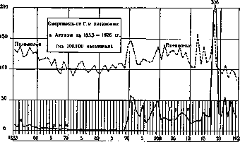
Figure 3. 193 and 322, in Portugal 174 and 246 (Fig. 3 shows mortality from influenza and pneumonia in England from 1853 to 1926 by years). As a result, during the influenza epidemic, a sharp increase in overall mortality is observed. The number of deaths from the 'spanish flu' throughout Europe was about 3,000,000 people, in the United States 450,000 people died (in 1918 mortality from influenza was 299 per 100,000 population), in Japan 250,000 people, in British India about 6 million people. The total number of deaths from the 'spanish flu' on the globe is estimated by some authors (Laumonier) at 20 million people, i.e., in an amount 3 times exceeding the losses of humanity for 51 months of the world war (Mollers). Mortality from influenza in the USSR (Russia) can be judged by the following data relating to Moscow, Leningrad, Saratov, and Odessa (see Table 4). Table 4. Mortality from influenza in some cities of the USSR (per 100,000 population). Years Moscow Leningrad Saratov Odessa. In Moscow and Leningrad (see Figure 4) the highest mortality from influenza was observed in 1919, in Saratov and Odessa in 1918. In Leningrad it was significantly higher than in other cities. If we proceed from these figures and assume the average mortality from influenza for the USSR for 1918 at 720, and in 1919 at 990 per 1 million inhabitants, the total number of deaths from influenza for both these years (with 130 million inhabitants according to the 1920 census) will be about 222,000 people. The highest mortality from the 'spanish flu' was observed in American and European cities in the autumn of 1918 (in October-November), in the cities of British India in the second (autumn 1918) and third waves of the epidemic (in January-February 1919). Corresponding data for some cities are given in Fig. 5. Fig. 6 shows mortality from influenza in the epidemic of 1889-90 for the same cities (according to Vaughan). A comparison of Figs. 5 and 6 gives an idea of the difference in the character of these epidemics. Mortality in the 1918 epidemic was 2 times higher in European cities than in the 1889-90 epidemic, and 6 times higher in American cities. At the end of 1926 and the beginning of 1927, a rise in influenza was again observed in a number of European countries. A comparison of mortality from influenza for 1925-26 (from July 1, 1925 to June 30, 1926) and for 1926-27 (from July 1, 1926 to June 30, 1927) gives the following figures (see Table 5).
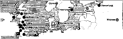
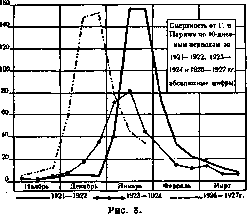
Table 5. Mortality from influenza in some countries and cities of Europe in 1925-26 and 1926-27 (per 100,000 population). States and groups of cities 1925-26 1926-27. Czechoslovakia... Scotland... 107 cities of England... 49 of Germany... 14 of Netherlands... 49 of Spain... An increase in mortality from influenza in 1927 is noted in almost all major cities of Europe (see Figure 7). Among non-European cities, an increase in mortality from influenza in 1927 was noted in the cities of Japan, Africa, and some cities of South America; in the latter, mortality from influenza has generally been registered at a very high level in recent years. In Moscow and Leningrad, the influenza outbreak began in the spring of 1926, and mortality from influenza in both these cities for 1926 and 1927 was significantly higher than in previous years (see Table 4). On the character of the movement of the 1927 epidemic in some cities, see Figure 8 (mortality from influenza by ten-day periods in Paris).
Mortality from influenza in cities and rural areas is not the same. The corresponding mortality in the cities of the United States, according to Vaughan, is presented in Table 6. Mortality from influenza in rural areas in individual years exceeded that in cities by up to two times; in 1918, no noticeable difference in mortality between cities and rural areas was observed (see Figure 9 - mortality from influenza in cities and rural areas in England). In the Netherlands, in populated areas of various types, mortality from influenza was as follows (see Table 7). Table 7. Mortality in various populated areas of the Netherlands (per 100,000 population). Types of populated areas Years. As a rule, with an increase in population in cities, mortality in them decreases. Mortality by age and sex. Mortality by age during influenza epidemics and in years free from it varies. It is also different in different epidemics. Table 8 shows what number of deaths from influenza (per 1,000 deaths from influenza at all ages) fell to individual age groups in different years. While in non-epidemic years the main part of deaths from influenza falls on older ages, in 1918 the highest mortality...
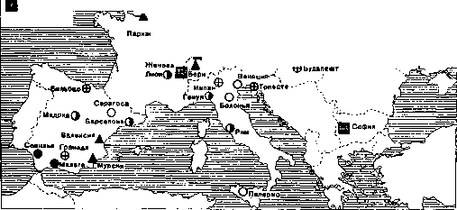
Figure 7. Mortality from influenza in major cities of Europe from July 1926 to June 30, 1927.
Table 8. Mortality from influenza by age in England and France (per 1,000 deaths at all ages). England 1909 y. 1910 y. 1918 y. 1920 y. 16-35 »...... 36-55 »...... 56-75 »...... 75 years and older . . France 1912 y. 1913 y. 1918 y. 1920 y. 0- 1 years..... 1-19 »...... 20-39 »...... 40-59 »...... 60 years and older . . occurred in the age groups from 16 to 40 years. The difference in mortality by age groups in the epidemics of 1889-90 and 1918 can be seen from the data given for Germany (see Figure 10). While in the 1889-90 epidemic the highest mortality occurred at an older age, with no mortality observed at all among the insured under 30 years of age, in 1918, conversely, this group yielded the highest mortality (excluding the group over 80 years). Mortality from influenza by age and sex in 1918 can be characterized by the following figures for Switzerland (see Table 9). Table 9. Mortality from influenza by age and sex in Switzerland in 1918 (per 100,000 population of the corresponding group). Age Males Females 0-14 years...... 15-19 »....... 20-29 »....... 30-49 »....... 60 years and older . . . The highest mortality in both sexes occurred at the age of 20 to 29 years. Men, with the exception of the children's age, showed higher mortality than women. Mortality in Moscow by age and sex can be illustrated by the following figures (see Table 10). Table 10. Mortality from influenza by age and sex in Moscow (per 100,000 persons of the corresponding group). Years Age m. Ht. both sexes 0- 4 years. . 7,9 9,8 22,1 18,2 13,6 15,9 5- 9 » 0,2 0,6 2,6 10-14 » 0,5 0,1 15-19 » 0,1 0,7 1,8 0,2 0,1 20-29 » 0,1 1,4 2,3 0,2 0,2 0,2 30-39 » 0,1 2,1 3,7 0,1 0,3 0,2 40-49 » 0,4 2,1 4,9 0,8 0,3 0,5 50-59 » 0,6 3,4 9,7 1,3 1,2 1,2 60 y. and olde r 4,8 10,9 23,9 2,2 5,9 5,1 The highest mortality from influenza during these years was observed in early childhood. In 1918-19 it increased particularly in the age groups over 30 years. Influenza in 1926 presents a mortality pattern approaching that of 1917 (see Figure 11). No noticeable difference in mortality by sex is observed according to Moscow data. ranged from 4.6% (Müller) to 10% (Brasch), and in Giessen—20% (Bechner). In cases complicated by pneumonia, the case fatality rate reached 65% and 80% (Mayer). Morbidity. Registration of influenza cases is conducted in a small number of countries. The number of cases in some of them since 1919 is expressed by the following figures (see Table 11). Table 11. Number of registered cases of influenza in various countries (absolute figures). States 1919 y. 1920 y. 1921 y. 1922 y. 1923 y. 1924 y. 1925 y. 1926 y. 1927 y. Norway (urban) . . Czechoslovakia . . . Switzerland..... 86.058 210.000 814.149 219.018 20.153 2.058 20.569 37.721 537.966 237.636 1.098.443 79.648 26.334 1.407 2.150 18.032 366.118 1.879.170 73.098 30.292 30.519 80.998 4.902.365 263.993 28.725 310.806
Fatality (mortality rate). The incompleteness of influenza registration makes it difficult to calculate the fatality rate from it. In Switzerland in 1918, the number of influenza cases is estimated at 2 million people (see below). Died during this year

from influenza 21,000 people, i.e., the fatality rate is about 1%. In the entire French army in 1918, 30,382 people died from influenza out of 408,180 registered patients, i.e., the fatality rate relative to the number of registered patients is about 7%. Mortality from influenza in cities and rural areas of England (per 100,000 population). rural areas. 191|| 12 | 13 | U | 15 | 16 } 17 | 18 | 19 | 20 | 21 ) 22 | 23 | 34 [>925|l926i Figure 9. patients is about 7%. Prein calculates the fatality rate from influenza in 1918 for the garrison of the city of Schwerin to be 6.8%. In hospital institutions, where the most severe patients ended up, the fatality rate from influenza in 1918 was significantly higher: in Munich it was estimated Morbidity per 10,000 population in recent years by individual countries is as follows (see Table 12). Table 12. Countries Years Norway (urban) Czechoslovakia Switzerland . . Sweden .... 64,0 705,1 649,2 154,6 155,2 103,0 236,0 330,0 1,0 5,5 30,0 140,0 216,5 130,8 77,6 51,0 203,2 173,1 15,8 107,1 5,9 13,6 67,4 319,4 209,6 46,1 195,9 3,1 39,6 17,4 325,7 756,2 361,3 223,4 1555,1 These tables give in particular some idea of the rise of influenza in 1922 and 1927. Registration of influenza hardly reflects the actual morbidity. On the one hand, ?6 27.13 / 1889- 1890 gg. 1918 g / ?4 / Г> / Mortality from influenza by age groups in Germany in 1889-1890 and 1918 (per 1,000 insured). No. p io.so/ 47Vl9 4 42/ ^W! 16?^^^. ^^08 g.p^-' .^-"gvb 1.01 | Ш9-1S90153 °IS2 15-30 p. 31-40 41-50 Ы-60 ) 61-70 81-90 Figure 10. this infection group includes, especially during epidemics, many unclarified cases (according to the observations of pathomorphologists, the number of erroneous diagnoses of influenza and abuses of this diagnosis is very large); on the other hand, many patients with mild forms of influenza do not consult a doctor and therefore do not get registered. Official registration figures should therefore be regarded as approximate. Any complete information about the morbidity
of influenza in 1918 is not available. According to official data, 644,463 cases were registered among the civilian population in Switzerland in that year, and 33,721 among military units. If for 1 registered patient one counts 2 unregistered ones, the total number of influenza cases in Switzerland for that year will be about 2 million people, i.e., about half of the entire population. In Vienna, about 1/3 of the population fell ill with influenza during the second half of 1918. In Oslo, according to official data, 10.6% of the entire population fell ill with influenza during the same year. In
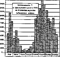
Figure 12.
the French army, 12.6% of patients with influenza were registered (see Figure 12; the figure gives an idea of the difference between the first and second waves of the "Spanish flu"). In the city of Schwerin (Germany), among the garrison, 6.1% fell ill during the first wave of the epidemic, 13.8% during the second wave, total 19.9%; among members of the sick fund, 3.4% during the first wave, 15.4% during the second, total 18.8%. These figures give an approximate idea of influenza morbidity in the 1918 pandemic. More or less exact information on influenza morbidity in 1918 in the USSR is not available. 1,238,967 patients were registered across 34 gubernias. This figure gives only some idea of the true morbidity of influenza. For the period from 1908 to 1915, from 2.5 to 3.5 million cases of influenza were registered annually in Russia, or from 200 to 250 cases per 10,000 population. For the period from 1917 to 1921, morbidity figures are very incomplete, and only from 1923 do they begin to arrive more completely. Gradually increasing, the number of registered influenza cases in the USSR reaches 5 million in 1927. In terms of the number of registered cases, influenza significantly surpasses all other infections. In Fig. 13
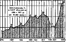
Figure 13.
morbidity in the USSR is presented for the period from 1896 to 1927 (1917–1921 are omitted). The number of registered patients by individual republics of the Union in recent years is as follows (see Table 13). Table 13. Influenza morbidity by republics of the USSR (per 10,000 population). Republics 1924, 1925, 1926, 1927. Ukraine... Belarus... Azerbaijan... Armenia... Georgia... Turkmenistan... Uzbekistan... Influenza is registered most highly in the RSFSR, followed by Belarus, Ukraine, the Transcaucasian and Central Asian republics. Morbidity by individual regions of the RSFSR (per 10,000 population) is shown in Table 14. The highest morbidity in recent years is shown by Crimea and the Central Industrial Region. The growth of influenza in 1926–1927 is noted in all regions of the RSFSR. Morbidity by age and sex for Moscow Governorate and the city of Moscow (per 10,000 population) can be seen from Table 15. The highest morbidity is observed both in the city of Moscow and in the governorate at the age under 1 year. The next groups giving higher morbidity are groups from 1 year to 4 years and from 20 to 49 years. Morbidity in 1926 is higher than in the pre-war period in all age groups. Table 14. Regions: North-Eastern... North-Western... Western... Central-Industrial... Central-Chernozem... Mid-Volga... Lower Volga... Vyatka-Vetluga... Ural... Bashkir Republic... Crimea... North Caucasus... Dagestan... Siberia... Yakut Republic... Buryat-Mongol Republic... Kyrgyz Republic... Kazakhstan... Far East... Table 15. Influenza morbidity by age and sex in Moscow and Moscow Governorate (per 10,000 persons of the corresponding group). Moscow Governorate, Moscow, Age, 1906–1910, 1926, 1926. Table 16. Number of deaths from influenza by states and cities (absolute figures). England (107 cities)... Hungary... Germany (48 cities)... Netherlands... Spain... Switzerland... Scotland... Czechoslovakia... Japan (21 cities)... Berlin... Vienna... Leningrad... London... Moscow... New York... Paris... Rome... Seasonality of influenza spread. Epidemic influenza mostly begins in winter or spring. According to Hirsch, out of 125 influenza epidemics, 50 began in winter, 35 in spring, 16 in summer, and 24 in autumn. Endemic influenza also has its maximum development in the winter-spring months. When summing up the number of deaths from influenza by trimesters, one can see that in most countries the maximum of deaths falls on the first trimester. The corresponding figures for some countries and cities over the past two years can be seen from Table 16. This table is also of interest because it gives an idea of the scale of the 1927 epidemic. A sharp increase in mortality from influenza is noted everywhere in 1927, which falls almost exclusively on the first trimester of this year. No less characteristic is the distribution of influenza cases by trimesters, as can be seen from Table 17. The first trimester of 1927 in Western European states gives an exceptionally high morbidity compared to other trimesters. In the RSFSR, as in other republics of the Union, where the rise of influenza began in 1926, no noticeable difference between morbidity by trimesters of 1926 and 1927 is noted. The distribution of the number of registered patients by months of the year can be judged by the attached diagrams. Fig. 14 shows the monthly distribution of cases for 1925–1927 in Denmark, Sweden, the RSFSR, Ukraine, Leningrad, and Moscow. In Denmark, the maximum of cases falls in 1925 in April, in 1926 in May, in 1927 in January; in Sweden in 1925 in April, in 1926 and 1927... For the RSFSR and Ukraine, the maximum of cases falls in February (1925) or March (1926 and 1927). For Moscow and Leningrad, the maximum in 1925 falls in January, in 1926 in March; in 1927 in Moscow in March, and in Leningrad in April. The outbreak of the influenza epidemic in 1926, as can be seen from these diagrams, began in Moscow and Leningrad in February, and in Moscow it was expressed much more strongly. Influenza and occupation. According to the Central Social Insurance Administration, based on the processing of sick leaves and relating to cases of illness with loss of working capacity, in the USSR in 1925 influenza morbidity was expressed in the figures given in Table 18. The conclusions from this table are as follows: 1. Women in all branches of labor in 1925 gave a relatively greater number of cases of influenza with loss of working capacity than men, and a greater number of days of illness from influenza. The average duration of 1 case of influenza in most branches of labor among women is slightly longer than among men. 2. Among production groups, influenza serves as a cause of loss of working capacity significantly more often than among non-production ones. The number of influenza cases reached 30.17 per 100 insured in the match industry, 34.84 in the production of lead paints, and dropped to 3.0 per 100 insured for persons engaged in stage activity, and to 2.33 for pedagogical and scientific-literary workers. 3. The average duration of one influenza illness among non-production groups is slightly higher than among production groups. In relation to all cases of illness with loss of working capacity, influenza in 1925 occupied among the insured...


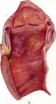
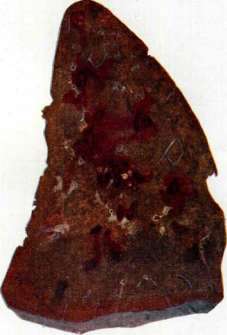
Fig. 1. Gingivitis catarrhalis s. simplex. Fig. 2. Gingivitis hypertrophica. Fig. 3. Acute hemorrhagic-purulent tracheitis. Fig. 4. Hemorrhagic (mottled) influenzal pneumonia. (Figures 1 and 2 orig.; figures 3 and 4 from specimens of the Pathologo-Anatomical Institute of the 1st Moscow State University.) To art. Gingivitis. Influenza. Table 18. Morbidity of influenza by various branches of labor in 1925. Mining and processing industry. Railway transport. Water transport. Public and scientific educational institutions. Supply. Public health. Soviet and administrative institutions. Number of cases (per 100 insured). Number of days of illness (per 100 insured). Average duration of one illness (in days). ... [The table data continues with statistical figures for men and women across various branches of labor].
i.
Dobreitzer. Pathological Anatomy. Pathologo-anatomical changes in mild forms of banal influenza boil down to acute catarrh of the mucous membrane of the nose, nasopharynx, larynx, trachea, bronchi, and sometimes the accessory sinuses of the skull. The mucous membrane appears hyperemic, swollen, secreting first a serous and subsequently a mucous and mucopurulent exudate. When such cases are complicated by pneumonia, autopsy usually reveals catarrhal bronchopneumonia, often confluent, or pneumonia of a catarrhal-fibrinous character; more rarely, typical croupous pneumonia. Involvement of the pleura is frequently observed, sometimes of the pericardium (fibrinous and serous-fibrinous pleurisy and pericarditis). In severe forms of influenza, namely those which from the very beginning have the character of a severe disease and are frequently encountered during the epidemic spread of influenza (epidemic influenza, Spanish disease) and lead to the death of young and robust subjects, various pictures can be found at autopsy. In general, pathological anatomy establishes two main types of changes. 1. In the rarer "acute influenzal toxicosis" (Oeller), or "purely toxic form" (Korach), in which death occurs in the very first days (sometimes hours) of the disease, the changes are characterized by sharply pronounced blood stasis and stasis in internal organs, especially in the brain, multiple petechial hemorrhages (often in the adrenals), degenerative changes in parenchymatous organs, hyperplasia of lymph glands and the spleen, degenerative-inflammatory changes in sympathetic nerve ganglia, and waxy degeneration of muscles. Death in such cases is the result of toxic damage to vegetative centers and ganglia (possibly also the adrenals) and subsequent vasomotor paralysis with a drop in blood pressure. 2. In more frequent cases with damage to the respiratory organs, ending in death no earlier than the 4th-5th day of the disease, various pictures are discovered on the dissection table depending on whether: a) there is inflammation only of the respiratory tract without involvement of the lungs, b) the lungs are involved, c) there is a complication in the form of suppuration. At the autopsy of cases ending in death with damage only to the respiratory tract, one may encounter the picture of catarrhal or catarrhal-hemorrhagic, and sometimes fibrinous-hemorrhagic inflammation of the larynx, trachea (see sep. table, fig. 3) and large bronchi, along with general changes characteristic of influenzal toxicosis, or the picture of widespread bronchitis and bronchiolitis with catarrhal or catarrhal-fibrinous exudate, sometimes with acute diffuse bronchiectasis, is discovered. In protracted cases of the latter kind in the small bronchi, metaplasia of cylindrical epithelium into squamous is observed, and with damage to the epithelium, overgrowth of the bronchial lumen with connective tissue (bronchiolitis obliterans). In cases with widespread damage to the small bronchi, death frequently occurs from asphyxia. Inflammatory changes in the lungs in severe forms of influenza are extremely motley. In general, one can observe: diffuse serous-hemorrhagic pneumonia, multiple focal bronchopneumonia, confluent catarrhal pneumonia, fibrinous pneumonia, and finally dissecting pneumonia (pneumonia dissecans), consisting of leukocytic (purulent) infiltration spreading along the interlobular connective tissue. Sometimes the lungs are captured to one degree or another by an inflammatory process of one of these types, but more often one has to observe a very motley combination of lung changes, which produces the very peculiar and extremely characteristic picture of lung changes for severe influenza, which Glaus and Fritzsche so aptly designated as the "large mottled lung" (see sep. table, fig. 4). In such a lung on section, areas of bloody imbibition alternate with dark brown granular regions and gray lobular foci. This motleyness is supplemented by the emergence of atelectases, as well as lobular necroses of the lung tissue, appearing in the form of gray-yellow multifaceted foci on the lung section and under the pleura, covered with a delicate coating of fibrin. Suppuration is one of the frequent complications of influenzal pneumonia. Sometimes it proceeds along the bronchial walls, eroding the latter and forming peculiar branching bronchiectatic cavities. Suppuration often develops in the center of the pneumonic focus with the formation of an abscess. Finally, suppuration of necrotic foci can occur, especially those located subpleurally. These suppurations can be the cause of purulent pleurisy and empyema. Among complications from other systems at the autopsy of cases of severe influenza, one sometimes finds: hemorrhagic encephalitis, meningitis, endocarditis, thrombosis of peripheral vessels with subsequent gangrene of a limb, purulent otitis and mastoiditis, and damage to the urinary tract (especially in early infancy); in the kidneys, pictures of nephrosis and glomerulonephritis, sometimes hemorrhagic. From the pleura, inflammation can pass to the mediastinum, pericardium, peritoneum (purulent mediastinitis, pericarditis, peritonitis). Finally, suppuration in the lung is frequently a source of septico-pyemia with metastatic abscesses in various organs. However, in such cases it is more correct to think that almost from the very beginning of the disease it was a matter of a septic disease. In the early death of patients with damage to the respiratory organs, the general changes found at autopsy are the same pictures as in toxic forms, but less intensely expressed. Upon death in later periods, these changes are even weaker. Finally, in cases complicated by suppuration, the changes in the remaining organs have a banal septic character. As remote consequences of past influenza, the possibility of the development of bronchial cancer (see Bronchi) is pointed out, as well as the development of general amyloidosis in connection with the long-term existence of post-influenzal bronchiectasis (Davydovsky). In conclusion, it should be noted that the clinical diagnosis of influenza very often finds no anatomical confirmation (Lubarsch).
A.
Abrikosov. Clinical picture. The causative agent of influenza invades the body through the nasopharynx and upper respiratory tract, actively multiplies, and then rapidly perishes, producing a general intoxication which underlies all pathological phenomena. The pure influenza virus by itself usually does not cause severe disease and apparently persists in the body of the patient for a relatively short time. It is characterized by the property of suppressing the body's defense functions, thereby creating conditions for the invasion of secondary infection agents, which extremely complicate the picture of the disease and worsen its course. Some authors base the pathogenesis and explanation of the severity of influenza on various reactive features of the organism itself, pointing to the significance of the intensity of reactive processes or their peculiarity (allergy, hyperergy): the more intense this reaction, the more severe the general picture of the disease. This is also used to explain the paradoxical fact of greater severity of influenza and mortality in individuals of good build and nutrition. The incubation period in influenza does not exceed 1–2 days; in some cases, it is equal to only a few hours. According to Hamburger (F. Hamburger), the disease can also develop without a latent period. Prodromal phenomena, not always clearly expressed, amount to malaise, loss of appetite, and chilliness. The onset of the disease is as a rule stormy: chills, fever, severe pains in the head and lower back are frequent. Sometimes the first symptom of influenza is profuse epistaxis or, as noted in 1889, a syncopal state. The patient is depressed, complains of body aches; at the same time, catarrhs of the conjunctiva of the eyes, nose, nasopharynx, and sometimes the larynx and bronchi appear. The further course of the disease develops very diversely depending on the particular localization of the infection and on the accompanying complications, which are characterized by extreme diversity in the sense of their localization and character. From this point of view, the isolation of special forms of influenza by localization or by the predominant manifestation of infections (see below) may encounter objections, since in the corresponding processes one can see not this or that localization of the primary infection, but the manifestation of a secondary infection. However, such a subdivision, for all its conventionality, is justified by the clinical significance of those diverse pathological processes by which influenza manifests itself. In any case, drawing a line between pure and complicated forms of influenza is not always easy. These circumstances explain the variety and multiplicity of proposed clinical classifications. Glinchikov, for example, distinguishes the following forms of influenza. 1. Pulmonary form, with the following subdivisions: a) lobular forms, b) pseudolobar forms proceeding without phenomena of hepatization of the lung tissue, c) pseudolobar forms proceeding with clearly expressed phenomena of hepatization, d) splenopneumonic forms (Transche's form). 2. Catarrhal form: a) with involvement of the upper respiratory tract, b) with involvement of bronchi down to the smallest. 3. Intestinal form: a) catarrhal, b) with ulcerative lesions. 4. Nervous form: a) with involvement of the central nervous system, b) with involvement of the peripheral nervous system. Strümpell distinguishes 4 forms of influenza: 1) toxic, or typhoid, 2) catarrhal, 3) rheumatoid, and 4) gastrointestinal, emphasizing at the same time the presence of numerous transitional and combined forms. Strümpell does not single out a special pulmonary form, referring pneumonia to the number of complications of influenza. Enriquez and Carrier, as well as Lereboullet, divide it into 1) uncomplicated influenza, 2) complicated influenza, and 3) complications of influenza, understanding by the latter diverse processes not always characteristic of influenza as such and caused both by the influenza virus itself and by the agents of secondary infections. In view of the practical value of such a subdivision (which by no means can serve as a classification of the forms of influenza), the following will be considered: 1) uncomplicated influenza (including here the hypertoxic form of influenza), 2) respiratory form of influenza, 3) intestinal form of influenza, and 4) nervous form of influenza (the last three forms in the scheme of Enriquez, Carrier, and Lereboullet belong to "complicated influenza"). 1. Uncomplicated influenza. The temperature rapidly rises to 39.5–40° and higher and persists for 2–3, often 5–6 days. French authors give the name "bell-shaped" (en cloche) (see Fig. 15) to a regularly rising and soon falling curve. Temperature 2 3 4 for 36.5 Figure 15. Mild form of uncomplicated influenza. Figure 16. Normal course of influenza. Often on the 2nd–4th day of the disease, a drop in temperature occurs almost to normal, replaced however after 12–14 hours by a new rise to the previous level (bileptic curve, v grippal of the French) (see Figure 16). The temperature usually gives significant fluctuations, accompanied by chills during rises and sweats. Redness of the face and conjunctivitis are often observed, suggesting measles, and sometimes typhus, in connection with catarrhal phenomena. Enlargement of the spleen and liver is rarely observed and is by no means characteristic of influenza. The tongue is usually coated; some authors describe a "porcelain tongue" covered with a white coating, sometimes with red edges, and even attach diagnostic significance to it. The pulse is usually accelerated, less often slowed, always very labile. Catarrh of the upper respiratory tract usually does not reach large degrees; runny nose is not sharply expressed, but cough, dry at first, is sometimes very agonizing. Diffuse bronchitis soon joins laryngitis and tracheitis. Sputum is mucous or mucopurulent. Shortness of breath in influenza strikes the eye sharply and does not correspond to the degree of damage to the respiratory apparatus. Urine is concentrated, sometimes contains traces of protein; the diazo reaction gives a positive result only in more severecases. Nosebleeds and premature appearance of menstruation are frequent. On the part of the blood, leukopenia is constantly observed (after the initial neutrophilic leukocytosis on the 1st day of the disease), which persists until the end of the fever. The number of lymphocytes drops especially sharply, which however already by the 4th day return to the normal level (Schotter). Sometimes influenza is accompanied by rashes (measles- and scarlatiniform). The disease ends with a lytic drop in temperature over 2–3, less often up to 7 days. Uncomplicated influenza proceeds easily and does not create an immediate threat to life. The hypertoxic form of influenza proceeds with phenomena of general severe intoxication. The temperature reaches 41–42° and does not decrease until death occurs (see Figure 17). Leaden coloration of the face and extremities, tremor of the fingers, twitching of facial muscles, an excited state reaching delirium, or, conversely, prostration turning into coma—such are the external manifestations of this form of the disease, depending on severe intoxication of the central nervous system and on sharply expressed heart failure and peripheral circulatory disorder. Jaundiced coloring of the skin and mucous membranes often appears, sometimes accompanied by enlargement of the liver. Protein is constantly detected in the urine in large quantities; the amount of chlorides decreases, while the urea content in both urine and blood reaches high figures. Catarrhal and inflammatory phenomena on the part of the respiratory apparatus may be completely absent or expressed very insignificantly. When coughing, scanty sputum is released (mucous or sometimes bloody). Shortness of breath (mainly of central nervous origin) reaches extreme degrees. Death usually occurs on the 4th–5th day from asphyxia or with phenomena of liver and kidney failure. Fulminant forms ending in death within the first day are very rare. 2. The respiratory form, most frequently encountered, presents at the same time the greatest variety of clinical and anatomical phenomena. A. Involvement of the upper
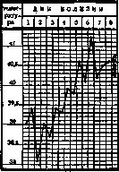
Fig. 17. Hypertoxic influenza. 113 respiratory tract. Almost every case of influenza begins with or is accompanied by inflammation of the nasal mucosa, which in itself is not of any serious significance, but often leads to involvement of the accessory cavities (Highmore's cavity, frontal sinuses). Inflammation of these cavities already affects the course of the disease in a noticeable way, sometimes taking on a purulent character and causing in many cases stubborn and distressing pains in the forehead and face. Nosebleeds are very frequent in influenza. Inflammation of the middle ear is frequent (especially in children); despite the relative benign nature of these otitis media, they can serve as a starting point for the development of meningitis or cerebral sinus thrombosis. Among other lesions of the upper respiratory tract, laryngitis and tracheitis should be noted, accompanied by sharp pains in the larynx and behind the sternum, especially with coughing fits. B. Grippal bronchitis (usually purulent) is a frequent complication that significantly worsens the prognosis, especially in the case of the process spreading to the small bronchi (bronchiolitis). Rales of various calibers, initially scattered and changing from day to day, sometimes spread throughout the lung, while shortness of breath sharply increases and cyanosis appears. The disease can end in death, especially in children and the elderly. Sputum is mucopurulent, often containing blood in the form of streaks or lumps. Sometimes bronchitis leads to the formation of bronchiectasis or to bronchiolitis obliterans. C. Pneumo_
nia. A typical feature of all influenzal pneumonias is the multiplicity of small scattered pneumonia foci (bronchopneumonia) and their tendency to coalesce (confluent pneumonia). The course of influenzal pneumonia can exhibit great fluctuations. In some cases, the tendency to spread circumferentially, characteristic of influenzal pneumonias, stands out particularly sharply, with large sections of the lung becoming involved in the affliction, up to an entire lobe (pseudolobar pneumonia). The process usually affects both lungs to one degree or another. Pneumonia usually joins on the 3rd to 5th day of the disease; late pneumonias are also observed on the 2nd to 3rd week, already after the fall of the temperature. The onset of pneumonia is marked by chills, a rise in temperature by 1--2° (see Figure 18), a sharp deterioration in the patient's well-being, and an increase in cough. Auscultation and percussion initially reveal a localized focus of crepitation with weakened respiration and a clear percussion tone. Sputum is mucopurulent, often admixed with blood, foamy, which indicates a certain degree of pulmonary tissue edema. The general condition of the patient is severe. Dyspnea does not correspond to the size of the focus; the pulse is weak, frequent. As a rule, cyanosis is observed. Depending on further development and the intensity and spread of the process, the auscultatory and percussive phenomena correspond to diffuse catarrhal, nodular, or confluent bronchopneumonia, or closely approach the phenomena characteristic of croupous pneumonia (dullness, bronchial breathing, increased bronchophony, etc.). A special form of influenzal pneumonia is described — splenopneumonia (Grancher, Glinchikov), in which objective symptoms (solid dullness in the lower parts of the lungs, sharply weakened respiration in this area, weakening or absence of vocal fremitus, sometimes aegophony) simulate pleurisy. This form is based on inflammation of the pulmonary parenchyma with an accumulation of liquid exudate poor in fibrin, in which the degree of density characteristic of croupous pneumonia ("hepatization") is not achieved, but a lesser one, which Grancher compares to the density of the spleen (see Grancher disease). The course of influenzal pneumonia is protracted. In cases showing no tendency to migration, the duration of the disease is about 14 days; cases lingering up to 1½-2 months are not uncommon. The 1918 epidemic was particularly characterized by an extremely severe course of pneumonias, obviously explained by the peculiarity of secondary infections (diplostreptococcus of Ivashentsov, Manukhin and Sokolova, etc.). In many cases, the general picture of these pneumonias was so severe that it led to the erroneous diagnosis of pulmonary plague (Armand-Delille). According to the observations of some authors, in pulmonary forms of influenza, enlargement of the spleen is often observed (Obolensky, Glinchikov, Widal). — Influenza edema of the lungs of a greater or lesser degree sometimes accompanies influenzal bronchitis and pneumonia, imparting a characteristic imprint to the picture of these processes (Henriques and Carrier, Lerbuillet). Sometimes it comes to the foreground and concludes the course of the disease. Sometimes, finally, after several days of indisposition, with insignificant or almost absent phenomena on the part of the respiratory apparatus, the picture of acute pulmonary edema rapidly develops, and the patient dies in a few hours or at most in 2-3 days. Cases of recovery are rare. Pulmonary edema can be the result of both acute heart failure and progressive circulatory disorders under the influence of general intoxication and damage to the chromaffin system. — D. Pleurisy, often bilateral, frequently joins influenzal pneumonia. Dry pleurisy has relatively little effect on the course of the disease. Effusive pleurisy (serofibrinous, purulent, hemorrhagic), which occurs more frequently in influenza than dry pleurisy, deserves much greater attention. With a small amount of exudate, diagnosis presents difficulty, as respiratory sounds and vocal fremitus may be preserved. Recognition is facilitated by trial puncture, and in interlobar pleurisy by X-ray. 3. Intestinal form. Not to mention the fact that certain phenomena from the gastrointestinal tract are quite often observed in all forms, they sometimes predominate so much in the clinical picture of the disease that some authors single out a special intestinal form. In any case, it occurs extremely rarely, and moreover not in every influenza epidemic. Many cases of this form proceed with a normal or subfebrile temperature. Clinically, this form appears in the form of catarrhal (sometimes hemorrhagic) enteritis and colitis. Cholera-, dysentery-, and typhoid-like cases are observed, and the pathoanatomical picture resembles to a certain extent the picture of the corresponding infections. Sometimes, conversely, the disease is accompanied by constipation. Respiratory catarrhs sometimes accompany the intestinal form. 4. Nervous form. Phenomena on the part of the nervous system accompany almost every case (headache, body aches, dizziness, clouding of consciousness, delirium, neuralgia, cutaneous hyperesthesia, etc.), but sometimes nervous symptoms sharply come to the foreground. Among affections of sensory nerves, neuralgia and various sensory disorders are frequent. Neuralgia is usually localized in the region of the trigeminal nerve (pain in neuralgia of the upper branch should not be confused with pain in inflammation of the frontal sinuses!), sciatic, and intercostal nerves. Hyperesthesia, as well as paresthesia and anesthesia (especially in the sense of loss of taste and olfactory sensations), are frequently observed. In the region of motor nerves, neuritis and polyneuritis are encountered, leading to paralysis of individual muscles or groups of them (paralysis of the extremities, accommodation, palate, hypoglossal and facial nerves, etc.). Rapidly developing cases of polyneuritis can present the picture of Landry's ascending paralysis. Paralyses in influenza appear in the recovery period and generally give a favorable prognosis. On the part of the brain membranes, apart from mild phenomena of their irritation, meningitis (serous and purulent) is encountered, especially when influenza is complicated by pneumonia. Their development depends on the carriage of pathogens through the blood or on the transition of the purulent process from the accessory nasal cavities or from the middle ear. In addition to pneumococcal and streptococcal flora, Pfeiffer's bacillus is often obtained in cultures in influenzal meningitis. Spinal cord lesions in influenza are rare (paraparesis of the lower extremities, paralysis of the urinary bladder). Conversely, brain lesions are not particularly rare. An acute form of hemorrhagic encephalitis is observed during the febrile period of the disease; the onset is sudden, with high temperature, accompanied by loss of consciousness, with convulsions and hemiplegia. Another form of influenzal encephalitis is later (sometimes a few weeks after influenza), developing slowly; initially it manifests only with general symptoms, and subsequently leads to paralysis in the region of individual nerves. Influenzal encephalitis must be distinguished from epidemic encephalitis, which clinically largely resembles the former. Epidemic encephalitis stands in some still unclear relation to influenza epidemics, but is caused by a different pathogen. In influenza, acute psychoses are encountered more often than in other infections, and in drunkards, delirium tremens. Psychoses in influenza — see Infectious psychoses. Complications. The course of the most frequently encountered forms of influenza can be extremely varied depending on complications or joining secondary infections. The nature of complications is dissimilar not only in various cases, but even more so during various epidemics. It is almost impossible to consider all complications of influenza, since for this it would be necessary to review almost the entire special pathology. One can limit oneself here to listing those of them that practically must not be overlooked when attending to a patient. These include (apart from those mentioned above in the description of the main forms of influenza): among heart complications, according to a few authors (Lerbuillet) — endocarditis and pericarditis, according to the majority — myocardial lesions, the presence of which explains a whole series of clinical phenomena (tachycardia or, conversely, bradycardia, arrhythmia, pain in the heart region, and attacks of cardiac weakness sometimes leading to a fatal outcome); among other complications — thrombophlebitis, abscess and gangrene of the lungs, parotitis, thyroiditis, nephritis (more often as a result of exacerbation of an old process), pyelitis, cystitis, periostitis, ostitis, osteomyelitis, arthritis (sometimes purulent), abscesses and hemorrhages in the muscles, iritis, keratitis, panophthalmitis, hemorrhages into the retina and vitreous body, gangrene of the skin and whole parts of the body (scrotum, extremities). Among the infections joining influenza, erysipelas, exacerbation of gonococcal focal infections, etc., should be noted. — Anergy in influenza. Influenza is accompanied by a peculiar decrease in the reactive capacity and defense forces of the organism: a positive Pirquet reaction is weakened or becomes negative, vaccination with smallpox vaccine does not cause the usual reaction (Netter and Porak), and the agglutination titer in those vaccinated against typhoid fever drops (Cayrel, Fontaine, and Descoffre).
Obviously this explains the frequency of secondary infections in influenza, especially in general hospital wards. As for tuberculosis, according to many authors, pulmonary forms of influenza have a very adverse effect on the course of tuberculosis, especially in pronounced cases of the latter. On the course of tuberculosis of the upper lobes in the initial stage and on benign cirrhotic processes, influenza, according to Hegler, usually has no effect. In any case, mass statistical data on mortality indicate an increase in mortality from tuberculosis during and after influenza epidemics (Lindhagen). Frequently, after suffering from influenza, the patient continues to run a low-grade fever, and this condition drags on for many weeks and even months. Small scattered bronchopneumonic foci are found in the lungs, X-rays give a mottled picture; such cases often have to be regarded as an exacerbation of the tuberculous process. But apparently some of these cases should be attributed to chronic complications of influenza—inflammatory foci in the pulmonary parenchyma, small empyemas, bronchiectasis (Ivashentsov, Shotter). Whether true chronic influenza exists is unclear and doubtful, although a number of authors [Filatov, Hellpach (Hellpach disease) and others] put forward this concept as a special form of the disease, in which for several months there is a remittent rise in temperature, accompanied by chills, lesions of the respiratory tract, and general weakness. In other cases, lesions of the nervous system are observed in the form of neuralgia or paresthesia or a lesion of the motor apparatus. Among individual phenomena characteristic of chronic influenza, the so-called "grippal knee" (sharp pain upon pressing the femoral condyles), bone pains, muscle and joint pains are noted. Prognosis and diagnosis. The prognosis in influenza largely depends on the form of the disease and the state of the organism. Uncomplicated influenza runs a mild course; however, one must always bear in mind the possibility of late complications, sometimes joining already in the convalescence period at a normal temperature. Intestinal forms also show a relatively mild course. Grippal pneumonias always present a serious threat to the patient, yielding an average of 20-30% mortality. Grippal encephalitis and meningitis give a bad prognosis. A good general state of the organism does not guarantee a mild course of influenza. In 1918-19, the highest morbidity and mortality were precisely in the prime of life. Influenza in pregnant women takes a particularly severe course (50% mortality and even more—Malartic and Saloz). In view of the frequency of myocardial damage in influenza, preceding heart diseases make one take the prognosis more seriously; lung diseases (emphysema, chronic bronchitis), liver, kidney, and thyroid diseases also worsen the prognosis. Finally, the prognosis worsens in heavy drinkers (potatores) and nervously burdened subjects. The diagnosis of influenza during an epidemic usually presents no difficulty, although pathologists point to significant abuses of this diagnosis. The acute onset, sharp depression, incomparable with the malaise in ordinary, so-called cold diseases, the tendency to give complications—these features make it possible to recognize the disease. Blood examination can serve as a subsidiary diagnostic method (see above). However, one must not lose sight of the fact that many infectious diseases in the first days can be mistaken for influenza and require differential diagnosis. Thus, it is sometimes necessary to exclude measles, typhoid fever, typhus (in the first days of the disease), epidemic encephalitis, and acute miliary tuberculosis, as well as acute articular rheumatism, malaria, and cerebrospinal meningitis. Treatment. There is no specific treatment for influenza. From the very beginning of the disease until a few days of the afebrile period have elapsed, the patient must remain in bed and be protected from sharp temperature fluctuations—from drafts and cold. By this, the development of many complications can be prevented. At the beginning of the disease, with high temperature and muscle pain, small doses of aspirin, pyramidon, phenacetin, and the like in combination with small doses of caffeine are appropriate. Quinine preparations are also recommended. A significant relief of the general condition, and sometimes a shortening of the duration of the disease, is provided by the diaphoretic method: abundant hot drink (infusions of raspberry, elderberry, linden blossom, tea with cognac, etc.). From the first hours of the disease, the state of the respiratory apparatus and heart must be monitored. In case of cardiac failure—oleum camphoratum, strophanthin, digalen, adrenaline. For severe shortness of breath, oxygen is useful. Recently, in the treatment of influenza, inhalation of chlorine has been used, usually obtained by adding potassium permanganate to hydrochloric acid. Otherwise, symptomatic treatment is carried out. The treatment of complications is conducted according to general rules (see corresponding diseases). In severe cases of pneumonia—anti-pneumococcal and anti-streptococcal sera; autohemotherapy and convalescent serum were also used. German authors recommend "Grippe-serum" (serum of a horse immunized against pneumococci and streptococci). Attempts at vaccine and protein therapy, which have not yet given convincing results, are permissible only in a clinical setting and require great caution. Undoubtedly useful, in the opinion of some authors, are injections of large doses of 20% oleum camphoratum (10 cm3 several times a day). German authors recommend optochin and eucupin in view of their specific action on pneumococci. With threatening pulmonary edema—bloodletting (200-500 cm3) and cardiacs. In the recovery period—caution in the general regimen of the patient (danger of late complications: pneumonia, encephalitis). Prophylaxis. Every patient must be isolated from others (by placement in a hospital or in a separate room; if neither is possible, the patient should be separated by hanging sheets around the bed). However, the effectiveness of these measures is weakened by the fact that during an epidemic, along with more severe, bedridden patients, there is a large number of patients who carry the disease on their feet and spread the infection. For the disinfection of objects that have been in contact with patients, boiling or washing with ordinary disinfecting solutions is sufficient. Among personal prophylactic measures, along with sensible hardening of the body, the importance of thorough hand washing before meals should be emphasized. Among public health measures during an influenza pandemic, it is necessary to point out the desirability of limiting crowds of people (theaters, festivities, etc.). Preventive vaccinations against influenza were tested quite widely in America and the West during the last pandemic. In view of the unclear question about the causative agent, polybacterial vaccines were usually used, into which were introduced in various combinations and relative amounts Bacillus influenzae, Pneumococcus (types I-III), Streptococcus haemolyticus and viridans, Staphylococcus albus and aureus, Bacillus friedlanderi, Micrococcus catarrhalis, Bacterium pneumoniae, etc. In a number of observations, a certain success was outlined both in terms of reducing morbidity and reducing mortality of the vaccinated (obviously due to the limitation of secondary infections and complications).
A. Sadov. Influenza in childhood. Influenza in childhood, just as in adults, is not a process that is sharply defined in etiological, clinical, and epidemiological respects. The epidemiology and clinical picture of the process present peculiar features in children, due to which one must consider the process in children, as some authors do (Filatov, Comby), in two varieties--in the form of so-called pandemic influenza and in the form of endemic influenza. Pandemic influenza. During pandemics that have repeatedly struck the population of the globe, children were also involved in this process. The last two influenza pandemics (1889-90 and 1918-19) left a number of descriptions of this disease in children (Comby, Weil, Dufourt, Filatov, Koltypin). Children of older ages are affected; infancy is affected very little (which distinguishes this disease from endemic influenza). The disease, as in adults, is characterized by extreme rapidity of spread. In its severity, the process varied significantly in different epidemics; thus, in the epidemic of 1889-90 (Filatov, Comby) the disease was not very severe, and mortality was insignificant, which strongly distinguished this outbreak from the epidemic of 1918-19, which was characterized by significant severity, a large number of complications, and high mortality [11% overall, 28.5% in those complicated by pneumonia (Koltypin)]. In children, three forms in which the disease manifests can be distinguished: toxic, toxic-catarrhal, and complicated. In the toxic form, phenomena of general poisoning (damage to the nervous and vascular systems) come to the fore. In the toxic-catarrhal form, lesions of the upper respiratory tract--laryngo-tracheo-bronchitis--are added. Forms complicated by various kinds of inflammation of the lung and pleura run a particularly severe course. The onset of the disease is rapid, temperature rises to 39-40° and, making fluctuations of various magnitudes, remains as such for 5-10 days, falling by irregular short lysis. Nosebleeds, characteristic pains in the arms and legs, hyperesthesia, and general weakness are frequent. Upon examination, the skin is clean, only in rather rare cases do we encounter transient erythema; the tongue is moderately coated, there is hyperemia of the fauces and pharynx. Cough, if present, is often dry and barking. In the toxic-catarrhal form, auscultation reveals a large number of dry and often small moist rales concentrated in the posterior lobes of the lungs; emphysema. Sputum is sometimes mixed with blood. In severe forms, cyanosis of the lips and extremities, and shortness of breath are often noted. A characteristic sign on the part of the cardiovascular system is bradycardia (despite high temperature, the pulse is slowed down to 60 beats per minute). In more severe cases at the height of the process, a drop in blood pressure, expansion of the heart borders, and muffling of the tones are observed. In the urine, there is sometimes protein and casts. Leukopenia with a predominance of lymphocytes is noted in the blood; even in cases complicated by pneumonia, leukocytosis is insignificant. These peculiar features of the process--cyanosis, early drop in blood pressure, bradycardia, emphysema, adynamia, leukopenia, and among complications, the predominance of respiratory tract lesions--gave the right to some authors (Elistratov, Andreyev, Koltypin) to express the opinion that the endocrine-vegetative apparatus (sympathoadrenal function) suffers severely in this process, which is confirmed by the changes that were actually found histologically in the ganglia of the sympathetic system in this disease by Abrikosov and Mogilnitsky. Among complications, lesions of the respiratory tract predominate: laryngitis, bronchitis, pneumonia, serofibrinous and purulent pleurisy. The so-called Spanish croup is peculiar--a severe lesion of the larynx leading to stenosis, with phenomena of barking dry cough, noisy breathing, and retractions of the compliant spaces of the chest. The picture very much resembles true diphtheritic croup, developing only much faster and proceeding much more severely, often ending in death despite surgical intervention (intubation, tracheotomy). Anatomically, fibrinous-necrotic lesions of the upper respiratory tracts are found, often with hemorrhages. Bacteriologically, the influenza bacillus was sometimes found, but more often the catarrhal micrococcus and nonspecific flora. Pneumonia is most often of the lobular type with various admixtures of fibrin and hemorrhagic infiltration of the tissue. The pleura is very frequently involved (Kuskov). Bronchiectasis, cavities, and multiple lung abscesses were noted. Other noted complications include stomatitis, sometimes gangrenous, bone necrosis, colitis, otitis, nephritis, synovitis, meningitis. Treatment. There is no specific treatment. Among symptomatic measures, it is important to provide a large amount of air (ventilation of the room), warm baths, administration of alkaline waters, proper and sufficient nutrition. For phenomena of croup--hot baths (36-40°). In the convalescence period--fish oil and kefir. In terms of prevention, it is extremely important to prevent crowding, wide ventilation of rooms, and prolonged stay of children in the air. Endemic influenza is encountered constantly, especially in large cities, intensifying in the cold season. Small children are affected mainly, with a significant number of cases falling into early childhood (starting from 3 months). Clinical picture. The onset is not as acute, the temperature rises in several swings, the temperature curve is irregular. Catarrhs of the mucous membranes appear very early (runny nose, cough, conjunctivitis). The cough is dry, persistent, sometimes barking. In older children, headaches, general malaise; in small children--capriciousness and restlessness. Upon examination, bronchitis, moderate emphysema. Cardiac disorders are not sharp (rhythm disturbance). Phenomena of parenteral dyspepsia are frequent in small children. Among complications, bronchitis, bronchopneumonia, and sometimes empyema are noted. Otitis is frequent. Meningitis and meningoencephalitis have been described. A frequent and peculiar complication is the so-called pseudocroup, more often encountered in spasmophilic children. The process resembles diphtheritic croup by phenomena of stenosis, but develops faster and usually ends in recovery, without requiring surgical intervention. Pyelitis is frequent in infants. In the anatomical picture of respiratory tract lesions, the presence of peribronchitis is characteristic, bringing the influenza process closer to measles and pertussis. As prevention, proper breastfeeding, prolonged stay of children in the air, especially in the autumn and winter time, administration of vitamins, combating the influence of chronic disease (tuberculosis, rickets) are important. Treatment. General measures of care and nutrition, room ventilation are important. For complications on the part of the respiratory tract--hot baths and mustard wraps. Internally--alkalis. For diarrhea in infants--breast milk, protein milk, buttermilk. D. Koltypin.
Related articles
Cite this page
“Influenza.” Soviet Medical Encyclopedia. English translation of Bolshaya Meditsinskaya Entsiklopediya, 1st ed. (Moscow, 1928–1936), ed. N. A. Semashko. https://sovietmedicalencyclopedia.pages.dev/article/influenza/