Cavities
Historical document, translated for reference. It reflects medical knowledge of the 1920s–30s and is not medical advice.
Summary
An overview of cavities (caverns) in pathological processes, detailing their formation in organs such as the lungs, kidneys, and liver, with a primary focus on tuberculosis.
Encyclopedia article (1928–1936)
CAVITIES (from the Latin caverna - cave, grotto), cavities that arise in an organ as a result of the destruction of its tissue by a pathological process. The conditions for the formation of cavities are: the development of a pathological process in the organ tissue that significantly disrupts its normal state, the death of a certain part of the area affected by this process, sequestration or liquefaction of the dead mass; in this case, a cavity is formed, filled with dead masses of a certain consistency, designated by some as a "closed" cavity. However, a typical cavity is observed in cases where the area of necrosis comes into contact with some tubular system, and the aforementioned sequestered or liquefied dead mass is emptied into this tube or duct; in this case, the cavity remains in communication with this tubular system. In the majority of cases, the concept of a cavity refers precisely to the latter kind of "open" cavity. Of the pathological processes that can underlie the formation of cavities, tuberculosis holds the primary significance; various necrotic (gangrenous) processes and suppurations play a significantly lesser role. These latter ones can produce cavities in cases where the dead mass that has undergone sequestration or liquefaction, or the formed abscess, is emptied into one or another duct, as a result of which a cavernous cavity open into the duct is observed. Cavities on the basis of tissue necrosis are observed most often in the lungs during their gangrene in cases where the gangrenous process stops, the dead mass is sequestered and subsequently, in parts or after liquefaction, is thrown out through the bronchus. Small cavities of this kind may subsequently undergo complete healing (see below). Purulent cavities, besides the lungs, can occur in the liver, where they are open into the bile ducts, and in the kidneys upon the opening of an abscess into the renal calyces. Besides abscesses as such, here (for example in the liver) it may be a matter of actinomycosis, or suppuration of alveolar echinococcus, which sometimes produces cavities of enormous sizes. Tubercular cavities can form in the liver and kidneys, in which the mechanism of their development is close to that indicated above in relation to purulent cavities; then in the vertebral bodies during tubercular spondylitis, when the melted caseous mass is released from the vertebral body into the surrounding soft tissues; but tubercular cavities are most frequently encountered in the lungs. It can be said that the word cavity in the vast majority of cases implies a tubercular cavity of the lungs. It should be noted that in recent years, in connection with the revision of everything concerning pulmonary tuberculosis, the question of pulmonary cavities was also re-examined, and the theme of the "cavity problem" ("das Kavernenproblem") was put forward as a programmatic theme at the II Congress of the German Tuberculosis Society in 1927. The "cavity problem" includes questions of the pathogenesis of cavities, their relation to the immuno-biological state of the organism, and the question of the healing of cavities. Regarding pathogenesis, it can now be considered established that in the majority of cases, a cavity develops in the lung from a focus of an exudative tubercular process, in which caseous necrosis affects, in addition to the exudate lying in the alveoli, the lung tissue itself, i.e., the alveolar septa. Foci of productive tuberculosis can produce cavities upon the addition of an exudative moment. Subsequently, liquefaction of the dead focus occurs due to the absorption of moisture by the dead mass and enzymatic softening of the dead substrate. This liquefaction proceeds either by the type of sequestration of dead areas, with free, more or less large pieces of dead mass being visible in the forming cavity at first, or by the type of continuous liquefaction and transformation of the dead substrate into a purulent, liquid mass. The subsequent emptying of the liquefied mass through the respiratory tract gives a typical "open" cavity. This most frequent type of cavity, formed from an area of tubercular pneumonia, is called a pneumoniogenic cavity (Schmincke). Another, rarer type of cavity, which has as its basis a tubercular lesion of the bronchial wall with caseous necrosis and its ulceration, is designated as a bronchogenic cavity. It should also be pointed out that in cases where the inner surface of a bronchiectasis (see) undergoes ulceration and decay (which can be caused, apart from the tubercle bacillus, by another infection), they speak of a bronchiectatic cavity. Regarding the relation of cavities to the immuno-biological state of the organism, Hübschmann's point of view, who spoke out in favor of the fact that a cavity is a manifestation of a hyperergic reaction or an expression of the highest degree of increased sensitivity of the organism to the tubercular virus, has not received universal recognition. It is pointed out that there is no reason to consider the formation of cavities as a manifestation of a special reaction of the organism, especially since a cavity can be observed in any period of pulmonary tuberculosis; only the circumstance that a cavity is usually formed in the presence of an exudative moment can be evaluated from the point of view of the character of the organism's reaction. The anatomo-pathological characteristic of a lung cavity can be various. A cavity forming from a focus of a primary tubercular affect is usually no larger than a hazelnut in size and is located under the pleura of one or another part of the lung; the inner surface is caseous; tubercles are found along the periphery of the cavity. Cavities developing during the period of generalization of tuberculosis in children are usually localized in the lower lobes of the lungs, less often in the lower part of the upper lobe, and even less often in the apex, and represent sinuous cavities with an uneven inner surface, sitting among an area of caseous pneumonia. Cavities belonging to chronic pulmonary consumption in adults are the most frequently encountered type of cavity. Most often they occur in the region of the posterior part of the apex, where they are in connection with the posterior branch of the upper bronchus of the corresponding lung; with a more widespread tubercular lesion of the lung, cavities in the lower section of the upper lobe and in the upper part of the lower lobe are not uncommon. With an acute onset of pulmonary tuberculosis in adults in the form of the so-called infraclavicular infiltrate, a cavity is often formed very early in the area of the focus of the indicated infiltrate (the so-called "early" cavity). The sizes of cavities can be diverse: from the size of a pea to the volume of almost the entire lung. The shape is rounded, oval, in cases of bronchiectatic cavities—cylindrical. In fresh cavities, the inner surface has an uneven appearance with additional depressions-caves and consists of a caseous mass, which also forms the wall of the cavity. The contents of the cavity can consist of a semi-fluid or liquid purulent mass with lumps of a caseous appearance or without them; in open cavities, the cavity is filled with air, and the aforementioned contents are found only near the wall of the cavity. The contents of a cavity are usually very rich in tubercle bacilli; in the majority of cases, there are also various, predominantly coccal bacteria related to secondary infection. In older cavities, the wall begins to granulate, the inner surface of the cavity is cleared of caseous masses and acquires a smoother appearance; upon fibrous transformation of the granulation tissue of the wall, the walls of the cavity acquire a connective tissue character, with the connective tissue of the wall transitioning without sharp boundaries into the fibrous tissue observed in such cases in the parts of the lung surrounding the cavity. In cavities with a granulating or fibrous wall, very often on the inner surface one finds small (up to 0.5 cm) whitish or brownish lenticular lumps, which represent pure cultures of tubercle bacilli (the so-called "Koch lenses"). It should also be noted that sometimes the inner surface of a fibrous cavity turns out to be covered with an epithelial lining, usually in the form of stratified squamous epithelium; this epithelium develops as a result of the spread into the cavity of the epithelial cover that is in communication with the bronchus cavity and the metaplasia of this growing epithelium. On the inner surface of an old cavity [see separate plate (arts. 251-252), Fig. 4] large ridges usually protrude, and strands are often visible, freely thrown from one wall of the cavity to the other; both of these represent vascular bundles that remain undestroyed during the formation of the cavity; the lumen of blood vessels running in these bundles in the majority of cases is obliterated by the proliferation of the inner lining, and sometimes preserved; in the latter kind of cases, aneurysms (see) can develop from such vessels passing through the cavity of the cavern or located in its wall near the cavity. The number of cavities in pulmonary tuberculosis is various; sometimes there is only one cavity, sometimes 2-3 or more; a tubercular process accompanied by the formation of multiple cavities is sometimes called cavernous or ulcerative consumption; in these cases, it is usually a matter of acutely developing caseous pneumonia with numerous softening foci. In terms of significance for the patient, a cavity represents a very severe complication of the tubercular process; firstly because from the cavity with an abundant amount of tubercle bacilli contained in it, infection of new areas not only of the lungs, but also of other organs (larynx, oral cavity, intestines) originates; secondly in view of the hemorrhages that can arise in a freshly formed cavity due to the destruction of blood vessels, and in an old cavity as a result of the rupture of aneurysms; thirdly in view of the fact that a cavity located near the pleura can undergo rupture and give pleural empyema or, more often, pyopneumothorax.
The "cavity problem" put forward in recent years also includes the question of the h e a l i n g of cavities, wherein what is meant is the possibility of spontaneous healing of a cavity, and by no means its cure through the compression of the lung by artificial pneumothorax. The opinion of the majority of modern pathologists is that cavities, both tuberculous ones and those formed in pulmonary gangrene and suppuration within the lungs, can undergo healing, and this healing proceeds by various pathways: either by the gradual filling of the cavity with granulation tissue, which subsequently scars, or by the compression of the cavity by connective tissue proliferating in its surroundings, or finally by the transformation of an open cavity into a closed one and its gradual collapse upon the resorption of the air contained within it. It goes without saying that all these outcomes can take place only in relatively fresh cavities, the wall of which has not yet undergone fibrous transformation and coarsening, and in cavities of small size (rarely larger than the size of a cherry or hazelnut). However, observations by clinicians and radiologists indicate that "early" tuberculous cavities and cavities after pulmonary gangrene larger even than a cherry disappear without a trace in 10–15% of cases (Bacmeister et al.); pathologists approach the evaluation of these observations, which are based solely on radiological evidence, with great caution. In particular, Hübschman believes that in some of such cases it may be a matter of pseudocavities; by this term he means the formation of cavities in the lung that have no relation whatsoever to cavities (bullae of interstitial emphysema, bullous emphysema), which can simulate true cavities on radiograms and subsequently disappear as a result of the resorption of air.
A. Abrikosov. Physical diagnosis of a cavity is possible only for a pulmonary cavity. Inspection: Retraction during inspiration of the corresponding intercostal spaces (mainly the upper ones) makes it possible to suspect the presence of a thick-walled cavity surrounded by cirrhotic lung tissue and pleural adhesions. Upon palpation and pressure, patients often complain of significant pain in the area where the cavity is located (perifocal reactive inflammatory processes in the pleura). Percussion is of little help in recognizing and localizing a cavity. Textbooks still cite as classical percussion signs of a cavity: 1. A tympanic shade of the percussion sound (the cavity acts as a resonator giving a sound to high-register overtones); it is observed in 25–30%; but besides that, it also occurs in infiltrates and atelectatic Figure 1. Fresh cavity (three weeks old) in the center of the infiltrate (metastasis from apical foci)—postpartum disease. Physical signs—moist rales and "squeaks" in the area of the cavity. Artificial pneumothorax. Figure 2. Cavity of the upper lobe of the left lung with an already cirrhotic wall and "mirror"—due to unsatisfactory drainage. Almost complete absence of physical signs. Inconstant moist rales. Figure 3. Multiple cavities of both lungs in galloping tuberculosis: some cavities with abundant contents ("mirror"). Confirmed by autopsy. Figure 4. Cavity of the upper lobes of both lungs. Almost complete absence of auscultatory signs. Figure 5. Giant cavity of the middle field of the right lung, arising on the basis of the melting of one of the old calcified foci. Confirmed by autopsy. Figure 6. Cirrhotic cavity (Stranghöhle) in the middle field. Many years of standing. Almost without physical signs (scant moist rales in the axillary region). Artificial pneumothorax with a good result. Figure 7. Large cavity of the upper lobe of the left lung. Figure 8. The same case—cure within two years. The case was followed up after recovery for four years. To the illustrations of the article Cavities.
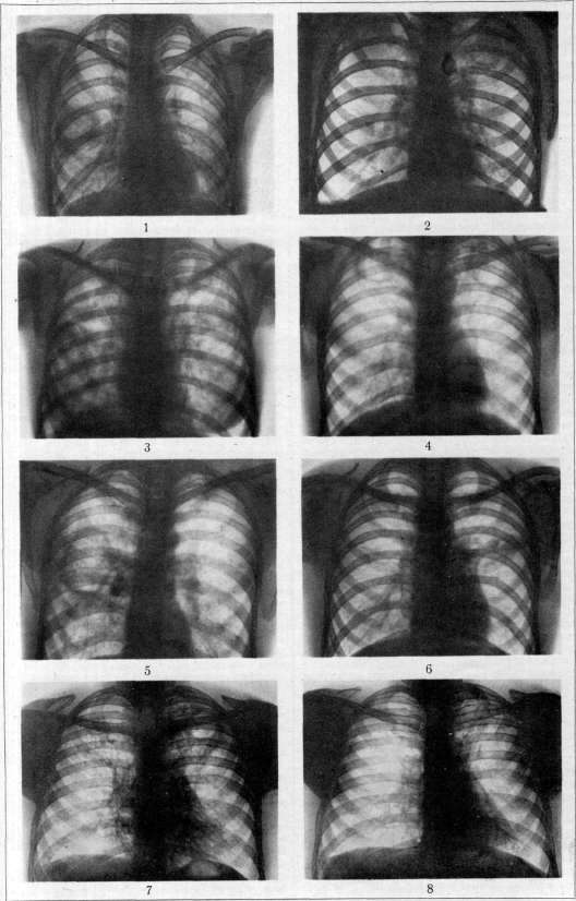
To the article Cavities.
changes in the lungs. 2. A metallic tone of the sound is observed only rarely (1-2% of cases) in large cavities (diameter > 6 cm) with smooth walls located close to the chest wall. 3. The "cracked pot" sound and "coin sound", described by Laennec, are observed in approximately 7-10% of cavities; but in addition, they are noted in pneumonic processes, at the border of exudates, in open pneumothorax, and even in healthy individuals with a thin, flexible skeletal framework of the chest and weak musculature. 4. Of the classical changes in percussion sound described by Wintrich, Gerhardt, and Friedrich, the first (lowering of the sound when closing the mouth) is observed in approximately 1/5 of all cavity cases and is explained by Bernoulli's law on the lowering of the sound of a tube with a blind end when narrowing its opening, but it can equally often accompany infiltrates, exudates (conduction of sound from the large bronchi and trachea), and open pneumothorax; the second (lowering of the sound when moving the patient to a lying position) is rare, inconstant; the third (lowering of the sound upon deep inspiration) is caused by a change in the tension of the cavity walls and can also be observed in various other changes in the lung tissue. Auscultation yields immeasurably more; 1. In 75% of cases, we hear large moist rales in the area of the cavity. If they are detected in areas where there are no large bronchi, and at the top, where the outflow of secretion goes from top to bottom, then they are an undoubted sign of a cavity. They are characterized by scarcity and low timbre. However, in a number of cases, well-drained cavities surrounded by air-bearing lung tissue do not give any auscultatory signs ("mute cavities"). 2. In approximately 20%, sounds resembling squeaking, hissing, and the creaking of wheels are heard; these sounds arise either in the bronchi draining the cavity or are caused by the movements of the cavity wall and are also pathognomonic for cavities. 3. Amphoric breathing (Laennec), sometimes with a metallic tint, is a sure sign of a cavity, observed also in approximately 18-20% and indicating that the cavity walls are smooth and tense. 4. Less commonly (7%), paracavernous rales described by Wallgren are auscultated—small sonorous rales arising in the infiltrated lung tissue and acquiring a particularly distinct tonality due to resonance from the cavity, the size of which can range from a pea to a walnut. In cases of significant infiltrates, the appearance of such rales with a constant localization is a formidable signal of incipient decay. 5. In approximately 10% of cavities, with the patient's mouth open, "oral" sounds (Ferngeräusche Dettweiler's) are auscultated at a distance—transmission and amplification due to resonance in the bronchi and trachea of cavernous rales. However, in a number of cases (according to Holtzman's observations in 15-20%, and according to some literary data in 30-40%), well-drained cavities surrounded by air-bearing tissue, or immovably walled up in dense fibrous tissue, or finally separated from the outside air due to bronchial stenosis, completely fail to give auscultatory signs (Fig. 4). In these cases, auscultation of the heart tones and large vessels in symmetrical areas of the chest in the region of the upper intercostal spaces helps. In this case, due to the displacement of the cardiovascular mass towards the cavity, the tones of the large vessels are auscultated more clearly and louder on the symmetrical areas of the affected half of the chest (Karpilovsky). The frequency of "mute" cavities and the insufficient specificity of physical signs give particular importance to X-ray examination. During fluoroscopy with a diaphragm, and even more sharply on the radiograph, cavities appear in the form of ring-shaped shadows surrounding a more or less clear internal field on which the pulmonary pattern is either not visible at all or its individual fragments are noticeable. The duration and nature of the origin of cavities are to a certain extent distinguished by the contours of the wall: a thick homogeneous wall merging with the diffuse shadow of a pneumonic infiltrate indicates the relative freshness of the cavity (Figs. 1 and 3). An angular, sharply contoured wall indicates a cirrhotic (Figs. 2, 5, 6), old cavity. In fresh infiltrates, "early" cavities are observed without a visible capsule, as if punched out in the lung tissue. Upon the fusion of many small cavities and incomplete sequestration, areas of cavernous decay can produce shadows resembling honeycomb cells and "leopard skin" (Sternberg). Under favorable conditions, small cavities the size of a cherry and even a pea can be recognized radiologically. But thick pleural adhesions, large infiltrates, and location in the deep layers of the lungs among healthy tissue can make cavities "invisible", often even in the presence of auscultatory signs. For the purpose of correct recognition of cavities, it is necessary both during fluoroscopy and on X-ray images to examine the patient in his various positions and with different settings of the tube and screen. Under these conditions, telestereoscopy and the use of contrast media (iodipin, lipiodol) are almost superfluous. Numerous studies (especially in America) are devoted to the issue of differentiating cavities from "annular shadows" formed by pleuritic adhesions, encapsulated pneumothoraces, and plexuses of bronchovascular shadows. Differentiation of these pseudocavities from true cavities is sometimes very easy (presence of a drainage bronchus, change in size during respiration and coughing in non-old true cavities), and sometimes requires very painstaking radiological research and careful comparison with clinical data. The above-described signs are common to all pulmonary cavities that form apart from tuberculosis in abscesses, gangrene, and tumor decay. A tubercular cavity is characterized by the presence of tubercle bacilli and elastic fibers in the sputum. The latter, however, are also observed in other destructive processes in the lungs. Sputum in a tubercular cavity is often secreted in compact spits that take on coin-like outlines in water. Despite the possibility of spontaneous cure of cavities (see above and Figs. 7, 8), the presence of a cavity in tuberculosis is a constant threat in the sense of the possibility of new outbreaks, hemorrhages, and dissemination of the process. In particular, this threat becomes serious when an exudative reaction predominates in the body (infiltrative processes with early cavities, the formation of "daughter" cavities and metastases, and dissemination of small foci). This feature of bronchogenic spread allows many to consider a cavity a kind of "second disease", giving a mortality rate of 70-90%. In view of this, the radical intervention at the first signs of the emergence of a cavity is the induction of artificial pneumothorax, and if necessary, even bilateral. The elective collapse of the affected area developing in this case leads to the blockage of the cavity and promotes its collapse and scarring. In 80% of artificial pneumothorax inductions in cases of fresh cavities, a stable clinical recovery is obtained (Holtzman). With the formation of a dense capsule and pleural adhesions in the area of the cavity, lung collapse may not occur, and in such cases, a stable effect of artificial pneumothorax is noted only in 40-44%. If the cavity does not collapse after artificial pneumothorax, it is urgently necessary to raise the question of more complex surgical interventions—phrenicectomy, thoracoplasty, and plombage with paraffin or transplanted fat; in case of abscesses, pneumotomy and drainage. Of casuistic interest is Burdenko's report on the excision of an isolated tubercular cavity from the lung tissue with complete healing per primam and practical recovery of the patient. In gangrene, the Brauer operation. In fresh pneumonic tubercular cavities, active therapy (tuberculin, helio-, phototherapy) should be avoided. On the other hand, in cirrhotic cavities and during periods of remission, correctly dosed irritation therapy (in particular, tuberculin, etc.) can give an excellent effect.
V. Holtzman.
Related articles
Cite this page
“Cavities.” Soviet Medical Encyclopedia. English translation of Bolshaya Meditsinskaya Entsiklopediya, 1st ed. (Moscow, 1928–1936), ed. N. A. Semashko. https://sovietmedicalencyclopedia.pages.dev/article/cavities/