Bronchiectasis
Historical document, translated for reference. It reflects medical knowledge of the 1920s–30s and is not medical advice.
Summary
This article from the 1928–1936 Soviet Great Medical Encyclopedia defines bronchiectasis as the dilation of bronchi, first described by Laennec in 1819. It details the classification of these dilations into cylindrical, saccular, and varicose forms, and discusses their origins, including congenital, atelectatic, inflammatory, and traction-related causes.
Encyclopedia article (1928–1936)
BRONCHIECTASIS (from the Greek bronchos—bronchus and ektasis—dilation), the dilation of the bronchi, was first described by Laennec in 1819. Bronchiectasis can vary in both form and origin. According to form, bronchiectasis is divided into cylindrical and saccular. The former, which are the result of diffuse dilation of a bronchus over a significant length (hence their other name—diffuse bronchiectasis), often involve very extensive sections of the bronchial tree, spreading predominantly to its small and medium branches. The general configuration of the bronchus in cylindrical dilation remains more or less regular, as a result of which the beginning of the bronchiectasis can often be noticed only because, at a certain place, one or both branches formed from the division of the bronchus retain the diameter of the trunk that gave rise to them or even expand. With extensive spread of cylindrical bronchiectasis, the corresponding part of the lung (usually one of the lower lobes) is often so permeated by them that its cut surface resembles a sponge or porous cheese. Saccular bronchiectasis forms with limited damage to one or another section of the bronchial wall; the cavities resulting here can have the most diverse size and shape, being located along the course of bronchi of any caliber. Saccular bronchiectasis arises most often in the upper lobes of the lungs. If a series of successive protrusions forms along the course of a bronchus, one speaks of varicose bronchiectasis. As an intermediate form between cylindrical and saccular bronchiectasis, some also distinguish fusiform bronchiectasis. All the listed forms can,
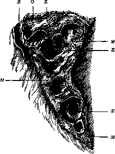
Middle lobe with numerous and large bronchiectases: V—bronchus; E—bronchiectases; M—densified, chronically indurated lung tissue; O—upper lobe; Z—connective tissue adhesion between the upper and middle lobes (according to Ribbert).
of course, be combined in the most diverse combinations. By origin, bronchiectasis can be divided into congenital and acquired. Congenital bronchiectasis, according to the generally accepted view, represents a complex developmental defect in which the most essential feature is that alveoli do not form at all on the terminal bronchial branches, thanks to which the entire pulmonary parenchyma of such a section turns out to consist only of air-conducting tubes. The latter, in these cases, as a rule, expand either diffusely throughout their entire length or only in their small ramifications (starting from the bronchi of the 3rd-4th order), where the dilations often take on a saccular character, forming a series of cysts following one another along the length of the bronchus. These cysts sometimes become detached, turning into completely closed cavities, covered more often than not not by cylindrical, but by stratified squamous epithelium, and containing either a liquid mucous or a thicker purulent-like mass. These changes can be limited to one or several lobules, but can also involve an entire lobe, and even the whole lung or both lungs (in the latter case, the fetus is, of course, non-viable). On cross-section, a lung with congenital bronchiectasis has a sharply honeycomb appearance, and the size of individual cells even in newborn children fluctuates within very wide limits (from a millet seed to a cherry and larger). Closest to congenital are the so-called atelectatic bronchiectases, which often develop over time in the area of congenital atelectasis (see). In view of the fact that the alveolar parenchyma in these areas gradually undergoes fibrous transformation and ceases to develop, the corresponding bronchi, partly due to continued growth, partly under the pressure of air, and also under the influence of stagnant contents, little by little expand, turning, in the end, into a whole system of very irregular wide cavities, lined for the most part by stratified squamous epithelium and sometimes even losing connection with the leading trunk. Cartilaginous plates, where they are present in the bronchial walls, often grow strongly in this process, take on an irregular shape, and are surrounded by adipose tissue. In general, the pictures of atelectatic and congenital bronchiectasis are very similar, so that it is possible to distinguish the former only as long as remnants of alveoli are still preserved around them. With the disappearance of the latter, any distinction is lost. These two forms differ very noticeably from all other types of bronchiectasis in that in the area of their spread, the lung tissue for the most part contains absolutely no coal pigment (due to the absence of respiratory function and normal airflow from birth). The development of bronchiectasis can also occur in acquired atelectatic areas if they exist for a sufficiently long time and lead to the desolation of the alveoli. Besides the just-mentioned atelectatic bronchiectasis, almost all bronchiectasis acquired in a later period of life has at its base inflammatory changes in the bronchial wall, mainly those associated with the destruction of the muscular-elastic layer, which plays a primary role in the bronchus maintaining its normal elasticity and contractility. Very important auxiliary factors in the pathogenesis of such bronchiectasis are: 1) cough impulses, since with them the expiratory air pressure in the bronchial system suddenly increases several tens of times compared to quiet exhalation (from 2-3 mm to 80 mm of mercury), and 2) abundant formation and stagnation of discharge. The combination of all these conditions can arise during many infectious diseases of the respiratory tract, but it is especially often realized in children with measles, whooping cough, and severe influenza with their protracted bronchitis and bronchiolitis, accompanied by deep infiltrative-proliferative and necrotic processes in the walls of the air-conducting pathways. Here, bronchiectasis can sometimes develop extremely rapidly (within 1-2 days) and obtain wide distribution (so-called acute diffuse bronchiectasis). The same changes arise with tuberculosis and other chronic infections of the respiratory tract (actinomycosis, aspergillosis, etc.), but usually in comparatively limited sections of the bronchial system, giving rise to either small cylindrical or saccular bronchiectases. Besides lesions of the bronchial wall itself, cicatricial processes in its vicinity can also give rise to the development of bronchiectasis; a necessary condition for this is the adhesion of the lung to the parietal pleura, since in a fully mobile organ, scarring and shrinkage of tissue always, of course, lead only to the narrowing or even complete compression of the corresponding air-conducting tubes. Conversely, with fixation of the pulmonary surface to the chest wall, the same processes cause stretching of the bronchial cavities (traction or cirrhotic bronchiectasis), which are predominantly saccular in character and especially sharply pronounced in cases where the bronchial wall is also damaged to one degree or another. If, at the same time, the fibrous process obtains wide distribution, then the lung on cross-section gives a very characteristic picture of cirrhosis with numerous bronchiectatic cavities embedded among the scar tissue—the so-called Corrigan cirrhosis of the lung (named after the author who first described it). Also deserving of mention are the so-called stenotic bronchiectases, which usually form during prolonged narrowing of the lumen of the respiratory tract (cicatricial stenoses, tumors, foreign bodies, etc.) behind the narrowed place. They arise under the combined action of three factors—the accumulation and stagnation of discharge, the development in connection with this of one or another inflammatory change in the bronchial wall, and an increase in expiratory pressure. Finally, it should be noted that with the death of significant sections of the pulmonary parenchyma, compensatory hypertrophy can develop in the remaining parts of the lung, accompanied by an increase in the size of all structural elements of the organ, including the bronchi, which in these cases give the impression of being ectasized. As for the state of the bronchial wall in bronchiectasis of inflammatory origin, its normal structure is always to a significant degree disturbed. Defects of the epithelial cover, edema, infiltrates of varying density and extent, the development of granulation tissue, always accompanied by greater or lesser death of muscular-elastic elements, partial necroses, ulcerations, and suppuration—these are the basic pictures that one has to encounter here in the most diverse combinations. Where the mucous membrane is preserved, it can be either thinned (atrophic bronchiectasis) or, conversely, thickened, succulent, and covered with polypous growths (hypertrophic bronchiectasis). If the processes of destruction go deeper, the entire wall of the bronchus can undergo atrophy over one or another extent, and then the peribronchial tissue or the adjacent alveolar parenchyma of the lung participates in the formation of the cavity. In this case, one speaks no longer of bronchiectasis, but of a bronchiectatic cavity (see).
M. Skvortsov.
Symptoms. Cough and characteristic sputum are constant symptoms in bronchiectasis. Coughing occurs in paroxysms, usually in the mornings and upon changing position after lying on the affected side. At this time, a rapid and copious discharge of sputum by the mouthful ("maulvolle Expectoration") is observed. After the discharge of sputum, temporary relief ensues until its new accumulation in the dilated bronchus. Upon the movement of sputum from a dilated bronchus with weakened mucosal sensitivity into a normal one (during a change in body position), a new paroxysm of coughing with copious sputum discharge appears. The daily amount of sputum can reach 500 g or more. It has an unpleasant sweetish odor, in some cases is fetid, and in its properties resembles the sputum in putrid bronchitis (see). The presence of erythrocytes in it is a common phenomenon, but more or less copious hemoptysis is frequently observed, depending on ulcerative processes in the bronchi and the rupture of dilated vessels in the walls of the bronchiectasis. Sometimes mucous sputum mixed with blood is discharged in the form of "raspberry jelly." Dyspnea is absent or weakly expressed and more often occurs during a coughing fit. In the presence of complications from the heart and lungs, it becomes constant. Physical symptoms are more or less pronounced in large bronchiectasis located superficially. The percussion sound is tympanic or dull, depending on the filling of the cavity with sputum or air; Wintrich's and Gerhardt's changes in sound. Upon auscultation—bronchial or amphoric breathing with moist resonant rales, sometimes with a metallic tinge. After emptying the cavity, physical symptoms can change significantly. The variability of phenomena during physical examination at the same site is an important sign of the existence of bronchiectasis. In small and deeply situated bronchiectasis, physical examination yields only signs of chronic bronchitis. In such cases, the periodic discharge of large quantities of characteristic sputum at once gives grounds to suspect the existence of bronchiectasis, and moist rales, heard over a long time in the same place, especially in the upper and middle sections of the lungs, can serve as an indication of localization. Sometimes changes in the configuration of the chest are observed in the form of retraction of individual parts, depending on the shrinkage of the lungs around the affected bronchi and on prolonged pleurisy. Course and complications. The course of the disease is prolonged and for a long time does not affect the general condition, although the appearance of patients (even with good nutrition) is sickly, with a pale-bluish skin coloration. Fever is usually absent, but with complications and accumulations of putrid sputum, it may appear. In many patients, club-shaped enlargements of the terminal phalanges of the fingers, and sometimes toes, gradually develop in the form of "drumstick fingers." Less frequently, more extensive chronic hyperplasias of the bones and joints ("osteo-arthropathie hypertrophiante pneumique" by Pierre Marie) are observed, as well as rheumatoid swelling of the joints. The most frequent complications: putrid decomposition of sputum, putrid bronchitis with subsequent pneumonia and gangrene, less frequently pyopneumothorax and metastatic brain abscesses. With a prolonged course, the disease is often complicated by impaired cardiac activity, amyloid degeneration of organs, especially the kidneys. Recognition of bronchiectasis is not always possible. Small and deeply situated dilations are usually not recognized; sometimes even with large dilations, complications mask the physical signs. The diagnosis is based on data from the anamnesis, the course and duration of the disease, and on the analysis of physical and other symptoms. In differential diagnosis, one must keep in mind the following diseases: pulmonary tuberculosis, cavity, lung abscess and gangrene, empyema, putrid bronchitis, etc. Fluoroscopy, and especially radiography, with the introduction of lipiodol or 40% iodipin into the bronchi, in many cases can provide a characteristic picture for the recognition of bronchiectasis [see separate table (col. 107 - 108), fig. 4)]. The prognosis depends to a significant degree on complications. Recovery is observed only in mild cases and in acute dilations. The prognosis is always doubtful. Treatment. Besides general hygienic measures (rest, nutrition, climate, treatment, elimination of harmful influences), it is necessary to strive for a reduction in secretion, better expectoration, and the prevention of putrid decomposition of sputum. Indicated are: inhalation of turpentine and other balsamic agents; internally—creosote preparations and standard expectorants. To eliminate sputum stasis in the cavities, mechanotherapeutic measures are applicable, consisting of placing the patient in such a position that facilitates the discharge of sputum, compression of the lower part of the chest with the hands according to Gerhardt, the oblique position according to Quincke, etc. In recent times, surgical methods of treatment have been proposed: artificial pneumothorax, pneumotomy, extrapleural thoracoplasty, resection of a lung lobe, etc. Surgery is indicated only with accurate diagnosis of bronchiectasis (radiography). The results are as yet not sufficiently satisfactory, A. Arutinov. Lit.: Wiese O., Bronchiektasien im Kindesalter, B., 1927; Handbuch der allgemeinen Pathologie und pathologischen Anatomie des Kindesalters, ed. by H. Brüning and E. Schwalbe, Vol. II, Chap. VII, Wiesbaden, 1914; Goerdt W., Über Bronchiektasien, Inaugural Dissertation, München, 1911; Kaufman E., Spezielle pathologische Anatomie, Vol. I, B., 1922.
Related articles
Mentioned in
Cite this page
“Bronchiectasis.” Soviet Medical Encyclopedia. English translation of Bolshaya Meditsinskaya Entsiklopediya, 1st ed. (Moscow, 1928–1936), ed. N. A. Semashko. https://sovietmedicalencyclopedia.pages.dev/article/bronchiectasis/