Emphysema
Historical document, translated for reference. It reflects medical knowledge of the 1920s–30s and is not medical advice.
Summary
Emphysema is a pathological condition characterized by excessive air in organs or tissues, most commonly affecting the lungs where it results in loss of elasticity and enlargement of alveoli. The article describes various types including true, senile, acute vesicular, interstitial, and subcutaneous emphysema, along with their pathological features and clinical manifestations.
Encyclopedia article (1928–1936)
EMPHYSEMA, emphysema (from Greek empliy-SaO-I inflate). This term denotes a pathological condition in which there is either an increased content of air in an organ (lung) or an unusual accumulation of air in a given tissue. Accordingly, one speaks of pulmonary emphysema, subcutaneous emphysema, and mediastinal emphysema. However, most often the concept of emphysema as a disease is associated with damage to lung tissue, the most characteristic feature of which is the loss of elasticity and enlargement of the pulmonary alveoli. Anatomically, pulmonary emphysema encompasses various processes, but of particular interest is true (substantive, genuine, vesicular) pulmonary emphysema, identified by Laennec. This disease has a chronic course and is accompanied not only by the expansion of alveoli but also by the death of lung parenchyma. Most often, men aged 40-60 years are affected. Upon autopsy of the chest cavity of those who died from true emphysema, a low position of the diaphragm and enlarged lungs are found; their edges cover the anterior mediastinum and touch each other; the lungs do not collapse, are bloodless, cut with a crunch, and contain very little pigment. The apices of the lungs are rounded. Depending on the predominant localization of the process, central and marginal (peripheral) emphysema are distinguished. From a patho-anatomical point of view, chronic emphysema is characterized by a combination of three processes: atrophy, anemia, and stretching. In some cases, atrophy is primary, in others, the loss of elasticity, but for many cases, the question of the role of one or another process remains unresolved. Histologically, in chronic pulmonary emphysema, a marked expansion of alveoli, alveolar ducts, and even bronchioles is found, thinning of the walls of alveoli, stretching of Kohn's pores, ruptures of thinned alveolar walls (see figure), death of elastic fibers, fusion of adjacent alveoli and sacs with each other, as a result of which cavities form, reaching the size of a large mandarin orange. Usually, these air-filled cavities are located along the edge of the lung (bullous emphysema - see separate table, fig. 2). In chronic emphysema, the stretching begins with the alveolar ducts, then gradually involves other elements of the acinus. In rare cases, expansion occurs only in the bronchiolar system of the acinus, and then one speaks of bronchioloectatic emphysema (Loschke). It develops as a result of destructive processes in the walls of respiratory bronchioles. With the stretching of alveolar ducts and alveoli, along with atrophy of the interalveolar septa, the obliteration of blood and lymphatic vessels occurs.
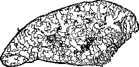
Pulmonary emphysema (schematic drawing). Field of resection (right side)-artificial atelectasis. On the right edge, the terminal bronchus is visible, branching into 3 alveolar ducts; the alveolar nature of these ducts is erased.
As a result of all these processes, the respiratory surface of the lung decreases. The distribution of blood in the emphysematous lung becomes irregular due to the obliteration of capillaries, and the most hyperemic areas are those with well-passable vessels, namely the bronchial mucosa. On the basis of prolonged hyperemia, catarrhal conditions of the bronchial mucosa and chronic bronchitis, a common companion of emphysema, easily develop. The obliteration of small vessels in the lung parenchyma leads to an increase in pressure in the pulmonary artery system and hypertrophy of the right ventricle of the heart. Subsequently, heart dilation may develop and the patient dies from signs of cardiac decompensation. The chest of emphysematics is little mobile, has a characteristic barrel-shaped form, and is in a state of inspiration. The dimensions of the chest are increased in the anteroposterior direction. Vicarious (secondary) emphysema appears in the lung either partially when part of the lung parenchyma is excluded due to old fibrous (tuberculosis, pneumoconiosis) processes, or when an entire lung is excluded by compression with exudate or air, and the other lung expands significantly and equalizes the act of breathing. Prolonged expansion eventually leads to atrophy, thinning, and rupture of the septa. Around limited scars, large air bubbles can often be seen. Senile emphysema (atrophic) occurs with senile impairment of nutrition of the elastic and fibrous elements of the lung, which leads to thinning of the alveolar walls, their rupture, fusion, and cavity formation. Sometimes the process is localized in the apex and along the edges, in other cases the entire lung is affected. At autopsy, the lung is pale and easily collapses. No hypertrophy of the heart is observed in senile emphysema. Acute vesicular emphysema (alveolar ectasia, lung distension, emphysema aquosum) arises &*s
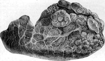
Figure 1. Erythema exsudativum multiforme. Figure 2. Bullous emphysema of the lung; numerous bubbles along the anterior-superior edge of the lung; sclerosis of interlobular septa. Figure 3. Schilde's disease; breakdown of myelin fibers and white matter of the brain. Figure 4. Chronic eczema of the shin. l.-tJf'iJs with enhanced inspiration. The changes in the lungs are insignificant and amount only to severe anemia due to compression of the vessel walls by stretched alveoli. Unlike chronic emphysema, there is no death of lung parenchyma here, and when the causing factor is eliminated, the lung returns to normal. Acute vesicular emphysema is found in those who died from suffocation, in pneumonia, in laryngospasm, laryngostenosis, during an attack of bronchial asthma. Prolonged and frequently repeated acute distension of the lungs, for example in asthma, laryngostenosis, can lead to a decrease in lung elasticity and the development of chronic emphysema. Interstitial (traumatic) emphysema differs from alveolar emphysema in that air accumulates in the interstitial tissue, where it penetrates only when the lung parenchyma is damaged. Here, gross ruptures with simultaneous hemorrhage are possible, but more often there are small tears without violation of the integrity of capillaries, but sufficient for air to penetrate into the interlobular tissue. This is observed with a sudden sharp increase in inspiratory or expiratory pressure, for example during a coughing attack in whooping cough, during acute laryngospasm, miliary tuberculosis. The occurrence of inspiratory emphysema during influenza is described. Air that has penetrated into the interstitial tissue spreads along the lymphatic vessels. Inspiratory emphysema is particularly often found at autopsy in children who died from croupous laryngotracheitis or whooping cough. The more frequent occurrence of inspiratory emphysema in children is explained by the loose structure of the child's lung and the greater development of interstitial tissue and therefore better possibilities for the spread of air in it. Patho-anatomically, lungs with inspiratory emphysema have a characteristic appearance: along the lobular septa (interlobular emphysema) and under the pleura (subpleural emphysema), air bubbles are arranged in a chain, which easily move when pressed. Spreading along the bronchi, air can reach the root of the lung, penetrate into the mediastinum and subcutaneous tissue of the neck (see below). When the access of air ceases, rapid resorption of it occurs and the lung returns to normal. In other cases (for example, in influenza pneumonia), infection of the tissue clefts may occur and the development of interstitial phlegmon of the lung and mediastinum. Air bubbles located under the pleura sometimes rupture into the pleural cavity and cause spontaneous pneumothorax. Subcutaneous emphysema occurs when air enters the subcutaneous tissue from the outside, accumulates and spreads in it, and increases its volume. This is observed after laparotomy surgery around the operative field, with sub fractures of the frontal sinuses, with open joint injuries when the sucking action of the joint promotes the injection of air into the subcutaneous tissue, with damage to the larynx and respiratory tract, esophagus with penetration to the subcutaneous tissue, during childbirth accompanied by ruptures of the birth canal. Universal subcutaneous emphysema can develop when a tuberculous lymph node ruptures into a bronchus, with each inspiration air rushes into the peribronchial tissue, then into the mediastinal and subcutaneous tissue of the neck. Subcutaneous emphysema reaches its greatest development in chest wounds with rib fractures and violation of the integrity of the pleural sheets and lung. Here air initially enters the pleural cavity and from there, without having a free outlet to the outside, it rushes into the subcutaneous tissue of the chest, and then the whole body; in such cases, subcutaneous emphysema becomes life-threatening. Subcutaneous emphysema can develop with the application of artificial pneumothorax. Often, especially in childhood, subcutaneous emphysema is a consequence of lung diseases and is preceded by interstitial emphysema (see above). Air first rushes into the mediastinum, where it forms large air cavities (mediastinal emphysema), and from here it enters the subcutaneous tissue of the supraclavicular fossa and gradually captures larger and larger areas of the body. With subcutaneous emphysema, there is significant swelling of the tissue, and palpation of the affected areas causes a characteristic, audible crackle from the bursting and movement of air bubbles. When the cause that caused the subcutaneous emphysema is eliminated, the air quickly resorbs and everything passes without a trace.
a.strukov.. Emphysema of the lungs. Etiology. Data from various authors regarding the frequency of genuine emphysema of the lungs diverge: according to Lebert, emphysema of the lungs constitutes about 5% of all diseases and is the cause of death in 2% or 3% of all fatal cases. Conversely, according to Virchow, in Berlin at the Charité Hospital, emphysema of the lungs constituted only 0.3% of all diseases and was the cause of death in 0.6-0.7% of all fatal cases. West gives figures close to Virchow's data. In Leningrad in 1933, emphysema of the lungs was found in 0.86% of all autopsies. Genuine emphysema of the lungs is observed predominantly in old age. Most authors indicate that men get sick 2-3 times more often than women, which may depend on differences in professional occupations and lifestyle and in part may be due to sexual differences. Old authors quite consistently indicated that emphysema of the lungs occurs more often in glassblowers, musicians playing wind instruments, and generally in people whose work requires great effort during exhalation. However, recent research refutes this opinion (West, Sherel, Harvier). Emphysema of the lungs develops in very many cases of bronchial asthma; it is often observed in chronic bronchitis, especially in dry bronchitis, in which, due to the difficulty of expectorating viscous sputum, the cough is stronger and longer than in bronchitis with more liquid sputum. In a certain number of cases, emphysema of the lungs is observed in the absence of the mentioned diseases. Pathological anatomy - see above. Emphysematous changes in the lungs are not always distributed equally. For example, in kyphosis of the upper part of the spine, according to Leschke, the upper part of the chest is in maximum inspiratory position, while the lower part of the chest is in maximum expiratory position, which makes expansion and collapse of the chest during breathing almost impossible. Moreover, the diaphragm descends and becomes flatter. Leschke's research showed that the line separating the upper part of the chest, which is in the inspiratory position, from the lower part, which is in the expiratory position, runs approximately from the angle of kyphosis to the lower part of the sternum. Depending on the higher or lower localization of kyphosis, the size of the parts of the lungs in the state of inhalation and exhalation also changes. Emphysematous changes in kyphosis of the spine develop only in that part of the lungs that undergoes sharp stretching, but not in the lower part of the lungs, which is in the state of exhalation. Often the upper, emphysematous and lower, normal parts of the lungs are separated by a clear linear boundary. At the same time, degeneration and ossification of the costal cartilages are also often observed. Photographs of the chest and lungs made by Leschke clearly show the changes in the chest and lungs that develop in kyphosis of the spine: the chest becomes barrel-shaped, the lungs are swollen in the upper part, the swollen anterior edges of the lungs cover the mediastinum. General acute distension of the lungs, or acute functional emphysema of the lungs, is observed with significant physical exertion, sometimes also with insufficient heart activity, because with increased breathing, inspiration occurs more completely than expiration. Acute distension of the lungs often develops in acute bronchitis and bronchiolitis accompanied by severe cough, especially in children. It is constantly observed in corpses in people who make deep breathing movements before death, i.e., in suffocation, in drowned persons. It is characteristic of acute distension of the lungs that it disappears 24-60 hours after the disappearance of the cause that caused it, for example, after cessation of heavy physical work, after recovery from bronchitis, whooping cough, etc. Finally, significant general distension of the lungs is constantly observed during, and often for some time after, an attack of bronchial asthma and in asthmatic bronchitis. Besides distension of the lungs, no anatomical changes are found in this condition. Local distension of the lungs, for example of one lobe of the lung, is observed in cases of development of valvular narrowing of one of the bronchi, for example, when a foreign body enters it, in neoplasms of the bronchi, etc. Pathogenesis. It is indicated above that emphysematous changes can be the result of various processes; undoubtedly, local and even widespread emphysema of the lungs can be the result of chronic inflammatory processes in the lungs, as is often observed in fibrous tuberculosis of the lung apices, in the vicinity of fibrous tuberculous foci, in pneumoconioses and sometimes even after prolonged bronchopneumonias. The frequency of emphysematous changes in fibrous tuberculosis of the lungs gave some basis, especially to French authors, to assert that emphysema of the lungs is the result of mild, attenuated tuberculosis of the lungs. Others, for example Antoniazzi, believe that even in genuine emphysema of the lungs, sclerosis with subsequent emphysematous changes develops on the basis of a chronic inflammatory process in the lungs. According to Rubel's theory, the development of emphysema of the lungs is associated with pneumosclerosis. It must also be considered established that emphysema of the lungs, under certain conditions, can be the result of stretching of the lungs, as is proved by the above observations of Leschke on emphysema of the lungs in kyphosis of the thoracic part of the spine. Then senile emphysema is also observed, different from genuine emphysema. Unfortunately, most authors who studied emphysema of the lungs did not distinguish its individual types, and this to a large extent explains both the contradiction of theories of the pathogenesis of emphysema of the lungs and the difference in statistical data on its frequency given by authors. Most authors to the present day believe that the main cause of the development of emphysema of the lungs is excessive, repeated over a long time stretching of the lung tissue and increased intrathoracic pressure. Stretching of the alveoli causes compression of the vessels passing in the interalveolar septa, the vessels become thinner and finally obliterated. The ischemia of tissues occurring in this process leads to atrophy of lung tissue and, in connection with the stretching of the lungs, to a decrease in their elasticity. At the same time, some authors attach greater importance to inspiratory stretching of the lungs, others to excessive and often repeated forced expiratory efforts, as occurs in severe cough, in bronchial asthma, etc. Difficulty in expiration and stretching of the lungs in bronchial asthma and probably in many bronchitides also depend on bronchospasm, which mainly hinders expiration. The question of the degree of decrease in elasticity of the lungs in emphysema of the lungs has not yet been finally resolved due to the lack of an impeccable technique. The pressure with which the lungs collapse after opening the chest cavity varies greatly in individual cases, and Leschke found no difference between normal or emphysematous lungs. The decrease in collapse of the lungs after opening the chest cavity may also depend on obstruction in the bronchi, for example, on the accumulation of viscous mucus in them. The decrease in elasticity of the lungs in emphysema of the lungs is definitely indicated by indentations in the lungs, formed from pressure and not disappearing after its cessation. In the question of the significance of the decrease in elasticity of the lungs for their stretching, it is necessary to take into account that the force of elastic retraction of the lungs (on a corpse = 40-60 mm H2O) is significantly less than the pressure that the chest can exert; thus, with deep inhalation, the pressure in the respiratory tract drops to -100 mm Hg, and with straining it rises to +160 mm. Therefore, the idea that a stretched emphysematous lung directly expands the chest is incorrect; rather, it should be thought that the chest gradually expands because the lungs, due to decreased elasticity, do not collapse sufficiently during expiration, and therefore inspiratory stretching of the lungs predominates over their expiratory collapse. Numerous observations speak against the mechanical theory of the origin of emphysema of the lungs, showing that in many cases emphysema of the lungs does not develop despite the prolonged existence of conditions and diseases in which there is significant stretching of the lungs and exhalation is performed with great effort; for example, in glassblowers and musicians playing wind instruments, emphysema of the lungs is not observed more often than in other professions; there are many cases of bronchial asthma where, despite the prolonged existence of the disease and frequent and severe attacks of suffocation, there was only simple expansion, increase in volume of the lungs, while the phenomena characteristic of emphysema of the lungs - atrophy and fusion of alveoli into larger cavities - were absent. Then there are patients with emphysema of the lungs who were not exposed to the harmful influences mentioned and did not suffer from bronchial asthma or other diseases accompanied by severe cough. In view of the impossibility of the mechanical theory to explain the origin of true emphysema of the lungs, even old authors, such as Laennec, Gerhardt, Virchow, and others, put forward the hypothesis that the main significance in the pathogenesis of emphysema of the lungs is the disorder of nutrition of the lung tissue.
It is undoubtedly the case that senile emphysema of the lungs, those cases of pulmonary emphysema that develop in individuals not subjected to harmful mechanical influences, and finally the numerous patients suffering from bronchial asthma or chronic bronchitis for many years without developing emphysema of the lungs, prove that emphysema of the lungs can develop only with a certain predisposition, in the presence of which harmful mechanical influences can only contribute to the development of emphysema. Nevertheless, the significance of mechanical conditions in the development of emphysema of the lungs cannot be completely denied. Their significance is indicated by the clearly described cases of emphysema of the lungs in kyphosis of the thoracic spine, in which emphysematous changes were present only in the upper, stretched part of the lungs, while the tissue of the lower parts of the lungs was normal. Freund had long ago pointed to the significance of immobile expansion of the chest for the development of emphysema of the lungs. He believes that primary ossification and lengthening of the cartilages of the upper ribs first occurs, as a result of which a barrel-shaped immobile expansion of the chest with flattening of the diaphragm occurs. Freund from the very beginning pointed out that not all cases of emphysema of the lungs, but only a certain part of them, develop on the basis of primary ossification of the cartilages of the upper ribs. The question of the significance of primary ossification of the rib cartilages and the frequency of these cases of emphysema of the lungs has not yet been resolved. It is undoubtedly the case that the changes in the rib cartilages described by Freund are often found in elderly people without emphysema of the lungs. Symptoms and course. Emphysema of the lungs always develops slowly and gradually. At first, patients suffer little and their capacity for work is not diminished. Many patients have long suffered from diseases accompanied by severe cough. Gradually, shortness of breath appears, which increases over the years, especially during periods of exacerbation of bronchitis. Shortness of breath sharply increases with physical exertion and during coughing attacks and only passes after a more or less prolonged rest. Exhalation is particularly difficult. Cough is observed in pronounced emphysema of the lungs almost constantly, as it develops in many cases on the basis of other chronic diseases of the bronchi or lungs, and on the other hand, emphysema of the lungs itself creates the soil for the development of chronic bronchitis. The general appearance of emphysematous patients in many cases is characteristic: the face is pale and slightly or markedly cyanotic, the neck is shortened, the jugular veins are filled with blood, the auxiliary respiratory muscles in the neck are tense. The chest is increased in volume, especially in the upper part, barrel-shaped, elevated, its anteroposterior diameter is increased, it is almost immobile during breathing. The force of exhalation is so insignificant that the patient sometimes cannot blow out a candle. The supra- and infraclavicular fossae are smoothed out. The ribs run more horizontally and the intercostal spaces are widened. Less frequently there is expansion only of the lower part of the chest. The act of breathing is difficult, the expiratory phase is prolonged. The percussion sound is louder and lower, usually with a tympanic tint ("boxy"). The lower borders of the lungs are lowered and immobile during breathing. The respiratory murmur is markedly weakened if there are no signs of accompanying bronchitis. Often the respiratory murmur has a special soft tone (respiration humee of the French); its inspiratory phase is short, the expiratory is prolonged. If there is also bronchitis, then dry and moist rales are heard, which drown out the respiratory murmur. The cardiac impulse is indeterminable. The cardiac dullness, especially the absolute, is markedly reduced due to the covering of the heart by the dilated edges of the lungs, the cardiac tones are weakened, on the pulmonary artery the second tone is intensified due to an increase in blood pressure in the small circle of circulation. In a later period of the disease, weakening of cardiac activity appears, which is expressed by an increase in shortness of breath and an expansion of the true right border of the heart, sometimes by the appearance of a systolic murmur of relative insufficiency of the valve flaps. When measured with a tape, the difference in chest circumference at maximum inspiration and maximum expiration is 4 cm and even 2 cm instead of 7 cm in healthy people. The respiratory capacity of the lungs instead of the normal 3 liters gradually falls to 2 liters and 1.5 liters. On radiography, the lungs are lighter than normal, the shadows of the hili are clearly visible, the diaphragm is lowered and little movable during breathing, the costodiaphragmatic sinuses form almost a right angle. When the patient attempts to breathe deeply, the chest remains immobile and expanded, the transparency of the lungs does not change during inspiration and expiration. Body temperature is normal. Blood pressure is often somewhat elevated. Emphysema of the lungs develops extremely slowly and gradually. Patients for a long time retain limited capacity for work: the latter is reduced mainly during periods of exacerbation of chronic bronchitis. Weakening of the respiratory excursions of the chest and the emptying of numerous pulmonary capillaries creates difficulty for the work of the right heart, which hypertrophies, eventually expands and becomes insufficient. Sometimes a huge congestive liver appears. The disorder of circulation is intensified by frequent severe cough, usually observed in emphysema of the lungs. At first, circulatory disorders yield to treatment. Over the years, however, the disease progresses, and patients become completely incapacitated. Death often occurs with signs of cardiac insufficiency. Diagnosis. Most authors hold the opinion that recognizing pronounced diffuse emphysema of the lungs usually presents no difficulties: they believe that the immobile expansion of the chest, the boxy sound, the weakening of respiratory murmurs, the prolonged exhalation, etc., provide sufficient data for diagnosis. However, at autopsies the diagnosis is not always confirmed. Leschke points out that often anatomically pronounced emphysema of the lungs is not recognized by clinicians, especially those emphysemas of the lungs that are not accompanied by bronchitis (senile emphysema of the lungs), and on the other hand, clinicians often diagnose severe emphysema of the lungs in cases where the pathologist finds only an inflated lung and a pronounced bronchitis. In reality, the immobile expansion of the chest, the boxy sound, the weakening of respiratory murmurs, dry rales with prolonged exhalation, the low position of the diaphragm and its immobility, etc., are also present during attacks of bronchial asthma and disappear with the cessation of the attack. This shows that all these symptoms may depend on bronchitis accompanied by bronchospasm. In cases of atypical bronchial asthma, with asthmatic bronchitis, such a condition can last for weeks and even months. It is known that non-asthmatic chronic bronchitis can also be accompanied by bronchospasm. This explains why the diagnosis of emphysema of the lungs is not always confirmed at autopsy, if under emphysema of the lungs one understands not only the swelling of the lungs but a disease expressing itself, besides the increase in volume of the lungs, by certain anatomical changes. It has already been pointed out above that senile emphysema of the lungs is not diagnosed in most cases. In pronounced cases, there is a decrease in lung volume, which is manifested by a decrease in chest volume, flattening and narrowing of the intercostal spaces, and a high position of the diaphragm. The superficial cardiac dullness may be increased. Percussion and auscultation of the lungs give the same results as in genuine emphysema of the lungs. Complementary emphysema of the lungs differs from the true one only by the absence of functional disorders and weakening of the respiratory murmur. Signs of bronchitis are usually absent. Complications occur in emphysema of the lungs rarely: a) due to the rupture of a subpleural emphysematous bubble, pneumothorax may occur; b) even more rarely, interstitial emphysema of the lungs develops due to the rupture of the alveolar wall and the entry of air into the interstitial pulmonary or subpleural tissue. This complication comparatively often leads to death. Prognosis. To judge the severity of emphysema of the lungs, it is necessary to pay attention not only to the general condition of the patient and to shortness of breath, which can significantly improve with the cure or reduction of bronchitis, but mainly to the condition of the heart and to the vital capacity of the lungs. The prognosis worsens with the onset of cardiac insufficiency and with a fall in the vital capacity of the lungs by half. The prognosis also worsens if the difference in chest circumference at maximum inspiration and expiration becomes less than 3 cm. Treatment. In view of the fact that the condition of patients sharply worsens with the intensification of bronchitis, treatment should be directed first of all against chronic bronchitis. To prevent exacerbations of bronchitis, patients should avoid colds and should live in areas with an even, mild climate with clean, if possible dust-free air; for winter stay, suitable areas on the coast of the Black Sea can be recommended, for summer, less hot forested areas in the central zone of the USSR are more suitable. If the air in rooms is too dry (central heating), it is necessary to take care of its humidification. Patients should not drink alcoholic beverages, should not smoke, for many even staying in a smoky room is harmful. Food should be light and not too abundant; it is necessary to take care of proper emptying of the intestines, as well as to limit muscular work; severe patients should have complete rest.
The same medications are used as in the treatment of chronic bronchitis; good results are often obtained from the long-term use of iodine preparations, most commonly potassium iodide. Since in emphysema of the lungs, in addition to catarrh, there is often also bronchospasm, anti-asthmatic remedies should be tried in suitable cases (significant dyspnea, especially when increasing in the form of attacks, eosinophilia of the blood) (smoking powders with Datura stramonium, adrenaline injections, etc.). Inhalation treatment is also recommended, preferably in special inhalation chambers. For inhalations, solutions of table salt, soda, alkaline-salt mineral waters, emulsions of balsamic substances such as creosote, turpentine, pine oil, etc., are used. To facilitate breathing, treatment by inhaling compressed air and exhaling into rarefied air with the help of special apparatuses is used (see Waldenburg's apparatus). Treatment with compressed air in pneumatic chambers is also used. To facilitate exhalation, it is also useful to apply a wide rubber bandage to the upper part of the abdomen and the lower ribs; by compressing the upper part of the abdomen, it slightly raises the diaphragm, which can then perform larger excursions during contractions. A bandage should not be applied to the chest, which in many cases of emphysema of the lungs is immovably fixed in an expanded state. The bandage should not be applied too tightly; it should only facilitate exhalation without hindering inhalation. In order to maintain the working capacity of the heart for as long as possible, it is necessary to establish a suitable regimen for the patient and avoid anything that may cause increased work and fatigue of the heart. When symptoms of heart failure appear, cardiac remedies are necessary—caffeine, digitalis, adonis, etc. In very severe dyspnea, temporary relief is given by extensive bloodletting (500-900 cm3). In cases where it can be assumed that emphysema of the lungs developed on the basis of primary immovable expansion of the chest, surgical mobilization of the chest is recommended. However, in most cases it is difficult to determine whether the immovable expansion of the chest developed primarily or secondarily. Due to the difficulty of establishing indications for surgical treatment of emphysema of the lungs, the results of surgical treatment vary: in some cases, resection of the cartilages of the II, III, and IV ribs brings definite benefit, in other cases it remains without effect. This question still requires further study. In any case, the good results obtained from surgical mobilization of the chest prove what a great influence the immovable expanded chest has in emphysema of the lungs. In cases of emphysema of the lungs that developed as a result of kyphosis of the spine, according to Leschke, the cartilages of all ribs in the inspiratory position should be resected. In the postoperative period, it is useful to conduct sequential treatment in the form of gymnastics and breathing exercises. The operation is contraindicated in cases with heart failure and severe arteriosclerosis. Emphysema of the lungs in animals. Among domestic animals, genuine emphysema of the lungs is observed mainly in horses; in cattle it occurs mainly in working oxen, among dogs—in old hunting dogs. In horses under five years of age, emphysema of the lungs occurs as an exception. It is assumed that the main etiological factor of emphysema of the lungs in animals are the continuing, long months and years of significant inspiratory and expiratory efforts during heavy work and chronic cough. The anatomical changes and symptoms in animals present a complete analogy to emphysema of the lungs in humans.
Related articles
Mentioned in
Cite this page
“Emphysema.” Soviet Medical Encyclopedia. English translation of Bolshaya Meditsinskaya Entsiklopediya, 1st ed. (Moscow, 1928–1936), ed. N. A. Semashko. https://sovietmedicalencyclopedia.pages.dev/article/emphysema/