Articular Menisci
Historical document, translated for reference. It reflects medical knowledge of the 1920s–30s and is not medical advice.
Summary
An overview of articular menisci (cartilaginous plates in certain joints), detailing their embryology, anatomy, and pathology in the temporomandibular, sternoclavicular, acromioclavicular, and knee joints based on 1930s medical literature.
Encyclopedia article (1928–1936)
ARTICULAR MENISCI (menisci articulares, cartilaginous menisci, fibrocartilagines interarticulares, interarticular cartilages, disci articulares), cartilaginous plates located between the articular surfaces of bones in certain joints. The surfaces of menisci are an exact replica of the bone surface in contact with them; with convex articular ends
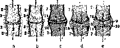
Fig. 1. Scheme of various stages in the development of diarthrosis: a-two chondrogenic layers in contact with each other after the disappearance of the middle layer, b-formation of the joint cavity, c-adult joint, d-joint with a meniscus occupying the entire joint cavity, e-joint with a meniscus occupying only the periphery of the cavity: 1-part of the skeleton in the cartilage stage, V-bone skeleton; 2-chondrogenic layer; 3-mesenchyme; 4-joint cleft; 5-joint cavity; 6-articular cartilage; 7-periosteum; 8-meniscus occupies the cleft; 9-meniscus occupies only the peripheral part of the joint cleft; 10-lateral ligaments; 11-synovial membrane.
of bones, menisci are most frequently biconcave. The shape and dimensions of menisci are very diverse: sometimes menisci occupy the entire joint, forming a complete disk (Fig. 1) (discus articularis) that completely separates the two articular surfaces of the bones, as for example in the temporomandibular joint; sometimes a meniscus has an opening in the center and presents a flat ring (Fig. 1), thereby allowing the bone surfaces of the joint to come into contact with each other in the center of this cartilaginous ring, which again is occasionally observed in the same temporomandibular articulation. In other cases, the interarticular cartilage has not only lost its central part but also part of its circumference; it then assumes the appearance of a crescent (semilunar), the convex edge of which is higher and fused with the surrounding tissues, while the inner thin edge lies freely inside the joint. An example of this type of meniscus is the menisci of the knee joint. Menisci have two smooth surfaces covered by a synovial membrane and moistened by synovial fluid. Menisci are anchored along their periphery, but nevertheless constantly follow the displacements of the joint bones during movements. Menisci consist of fibrous bundles and cartilage cells between them. Vessels and nerves of the menisci enter them from the outer edge, pass through a part of their thickness, and branch without reaching their inner edge; nerves penetrate into the menisci both with vessels and independently. Embryology of menisci. Various parts of the skeleton in the state of a cartilaginous framework do not come into direct contact with each other like in the adult; between them at the site of the future joint there is a more or less thickened zone called (Henke) the intermediate disk. This intermediate or intercartilaginous disk itself consists of 3 different layers: the middle (mesenchymal layer) and two marginal layers adjacent to the corresponding cartilaginous framework (chondrogenic layers). From the latter two
layers develops the cartilage of the articular ends of bones; from the mesenchymal layer develops the meniscus in joints where the articular ends of bones do not come into contact with each other at all points. Menisci are present in the temporomandibular, sternoclavicular, acromioclavicular, knee, and radiocarpal joints.
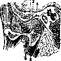
The meniscus between the articular process of the mandible and the articular fossa and articular tubercle of the temporal bone is close in shape to a biconcave transversely oriented plate (Fig. 2). Such a shape is necessary as a compensation for the two convex surfaces of the head of the mandible and the articular tubercle of the temporal bone (Batuyev). The joint cavity is divided by the meniscus into two non-communicating cavities. The meniscus is elongated in the shape of an ellipse and is located obliquely downward and forward. The anterior part of the anterosuperior surface of the meniscus, corresponding to the articular tubercle of the temporal bone, is concave, while the posterior part of this surface, adjacent to the articular fossa of the same bone, is convex; the inferoposterior surface of the meniscus, adjacent to the articular process of the mandible, is concave. The edges of the meniscus are fused with the joint capsule; by means of the capsule, the meniscus is connected to the upper portion of the lateral pterygoid muscle (Fig. 2). In all movements of the lower jaw, the meniscus follows the articular process of the mandible, but the meniscus and the lower jaw are not rigidly connected to each other; when opening the mouth, the meniscus follows the lower jaw only up to a certain limit; at maximum mouth opening, the meniscus, having shifted somewhat forward, stops, and the articular process of the mandible in its forward translatory movement already moves itself along the lower surface of the meniscus, approaching its anterior edge; with such displacement, the meniscus changes its position from oblique downward and forward to horizontal and finally to oblique downward and backward (Fig. 3). When closing the mouth, all
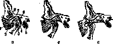
Figure 2. Sagittal section of the temporomandibular joint, mouth closed (left side, inner segment of the section): 1-articular tubercle of the temporal bone; 2-meniscus; 3-articular fossa of the temporal bone; 4-lateral pterygoid muscle; 5-articular process of the mandible.
Figure 3. Sagittal section of the temporomandibular joint; a-mouth closed, b-mouth half-open, c-mouth wide open: 1-meniscus; 2-lateral pterygoid muscle; 3-articular process of the mandible; 4-posterior ligament of the joint; 5-external acoustic meatus; 6-cell tissue behind the articular process of the mandible; 7-articular fossa of the temporal bone; 8-articular tubercle of the temporal bone.
movements occur in the reverse direction. In other movements of the lower jaw (forward, sideways), being of small volume, the meniscus participates to an insignificant degree. Pathology. The clinic knows no isolated disease of the mandibular meniscus; the meniscus is involved in pathological processes of the entire joint; likewise, treatment reduces to the treatment of the joint. Sternoclavicular joint: divided by the interarticular cartilage into two isolated cavities. The meniscus is of irregular shape, lying obliquely from top to bottom and from inside outwards; the center of the meniscus is thin, sometimes thinned to the formation of an opening. Anteriorly and posteriorly, the meniscus is firmly connected to the ligaments of the joint; the upper part of the meniscus is firmly bound to the interclavicular ligament; below, the meniscus usually extends onto the cartilage of the first rib near the sternum and occasionally reaches the costoclavicular ligament. In the articulation of the clavicle and the acromion of the scapula, the clavicle is often (in 34%, Testut) separated from the acromion by a meniscus. The meniscus in various cases is expressed differently. 8 types of menisci are described (Winslow, Weitbrecht, Testut) according to the degree of its expression-from a meniscus in the form of an insignificant cartilaginous separation from the articular cartilage of the acromion with a free edge in the joint to the degree of a complete meniscus partitioning the joint into two mutually independent cavities. Isolated diseases and injuries of these menisci are not known to the clinic. Menisci of the knee joint. The curvatures of the articular surfaces of the femur and tibia do not coincide with each other: the concavity
Figure 5.
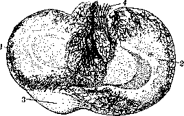
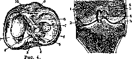
Figure 4. 1-internal meniscus of the knee; 2 and 10-attachment of the posterior and anterior horns of the internal meniscus; 3-posterior cruciate ligament; 4-Robert's ligament of the lateral (external) meniscus; 5 and 8-attachment of the posterior and anterior horns of the external meniscus; 6-external meniscus; 7-anterior cruciate ligament; 9-transverse ligament of the knee joint. Figure 5. Frontal section of the right knee, front view: 1-lateral condyle; 2-external meniscus; 3-internal meniscus; 4-anterior cruciate ligament; 5-medial condyle; 6-joint capsule. The articular facet of the tibia is small in comparison with the strongly convex surface of the femoral condyles. The depth of the tibial articular facet is increased by cartilaginous half-rings, the thickness of which decreases from the periphery to the center (Fig. 4). Menisci have two horns (anterior and posterior), two surfaces (upper and lower), and two edges (outer and inner). In adults, the upper surface is concave, the lower is flat; in children (according to Sirotkin's observations), the lower surface is also concave; in addition, the meniscus in children is twisted in a spiral along its long axis. Being triangular in cross-section (Fig. 5), the menisci with their inner sharp edge are wedged into the dihedral space (sinus condylotibialis) between the femoral condyles and the tibia. The shape of the menisci is related to the function of the lower extremities: they are more unfolded and straightened in leaping primates and close to a ring shape in crawling primates. In each knee there are two menisci: the internal in the shape of the letter C and the external in the shape of the letter O, forming almost a ring interrupted at the intercondylar eminence. Menisci are anchored by their horns to the tibia; the anterior horns are connected to each other by a transverse ligament. The horns of the menisci in relation to the cruciate ligaments of the knee are located as follows, going from front to back: attachment of the anterior horn of the internal meniscus to the tibia, anterior cruciate ligament, anterior horn of the external meniscus attached in front of the tibial spine, posterior horn of the external meniscus, and posterior cruciate ligament. The ligamentous apparatus of the internal meniscus is weaker than the external, but its fixation to the joint capsule is stronger than the fixation of the external meniscus. The internal meniscus is firmly fused with the medial collateral ligament of the knee; the external meniscus has no connection whatsoever with the corresponding ligament. Blood
Menisci receive blood through arteries extending from the capsule and cruciate ligaments. According to observations by Henschen (1928), the anterior and posterior horns are in the best nutritional conditions. A network of vessels penetrates into them with radial branches almost to the free edge of the meniscus. In the middle parts of the meniscus, however, it is distributed only in their outer fourth (Fig. 6). Nerve trunks enter the meniscus to the same depth along with the vessels. The presence of such a development of the vascular network of the meniscus resolves in the affirmative the previously disputed question of the possibility of inflammatory phenomena developing in the meniscus (hyperemia, infiltration, etc.), described by C. Roux in 1895 in the form of meniscitis chronica traumatica. The remaining mass of the meniscus has the synovial fluid with its chemical and biological
A
B
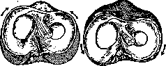
Figure 7. Displacement of the menisci (Mouchet and Tavernier): A - leg extended, meniscus displaced anteriorly; B - leg flexed, meniscus displaced posteriorly. properties, capable not only of nourishing but also of resorbing even the osteocartilaginous elements of joints that have undergone destruction (Tsanov). Physiology of the knee meniscus. When flexing and extending the knee, the menisci inevitably move together with the femoral condyles (Pirogov); upon extension, the menisci shift forward, and upon flexion, backward (Fig. 7). The menisci, located in the angle between the femur and the tibia,
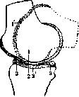
Figure 8. Sagittal section of the medial condyle of the femur, to show the movement of the femur during flexion and extension; 1 and 2 - points at which the femur and tibia touch upon extension; 1' and 2' - the same points upon flexion; 3 - meniscus.
do not resemble a support under the heel of a shortened leg, i.e., they do not lengthen the limb and the height of the joint space with their thickness. According to most observations, the removal of the meniscus does not affect the work of the joint in the least. The empty space formed due to the absence of the meniscus is occupied by synovial fluid. When the knee is flexed with a stationary lower leg, the femoral condyles roll from front to back and at the same time slide from back to front (Fig. 8). In addition to this, flexion in the knee is accompanied by outward rotation of the femur, and extension by inward rotation of the femur. During outward rotation of the lower leg, when damage to the inner meniscus most often occurs, the latter, together with the capsule, is pulled from behind and inwards forward and outward, while its posterior horn is pushed backward by the femoral condyle sliding in the same direction. As a result of strong traction on the meniscus, this may lead to detachment of the anterior horn or longitudinal splitting of the meniscus body.
D. Sirotkin. Meniscitis — damage to the semilunar cartilages of the knee joint — most often occurs in people who, due to their profession, are forced to work with bent knees or make sharp movements in the knee joints (coal miners, mineworkers, carpet layers, athletes). The etiological factor is trauma. Men are more frequently affected. The predominant age is youth and middle age. Regarding distribution, a high frequency of lesions is noted among inhabitants of mountainous regions. Pathologo-anatomical forms of meniscus injuries are divided into 1) detachments from attachment sites (at the anterior and posterior horns and from the joint capsule), 2) tears of the cartilage substance itself (transverse, longitudinal, and flap-like tears and crushing of the cartilage), 3) all kinds of combinations of these crushings, and 4) cases with abnormal mobility of the meniscus. The central part of the meniscus is often trapped by the articular ends of the femur and tibia, and as a result of repeated entrapments, in many cases it appears thickened with frayed edges and cicatricial growths of connective tissue. Histological examination reveals a great development of vascular tissue, traces of old hemorrhages, edema of the connective tissue, and the transition of part of the cartilaginous tissue into connective tissue. As a very rare phenomenon, one should also point out cysts occurring in the outer menisci and appearing several weeks or months after trauma. The mechanism of injury consists in excessive flexion and rotation of the knee joint, with the middle meniscus most frequently suffering (175 cases out of 214 according to Kroiss and 50 to 1 according to Morrison's statistics). Symptomatology reduces to the sensation of sharp pain either in the entire knee or in the part corresponding to the damaged meniscus, immediately after receiving the injury; soon significant swelling is noted, mostly of the entire joint; sometimes the patient is unable to straighten the limb; experiences difficulty walking, especially when descending stairs. The course of the disease is either such that conservative treatment yields no results and one has to resort to surgical intervention, or the patient recovers, but in the presence of damage to the semilunar cartilage, the meniscus undergoes recurrent entrapments. The occasion in this latter case is an awkward position or twist of the leg during work, walking, etc., and sharp pain in the damaged joint is felt quite unexpectedly, forcing the patient to keep the leg in a half-bent position. Such entrapments can recur, ranging from several times a day and month to rare cases within a year. To establish a diagnosis, it is necessary to exclude the following diseases: joint mouse, rupture of the internal lateral ligament, damage to the anterior cruciate ligament, habitual dislocation of the patella, insufficiency of the quadriceps muscle, and tuberculosis of the knee joint. The most reliable method for recognizing meniscus injury is X-ray examination. To obtain a clearer picture, the joint capsule of the knee can be filled using a twenty-gram syringe with oxygen in an amount of 100–250 cm3. In this case, the gas, penetrating into all parts of the joint, bypasses both menisci from all sides. X-ray data are necessary only to exclude other diseases; with respect to the diagnosis of meniscitis itself, the X-ray picture yields nothing. The phenomena of recurrent entrapment serve as the most characteristic diagnostic sign. Prognosis is generally favorable. Treatment in acute cases is conservative (reposition of the meniscus with application of an immobilizing bandage, rest, baths, massage, and other physical therapy procedures). In cases of lack of positive results, surgical intervention is resorted to, consisting either in partial or complete extirpation of the meniscus or in its fixation (meniscopexia). Access to the damaged meniscus is most rationally achieved by longitudinal parapatellar arthrotomy and transverse arthrotomy with section of the collateral ligament of the knee. Prophylaxis should be aimed at strengthening the selection in relation to special professions, and special attention should be paid to preventing persons with weakness of the ligamentous apparatus and osteoarthritis deformans from working. In addition, it is necessary to pay attention to the working conditions themselves, take breaks in work to return to normal statics, and periodically subject workers who have worked for a long time in a given production to special examination.
L. Lepilina-Brusyalovskaya.
Related articles
Mentioned in
Cite this page
“Articular Menisci.” Soviet Medical Encyclopedia. English translation of Bolshaya Meditsinskaya Entsiklopediya, 1st ed. (Moscow, 1928–1936), ed. N. A. Semashko. https://sovietmedicalencyclopedia.pages.dev/article/articular-menisci/