Pericardium
Historical document, translated for reference. It reflects medical knowledge of the 1920s–30s and is not medical advice.
Summary
This article from the 1928–1936 Soviet Great Medical Encyclopedia provides a detailed anatomical description of the pericardium, including its structure, layers, and relationship to surrounding organs. It also covers its development, blood supply, and pathological conditions.
Encyclopedia article (1928–1936)
PERICARDIUM (pericardium), the pericardial sac or heart covering, represents a sac or bag, one part of which directly covers the heart muscle on all sides—pericardium viscerale, s. epi-cardium—and the other part separates the pericardial cavity from the thoracic cavity and constitutes the true pericardium—pericardium parietale. The shape and size of the pericardium depend on the changes and development of the heart, as well as on age and typological peculiarities of the mutual arrangement of organs in the thoracic cavity.
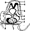
Figure 1. Development of the heart covering: 1- amnion; 2- spinal cord primordium; 3- pharynx; 4- mesocardium, dorsal part; 5- pericardium; 6- myo-epicardium; 7- endocardial cavity; 8- pericardial sac cavity; 9- amniotic cavity. (According to Braus.) (Fig. 1),
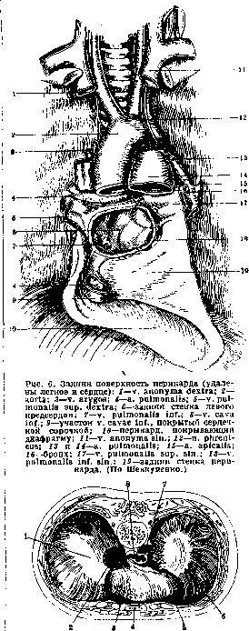
and also on age and typological peculiarities of the mutual arrangement of organs in the thoracic cavity. The visceral layer covers the entire surface of the ventricles and the greater part of the surface of the atria, except for the posterior surface of the left atrium and a narrow strip of the right atrium. Above, in the region of the vessels, the visceral layer folds over and transitions into the parietal layer (Fig. 2 and 3). The site of transition is not constant. On the right, the visceral layer covers the superior vena cava at the place of its entry into the right atrium and only in front and inside, and then proceeds along the anterior surface of the ascending aorta to a point one elbow's length below the a. anonyma; from there it transitions onto the anterior wall of the a. pulmonalis (under the Botalli ligament) and then onto the right side of the artery above the place of its exit from the right ventricle. At the point of contact of the pulmonary artery and the ascending aorta, the visceral layer covers them along the entire circumference and thus separates them from the anterior wall of the atria, forming a slit (sinus transversus pericardii) (Fig. 5), closed from below and above and open from right and left. This opening has great practical significance for performing an operation on the pulmonary artery in cases of embolism. The pulmonary veins are covered by the pericardium over a small area, forming depressions in the region of the lung hilum. Below, the transition of the visceral layer into the parietal layer is limited by the anterior surface of the inferior vena cava (Fig. 6). Behind the left atrium, the pericardium forms a blind sac (sinus retropericardiacus), directly adjacent to the esophagus. The pericardial cavity is almost completely of the same size as the heart, and between the visceral and parietal layers there is only a capillary slit containing a small amount of serous fluid (liquor pericardii). This cavity has the appearance of an obliquely cut cone, the base of which is directed backward and downward to the level of the cartilage of the fifth rib, upward to the level of the junction of the manubrium of the sternum with its body (Ludovici's angle), that is, to the level of the upper edge of the third rib, protruding to the right beyond the edge of the sternum by 1-2 cm, and to the left by 7-8 cm. Depending on the contact with various organs in the pericardial sac, the following parts are distinguished: 1) pars diaphragmatica, 2) pars sterno-costalis, 3) pars mediastinalis (dextra et sinistra), 4) pars oesophagea, s. mediastinalis posterior. Pars diaphragmatica in the lower part firmly fuses with the tendinous center of the diaphragm (Fig. 7, 8 and 9) and with its anterior muscular part; it represents a plane running obliquely from right and back to left and forward (planum cardiacum diaphragmatis). In this plane, the displacement of the heart occurs during contraction. With the pars sterno-costalis, this part forms a sinus in front, which is filled during diastole by the right ventricle and is emptied during systole. Pars diaphragmatica also participates in the respiratory excursions of the diaphragm and can be displaced when the stomach is full of food or gas. The anterior layer of the pericardium—pars sterno-costalis—goes only to the edge of the sternum due to the displacement of the pleural boundary. Therefore, it is more expedient to make a sternal incision rather than an intercostal one when opening. On the sides, the pericardium has the largest mediastinal parts, dext. and sin., loosely connected with the mediastinal pleura. Between the pars mediastinalis pericardii and the mediastinal pleura, from above to below, the phrenic nerves and the a. and vv. pericardiaco-phrenicae pass on both sides.
Figure 2. The relationship of the pericardial sac to the heart and vessels: 1- v. cava sup.; 2- diaphragm; 3- a. pulmonalis; 4- aorta ascendens. (According to Corning.)
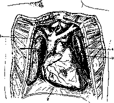
Figure 3. The posterior wall of the heart after removal of the pericardium: 1- n. vagus; 2- a. pulmonalis sin.; 3- bronchus sin.; 4- vv. pulmonales sin.; 5- v. cava inf.; 6- vv. pulmonales dextr.; 7- sinus transversus; 8- v. cava sup.; 9- aorta. (According to Braus.)
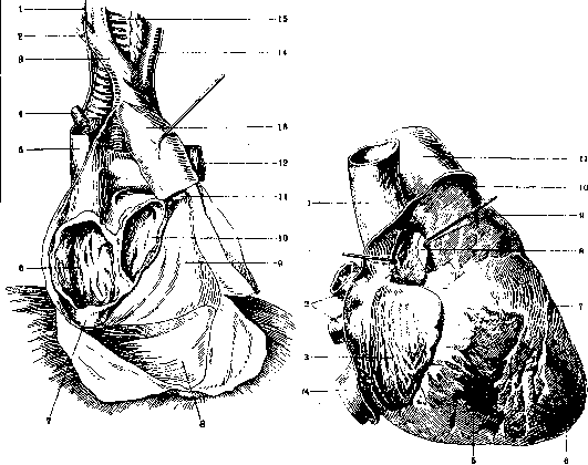
Figure 4. The right lung and heart have been removed, the right atrium is left, except for its anterior wall, and the left half of the left atrium. 1 and 14- n. vagus; 2- a. subclavia; 3- a. anonyma; 4- v. azygos; 5- v. cava sup.; 6- right atrium; 7- v. cava inf.; 8- floor of the pericardial sac; 9- posterior wall of the pericardium; 10- left atrium; 11- v. pulmonalis; 12- a. pulmonalis; 13- aorta; 15- trachea. (According to Shevchenko.)
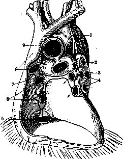
Figure 5. 1- v. cava sup.; 2- vv. pulmonales dextr.; 3- atrium dextr.; 4- v. cava inf.; 5- ventriculus dextr.; 6- apex cordis; 7- sulcus longitudinalis ant.; 8- sinus transversus pericardii; 9- a. pulmonalis; 10- site of transition of the heart covering; 11- aorta ascendens. (According to Spalteholz.)
The pars sterno-costalis directly adjoins the body of the sternum and the IV and V intercostal spaces of both sides. This boundary is extremely variable, and often the left edge extends beyond the sternum. The pericardium has the largest mediastinal parts, dext. and sin., loosely connected with the mediastinal pleura. Between the pars mediastinalis pericardii and the mediastinal pleura, from above to below, the phrenic nerves and the a. and vv. pericardiaco-phrenicae pass on both sides.
Figure 7. Diaphragm from above: 1- centrum tendineum; 2- v. cava inf.; 3- pars diaphragmatica; 4- sternum; 5- pericardium parietale; 6- pleura1 costalis; 7- aorta; 8- oesophagus. (According to Corning.)
Behind, the pars mediastinalis lateralis transitions into the pars mediastinalis posterior. The latter directly adjoins the esophagus, thoracic aorta, and v. azygos (Fig. 10). The blood supply of the pericardium comes from the a. pericardiaco-phrenicae (branches of the a. mammariae int.) (Fig. 11). The rich lymphatic network of the pericardium drains into the lymph nodes of the anterior and posterior mediastinum. The pericardium receives its nerve branches from the nn. phrenici.
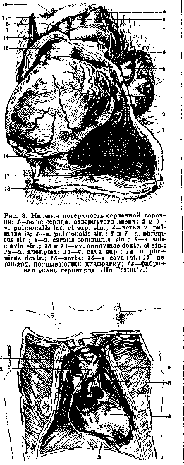
Figure 8. The pericardium and its vessels: 1- a. pericardiaco-phrenica; 2- v. cava inf.; 3- pars diaphragmatica; 4- pars sterno-costalis; 5- pars mediastinalis; 6- pars oesophagea; 7- a. pulmonalis; 8- a. anonyma; 9- v. thyreoidea ima; 10- v. mammaria int.; 11- v. cava sup.; 12- n. phrenicus; 13- n. vagus; 14- v. azygos. (According to Corning.)
Histologically, the wall of the pericardium consists of two layers: 1) the outer (tunica fibrosa) and 2) the inner (tunica serosa). The tunica fibrosa represents a plexus of connective tissue fibers forming a dense tissue incapable of stretching. The tunica serosa of the pericardium, like the epicardium, consists of a thin connective tissue layer with elastic fibers (tunica propria), without sharp boundaries, transitioning into the fibrous layer, and endothelial cells on the contacting surfaces of the parietal and visceral layers, serving to form serous fluid, protecting both layers from friction during the contractions of the heart and respiratory excursions of the pericardium.
A. Bakulev. Pathology. Of the developmental deformities of the pericardium, which are generally rare, defects of the parietal layer are observed mainly in connection with malformations of the sternum. In cases of ectopia of the heart, the pericardium may be completely absent. Diverticula of the pericardium are a very rare occurrence. Normally, the pericardial cavity contains no more than 30 cm3 of clear fluid; sometimes this fluid completely disappears, which is usually explained by post-mortem evaporation due to its proximity to the air-containing tissue of the lungs, especially in cases of emphysema of the latter; sometimes this phenomenon is associated with severe losses of fluid by the organism. (See Hydropericardium for an increase in the amount of fluid.) Small increases are often associated with the duration of agony. (See Hemopericardium for bleeding into the pericardial region.)
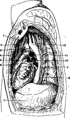
Figure 9. Topography of the pericardium parietale and vessels: 1- a. subclavia dextra; 2- truncus anonymus; 3- pericardium parietale; 4- the same—posterior surface; 5- n. phrenicus sin.; 6- n. vagus sin. (According to Corning.)
Under pneumopericardium is understood the state when the pericardial cavity contains air or other gases, which can be observed during perforations into the pericardial cavity from the side of the esophagus, stomach (cancers, foreign bodies), bronchi, in cases of hydatid pericarditis (see), etc. Free bodies in the pericardial cavity can be observed; usually these are hardened, hyalinized degenerated particles of fibrous exudate, less often—liberated polypoid adipose lobules of the epicardium with subsequent necrosis of them, sometimes ossification. Primary tumors of the pericardium are a rare occurrence. Lipomas, sarcomas, endotheliomas have been described, especially lymphosarcomas with their characteristic diffuse thickening of the entire sac and transition to the epicardium. Of the secondary tumors (metastatic or arising from adjacent organs), carcinomas are observed, usually accompanied by exudate, and more often hemorrhagic.
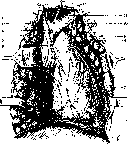
Figure 10. The relationship of the pericardial sac to the heart and vessels (from the side): 1- a. anonyma; 2- n. vagus sin.; 3- n. recurrens sin.; 4- a. pulmonalis; 5- vv. pulmonales sin.; 6- auricula sin.; 7- diaphragm; 8- n. splanchnicus major; 9- aorta thoracica; 10- bronchus sin.; 11- ramus sin. a. pulmonalis; 12- esophagus. (According to Corning.)
Figure 11. Heart covering with supplying vessels: 1- v. thyreoidea ima; 2- v. anonyma dextra; 3 and 9 a. pericardiaco-phrenica; 4 and 10- v. mammaria int.; 5- v. cava sup.; 6- n. phrenicus; 7- heart covering with vessels; 8- lymphatic glands; 11- v. anonyma sin. (According to Shevchenko.)
i. Davydovsky. Surgery of the pericardium. The pericardial sac is the object of surgical treatment mainly in traumatic injuries and inflammatory processes (see Pericarditis) of the heart region. Due to the intimate topographical relationships between the heart and the pericardial sac, anatomical changes spread simultaneously to both these formations. The nature of certain symptoms observed in traumatic and inflammatory processes of the heart region, as well as the effectiveness of surgical measures in this area, is determined by these relationships. Surgical intervention is most often undertaken for penetrating wounds of the heart region. The location of the external wounds in this case has only relative significance. The external wound can be located over a very wide range, from the 2nd to the 8th rib, from the left axillary to the right mammary line, often lying outside the projection of the heart figure on the outer surface of the body, whereas many wounds lying in the area of the heart figure projection do not penetrate the heart region. According to Bradbury, wounds of the pericardium correspond in appearance to external wounds of the chest wall. In stab wounds, the damage to the pericardium can be smaller than the external wounds. In gunshot wounds, the damage to the pericardium is round with uneven edges, and in stab wounds it is linear or fissured (Janelidze). According to Borchardt, the size of the wound usually does not exceed 1-2 cm. Kirschner, Fischer, and others consider that digital examination sometimes allows one to palpate the wound of the pericardium and the heart. Janelidze considers this method unreliable. After exposing the pericardium surgically, finding the site of its injury does not present difficulties; it is facilitated by the copious outflow of blood through the wound of the pericardium. Isolated wounds of the pericardium are extremely rare. The clinical picture of these wounds is determined by the signs of damage to the chest wall. According to Napalkov, opening the pericardium does not affect the function of the heart. A significant accumulation of blood (hemopericardium) in isolated wounds of the pericardium is possible with damage to the pericardiacophrenic artery. The pericardial sac is damaged as a rule in wounds of the heart (see Heart). Mackenzie considers the pericardium and myocardium insensitive to pain. According to Janelidze, 'even an inflamed pericardium can be cut without causing pain.' The clinical significance lies in pains irradiating towards the abdomen in some cases. These pains were observed by Naismith in slow intrapericardial bleeding. Percussion changes in the heart region in wounds of the heart may be absent. According to Janelidze, in half of all cases of heart wounds, cardiac dullness turns out to be increased due to the accumulation of blood in the cavity of the pericardium (hemopericardium). The rarely observed tympanic percussion sound in the heart region indicates the penetration of air into the cavity of the pericardium (pneumopericardium). Pathological murmurs observed in wounds of the pericardium and the heart are not constant and not typical. The pericardial friction murmur (sometimes masking the heart tones) is not encountered often and can be similar to the murmur in pericarditis. The Morel-Lavalée sign has long been considered a valuable sign, namely the murmur heard sometimes in the heart region from a distance and resembling the noise produced by a mill wheel when it strikes flowing water. Morel-Lavalée considers this murmur a sign of pericardial rupture and links it to pneumopericardium and the accumulation of blood in the heart region. Subsequent observations have shown that the 'mill wheel' murmur also occurs with changes in the mediastinum, in the pleura, in the lungs, and in the stomach. Isolated wounds of the pericardium are extremely rare. In the majority of cases, simultaneous wounds of the heart and pericardium are described; the clinical picture proceeds rapidly and requires rapid intervention. The wound of the pericardium is exposed by the same principles as wounds of the heart (see). The wound of the pericardium after applying a cardiac suture must be closed tightly. However, in each individual case, the question of the possibility of applying a primary suture is decided by the surgeon depending on the nature of the damage and the infection of the wound. To prevent subsequent accumulation of exudate in the cavity of the pericardium, it is recommended when closing the pericardial sac to apply rare sutures so that fluid can drain from the pericardium through the gaps between them. When applying a primary suture, some suggest suturing the pericardial sac near the xiphoid process so that in case of accumulation of exudate, the pericardial sac can be opened and drained. A drain (rubber tube) is introduced at the lowest point of the pericardial sac. Adhesive pericarditis and mediastino-pericarditis belong to frequent postoperative complications of cardiac suture (Hesse), which is conditioned by the susceptibility of the pericardium to infection. Janelidze points out that if timely application of cardiac suture succeeds in saving the patient from immediate death from anemia or compression of the heart, then in the future he is threatened with death from infection of the pericardium and pleura. This circumstance requires especially careful observance of aseptic rules during surgical intervention on the heart and pericardium.
Related articles
Mentioned in
- Adhesive Process
- Anomaly
- Aspiration Puncture
- Atelectasis
- Atrial Fibrillation
- Bamberger's Sign
- Body Cavities
- Bronchostenosis
- Cardiolysis
- Cardiovascular System
- Cavae Venae
- Cavum
- Diastole
- Endothelioma
- FOCHT ALEXANDER
- Heart
- Heart Defects
- Heinrich Curschmann
- Hemopericardium
- Hemorrhage (the accumulation of blood that has escaped from)
Cite this page
“Pericardium.” Soviet Medical Encyclopedia. English translation of Bolshaya Meditsinskaya Entsiklopediya, 1st ed. (Moscow, 1928–1936), ed. N. A. Semashko. https://sovietmedicalencyclopedia.pages.dev/article/pericardium/