Gastroscopy
Historical document, translated for reference. It reflects medical knowledge of the 1920s–30s and is not medical advice.
Summary
A historical overview of the development and technique of gastroscopy in the early 20th century. It details the evolution of rigid and flexible instruments, the optical systems used, and the clinical procedure for examining the stomach.
Encyclopedia article (1928–1936)
GASTROSCOPY (from Greek gaster—stomach and scopeo—I look), a method of examining the stomach by direct inspection of its cavity with the help of a special instrument (gastroscope) introduced through the mouth and esophagus. The idea of 'looking' into the stomach cavity first occurred to Kussmaul in 1868 and was inspired by the observation of a sword swallower, on whom the first experiment was performed. Kussmaul inserted a straight metal tube with a diameter of 13 mm into him and attached a Desormeaux illuminator to the outer end of the tube, but the weakness of the illumination and the accumulation of fluid in the stomach cavity did not make it possible to see the gastric mucosa at all satisfactorily. This attempt, which proved fruitless, was forgotten, and the idea of constructing a gastroscope only reached a practical footing after Nitze succeeded in realizing the idea of endoscopy by introducing a light source into the cavity of the organ itself. Nitze himself constructed the first model of a gastroscope in 1879, which consisted of 2 parts: a movable, curved tube assembled from individual links, introduced through the esophagus into the stomach, and an upper part curved at a right angle, reaching the pharynx. With the help of a special device, the entire system of the movable tube straightened out after introduction into the stomach, and light rays reflected by the mucous membrane were directed along the length of the tube, where, having passed through a system of prisms and spherical lenses, they reached the observer's eye. This instrument also did not yield satisfactory results. The first positive result was obtained only by Mikulicz, who built his own model of a gastroscope (1881): the latter consisted of a tube 65 cm long, 14 mm in diameter, curved at an angle of 150° at the border between the gastric and esophageal parts; the optical system was analogous to that of a cystoscope. Mikulicz succeeded in examining the stomach cavity and even diagnosing gastric carcinoma; therefore, Mikulicz should justly be considered the founder of gastroscopy, although his gastroscope did not receive wide practical application due to the difficulty of introducing it into the stomach, on the one hand, and the imperfection of the illumination and optics, on the other. The attempt to further improve the methodology and instrumentation continued, mainly by German authors (Rosenheim, Kelling, Kuttner, Loening and Stieda, and in later times Eisner, Schindler, Sternberg, Hübner) and Bensaude in France. Until recently, two systems competed in the construction of the gastroscope: flexible systems, which pursued, mainly, the goals of easier insertion of the instrument, and rigid ones, which set good optics as their main goal. Victory remained with the rigid systems; to facilitate their introduction into the stomach, various devices are added: 1) an elastic tip is screwed onto the end (Eisner, Korbsch); 2) the lower end of the gastroscope is given a beak-like curved shape (Sternberg), and this alone facilitates its passage through the most difficult places in the esophagus—at the beginning of the thoracic part, and especially in the supracardiac section, where the esophagus makes a bend to the left and forward; 3) special guides are used; thus, in Bensaude's gastroscope, a piano wire with an olive at the end is introduced into the stomach as a guide, which the patient swallows: the gastroscope itself is then introduced along this guide; 4) an outer metal tube is introduced into the stomach with an elastic mandrel (see Figure 1 B), which is then replaced by an inner tube with optics (Schindler, Hübner; see Figure 1 A). As for the optical system of gastroscopes, it is almost in all modern models of the Great Medical Encyclopedia Vol. VI. and
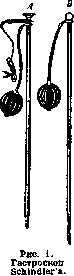
models uniform and analogous to the optics of a cystoscope: at the gastric end, a prism (Amici) is inserted into the metal tube (window), which refracts light rays at a right angle and then directs them along the length of the gastroscope through a series of spherical lenses to the eyepiece located at the outer end of the instrument. Illumination of the stomach cavity is produced by an electric light bulb located at the end of the instrument. To absorb red rays, light filters have recently been inserted into the optical system: green—at the prism, blue—at the eyepiece. The optical system of modern gastroscopes is already so satisfactory that with the correct and good setup of the apparatus, one can clearly see all the details. Light filters also provide a natural color of the mucosa and a more relief, plastic picture. Technique of gastroscopy. At the present time, gastroscopy remains the most difficult method of endoscopy. Previously, the patient should be accustomed to the introduction of elastic bougies or probes into the stomach. The examination is performed on an empty stomach; if there is no pyloric stricture, one can do without preliminary gastric lavage. 1/2–1 hour before, morphine is injected into the patient; local anesthesia is performed by smearing the pharynx, the root of the tongue, and the entrances to the larynx and esophagus with a 10% cocaine solution. The examination can be performed in different positions of the patient—sitting, lying, on the right or, better, on the left side, with thighs strongly brought to the abdomen, or in the knee-elbow position (Sternberg). The latter is advantageous in that negative pressure is obtained in the abdominal cavity, and, in addition, saliva flows out freely, but it is tiring for the patient. The most convenient is the position on the side. In this case, the patient's head is strongly tilted back and held by an assistant. Having inserted a finger into the mouth behind the root of the tongue, it is pushed anteriorly and the gastroscope is inserted along the finger, if it does not have other (above-mentioned) devices for insertion. Gastroscopes with guides, of course, have all the advantages when introducing the instrument. After the optical system is set in place, air is pumped into the stomach using a double balloon, which penetrates there through the space between the two tubes, the light is turned on, and the examination begins. A button on the eyepiece serves for orientation regarding which part of the stomach is being examined. Since with gastroscopy the instrument can be tilted to the side only to a very insignificant degree, it follows that the field of view can be changed only by turning the instrument along the axis and advancing it to a greater
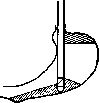
Figure 2.
or lesser depth, but in doing so, it is necessary to avoid touching the gastric mucosa with the prism, otherwise the clarity of the picture is lost. According to the design of the optical system, rays enter the gastroscope in a cone, at an angle of 50–60°, and parts corresponding to the base of the cone fall into the field of view. Everything that cannot fall into the region of this cone remains inaccessible to inspection; therefore, the area of visibility depends partly on the shape and position of the stomach. For two types of stomach—Holzknecht (see Figure 2) and Rieder-Gredell (see Figure 3)—the area inaccessible to inspection is different (shaded in the figure); with the Rieder form of the stomach, the pylorus cannot be set in the field of view. That is why in some gastroscopes (Sternberg) the beak is made curved. Thus, one of the disadvantages of the gastroscope is the impossibility of examining all sections of the stomach. True, in some models ('universal gastroscope') there are two additional prisms, which, by means of rotating the optical system along the axis, can be extended from the niches in which they lie, and, tilting over the main prism of the gastroscope itself, make it possible for light rays reflected from the region of the cardia or the greater curvature to penetrate (retrograde gastroscopy), but these additional parts are so fragile that using them is difficult. The examination is not easy for the patient, and it should not be prolonged. The examination is most hindered by the mobility of the stomach walls, depending on its contraction, on the partial escape of air and the need to pump it in, on respiratory and vomiting movements, and the tension of the abdominal wall. Mucus and gastric juice accumulate in the dependent parts of the stomach, covering part of its walls. Therefore, to set the pylorus in the field of view, the patient's position must be on the left side. Picture of a normal stomach. The gastric mucosa in the gastroscope has a color of various shades, from pale pink to red; with light filters, its color is close to normal. On the anterior wall, the mucosa is smooth because when the stomach is inflated, its stretching occurs mainly at the expense of the anterior wall; on the posterior wall, large folds and furrows between them are usually visible. The pyloric section (antrum) is visible in the form of a cone-shaped narrowing part, with a strongly protruding fold on the right and above (slope of the lesser curvature); in the depth, the black round opening of the pylorus is visible, rhythmically contracting until the ring closes completely with star-shaped folds (see separate table, Fig. 1 and 2). Pathological changes, as far as
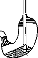
Figure 3.
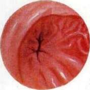
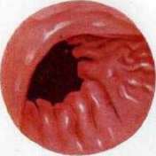
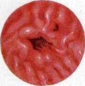
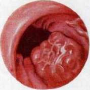
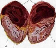
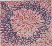
Plate 1. Fig. 1. Normal preparation. Fig. 3. Ulcer on the lesser curvature. Fig. 4. Carcinoma of the pylorus in the pyloric part (after Schindler). Fig. 5. Hematoma... Regarding gastroscopy, those [changes] which have been studied by the available experience of gastroscopy concern the appearance of the mucous membrane itself, ulcers, and neoplasms of the stomach. As for changes in the shape of the stomach, they can be determined more simply and easily by fluoroscopy. Diffuse processes—various forms of gastritis—are most accessible to diagnosis, and in this regard, gastroscopy makes it possible to perform differential diagnosis between neuroses of the stomach, its functional diseases, and organic processes. Schindler and Rachet established, on the basis of extensive experience, three forms of gastritis: 1) chronic catarrhal gastritis, characterized by mottled redness and increased mucus production, 2) hypertrophic form of gastritis, and 3) atrophic—the latter two are associated with changes in the appearance and color of the folds of the mucous membrane. Localized processes on the mucosa (ulcers, tumors) are more difficult to determine, as they do not always fall into the field of view and have to be searched for. The pictures of ulcers (see separate table, Fig. 3) are diverse, depending on the depth, properties of the edges, and inflammatory reaction in the surrounding area; erosions have the appearance of superficial defects of the mucosa, yellowish-pink in color, with a hemorrhagic rim; in deep ulcers, their base is covered with whitish deposits of fibrin, and the edges are outlined by sharper redness. Cicatricially degenerated parts of the walls have the appearance of whitish spots, disrupting the regular arrangement of the folds. Cicatricial strictures of the pyloric part are characterized, in addition, by the immobility of the walls, a whitish border along the edge of the ring, and smoothing and flattening of the pattern of the mucous membrane folds. Cancrous tumors (see separate table, Fig. 4), growing into the cavity of the stomach in the form of a mushroom or disintegrating, have a very characteristic appearance for recognition. A cancrous ulcer is characterized by uneven edges, but, of course, it cannot always be distinguished from a callous ulcer, just as when examining an anatomical specimen with the naked eye. In infiltrating carcinomas, the folds of the mucosa are smoothed out and take on a grayish-white color. The dangers of using gastroscopy are almost exclusively associated with damage to the esophagus during the insertion of the instrument, but not to the stomach itself. Hübner, from the literature and by means of a questionnaire, collected 9 cases of fatal outcomes in more than 3,500 gastroscopies (1/4%), but these figures relate to the first years of gastroscopy. Contraindications to gastroscopy should include: aortic aneurysms, severe arteriosclerosis, heart defects, fresh ulcers, recent gastric hemorrhages, and, finally, conditions that make the insertion of the gastroscope difficult: curvature of the spine, a thick short neck, and excessive obesity. Some of these contraindications are, of course, relative. It should be especially noted that in surgery, one has to resort to gastroscopy even during the operation on the stomach itself, for example, to find ulcers or the source of bleeding. For this purpose, Rovsing proposed a special instrument—a gastroduodenoscope, which is, in essence, an exact copy of a catheterizing cystoscope, only of a significantly larger caliber. Examination with the help of this instrument is performed as follows: the stomach is extracted through an abdominal wound, and an opening is made in its anterior wall sufficient for the insertion of the instrument; around the latter, the incision is tightened with a purse-string suture, after which the examination is performed without fear of contaminating the abdominal cavity. The stomach is inflated with air, but before proceeding to the examination from the inside, the walls of the stomach are examined by transillumination (diaphanoscopy), during which not only blood vessels but also muscle bundles are clearly indicated. Against a general pink background, tumors and ulcers shine through in the form of dark spots. Having noted suspicious places during diaphanoscopy, the prism of the gastroscope is then directed at them during direct examination of the stomach cavity. In Rovsing's gastroscope, there is, in addition, a device for passing a bougie through the cardia, and this instrument can be used for bougienage of esophageal strictures through gastrostomy fistulas. However, for this purpose, Rovsing's gastroscope can be successfully replaced by an ordinary catheterizing cystoscope.
Related articles
Mentioned in
Cite this page
“Gastroscopy.” Soviet Medical Encyclopedia. English translation of Bolshaya Meditsinskaya Entsiklopediya, 1st ed. (Moscow, 1928–1936), ed. N. A. Semashko. https://sovietmedicalencyclopedia.pages.dev/article/gastroscopy/