Gastrostomy
Historical document, translated for reference. It reflects medical knowledge of the 1920s–30s and is not medical advice.
Summary
Gastrostomy is a surgical procedure creating an artificial opening into the stomach for feeding patients with esophageal obstruction. The article describes various techniques for performing gastrostomy, including methods by Witzel, Stamm-Senn-Kader, Frank, and others, with considerations for tube size and patient nutrition.
Encyclopedia article (1928–1936)
Gastrostomy (from Greek gaster-stomach and stoma-mouth), or a gastric feeding fistula, is created for the purpose of artificially feeding patients in whom the esophagus has become impassable for various reasons (tumors and scarred strictures of the esophagus). In addition, gastrostomy is used as a preliminary operation in large operations on the pharynx and esophagus for the purpose of bougienage "without an end" according to the Hacker method in cases of scarred strictures of the esophagus, and for treating cancer of the stomach and lower part of the esadium with radium. The operation of creating a gastric fistula was first performed on animals by Basov in 1842. On humans, it was first applied by the French surgeon Sedillot in 1849. In its original form, the method of creating a gastric fistula consisted in that the edges of the stomach opened by incision were sutured to the abdominal wound, as a result of which the fistulous opening did not tightly fit around the tube, the stomach contents flowed past it, irritating and corroding the skin and wound. At present, the following requirements are made for a gastric digestive fistula: 1) the fistula must tightly fit the rubber tube inserted into the stomach and not leak when the stomach is full, 2) it must allow a sufficiently thick tube to pass so that the patient can

Figure 1. Course of the oblique channel in gastrostomy according to Witzel.
feed not only liquid but also thick food, 3) it must not allow food from the stomach to pass, even if the tube is removed for some time. The fistula created according to the Witzel method meets these requirements to a greater extent, but with it, tubes that are too thick cannot be used. The technique of its creation is as follows: the abdominal cavity is opened with a longitudinal incision through the uppermost part of the left rectus muscle, which is separated along the course of the fibers. The stomach is extracted. On its anterior wall, closer to the cardia, a sufficiently thick (catheter No. 22-24) rubber tube is placed in the direction from left and top to right and bottom, and is surrounded by a channel from the wall sutured over it for a distance of 4-5 cm. At the right end of the created channel, the stomach is opened for a very short distance and the end of the tube is inserted into it. The stomach wound is immediately sutured, thereby creating a channel in the stomach wall, lined with serous membrane, passing obliquely and tightly fitting the tube (see Figure 1). To prevent the tube from slipping out of the stomach, it is better to attach it to the upper end of the channel with a catgut thread, which is previously tied in a loop around the tube. The wall of the channel near the exit of the tube is sutured along its entire circumference to the parietal peritoneum and the posterior wall of the rectus sheath, and the wound is sutured in layers. The tube is fixed to the skin with a plaster. Before closing the abdominal wound, one should make sure that the end of the tube is in the stomach, since in unsuccessful cases it may be placed between the mucous and muscular membranes of the stomach. For this purpose, a certain amount of saline solution can be poured through the tube using a sterile funnel, which, with a properly created fistula, easily passes into the stomach. Dyakonov introduced a modification of this method, which was applied in his clinic. 3 cm to the left of the midline, an incision is made, as in the Witzel method. The anterior wall of the rectus sheath is incised. The medial edge of the muscle is isolated and retracted to the side. The posterior wall of the sheath is incised corresponding to the skin incision, but for a shorter distance. The anterior wall of the stomach is extracted and sutured at the lower end of the wound to the posterior wall of the sheath and the peritoneum. The end of the tube (catheter No. 22-24) is enclosed according to Witzel. At the level of the upper end of the peritoneal incision, the medial edge of the common cover and anterior leaf of the sheath is transversely incised to the midline. The upper end of the stomach wall channel is placed in this incision and secured with sutures. The rubber tube is attached with one suture to the stomach wall at the upper end of the channel. The rectus muscle is lowered and presses the protruding part of the stomach, which lies between it and the posterior wall of the sheath. The skin wound is sutured. To allow thicker food to be introduced, Dyakonov replaced the rubber tube with an aluminum one, which has thinner walls and therefore a wider lumen. According to the Stamm-Senn-Kader method, a straight channel is created in the stomach wall, but one that also tightly fits the tube (see Figure 2). The advantage of this method is that thick tubes up to 1 cm or more in diameter can be introduced into the stomach. The tube is inserted into the stomach through a small incision in its anterior wall and is secured with a purse-string suture around it. Then 1 or 2 more purse-string sutures are applied, thereby creating a straight channel around the tube, the external opening of which is attached to the parietal peritoneum. Hans considers it necessary to create a channel at least 5-6 cm long. With such a length, the tube can be removed from it, and the stomach contents do not leak out (Bulyginsky). Marwedel created the oblique channel somewhat differently. In his method, the serous and muscular membranes of the anterior wall of the stomach are incised down to the submucosal layer. At the lower end of the incision, a small opening is made in the mucosa and the end of the tube is inserted into the stomach. The tube is placed in the stomach wall incision, and the latter is sutured.

Senn-Kader. With such
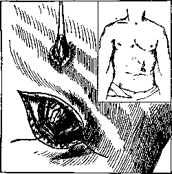
Figure 3. Gastrostomy according to Frank's method. The cone of the stomach wall with a thread at its apex is passed through a subcutaneous tunnel and sutured at its base to the parietal peritoneum (from Bier-Braun-Küttmell with modifications).
A more complex method is that of Frank, but it is designed for the active action of the muscle to compress the channel. The skin incision is made parallel to the left costal margin. The skin wound is stretched, and the muscle and peritoneum are incised longitudinally, as in the Witzel method. A cone of the stomach is brought out, and through its serous and muscular membrane a strong retaining thread is passed. The base of the cone is sutured in a circle to the parietal peritoneum with tension on the thread. With a blunt dissector, a tunnel is created subcutaneously upward beyond the costal margin at the upper edge of the incision. A small transverse skin incision is made over the end of the forceps, and through the retaining thread, the apex of the stomach cone is brought out here. With tension on the thread, the edges of the muscle are sutured so that the cone remains patent (see Figure 3). The skin wound is sutured. The apex of the stomach cone is opened and sutured into the skin wound. A rubber tube is inserted into the stomach through the created channel. Good functional results are obtained on the condition that the subcutaneous channel is sufficiently long and the rectus muscle fits tightly over the base of the cone. Of the methods presented, the Witzel and Stamm-Senn-Kader methods are most widely used. It is important that the tube be wide enough and that patients, using a funnel, can send chewed solid food through it into the stomach. If this is ensured, the nutrition of patients usually improves quickly. The Tavel method should also be mentioned - the creation of a channel from a segment of jejunum, one end of which is connected to the stomach, and the other to the skin wound above. Due to its complexity, this method cannot compete with the previous ones and is generally not used, but has historical significance, as it was the origin of the creation of an artificial esophagus from intestine.
Related articles
Mentioned in
Cite this page
“Gastrostomy.” Soviet Medical Encyclopedia. English translation of Bolshaya Meditsinskaya Entsiklopediya, 1st ed. (Moscow, 1928–1936), ed. N. A. Semashko. https://sovietmedicalencyclopedia.pages.dev/article/gastrostomy/