Preoperative Period
Historical document, translated for reference. It reflects medical knowledge of the 1920s–30s and is not medical advice.
Summary
The preoperative period is the time needed for research and preparation of the patient for surgery. This article discusses the importance of proper patient preparation before operations to prevent complications, including assessment of organ function, treatment of existing conditions, and psychological preparation.
Encyclopedia article (1928–1936)
PREOPERATIVE PERIOD, the time necessary for research and preparation of the patient for surgery. During the period when surgery was flourishing with enthusiasm for technical details of operations and development of techniques for new operations, little attention was paid to the pre- and postoperative periods. However, a series of unsuccessful outcomes of operations, technically brilliantly performed, led surgeons to the conviction that to achieve desired results it is necessary not only technically good execution of the operation, but also appropriate preparation and postoperative regimen. Subsequently, much attention was devoted to the postoperative period, and only in recent decades has surgery begun on the right path of prevention of postoperative complications through appropriate preparation of the patient for surgery. This preparation is especially necessary in the case where any organ is already pathologically altered and the operation may affect it particularly harmfully. The duration of the P.P. can vary depending on the nature of the disease and necessary measures; in urgent operations (ectopic pregnancy, perforated gastric ulcer, acute appendicitis, bleeding) its duration should be very limited; after a careful but possibly rapid examination of the patient, those or other measures are applied if necessary (temporary arrest of bleeding, combating shock in severe injuries, etc.), after which the patient is taken to the operating room, and sometimes the operation is performed under any conditions (arrest of bleeding, tracheotomy). In other cases, the P.P. can be lengthy. During this time, the patient is thoroughly examined and prepared for surgery by various measures. On average for ordinary operations (appendectomy, herniotomy) the P.P. equals several (2-3) days. During this time the patient not only prepares for the operation but also becomes accustomed to the surrounding environment. In many cases the concept of P.P. will be vague, but since for most patients admitted to the surgical department operative intervention is required, for them the P.P. begins from the moment of admission to the institution. In some cases, if the operation is not emergency, it has to be postponed due to concomitant acute diseases (angina, influenza, etc.). In particular, after influenza and angina, it is recommended to perform the operation no earlier than 25-30 days after establishing normal temperature. Patients with rashes on the body (scabies, furunculosis), feverish upon admission regardless of the main disease, cannot be subjected to operative intervention until the cause of the febrile state is clarified or the pathological phenomena are eliminated. Before admission to a surgical institution, the patient should take a bath, if there are no contraindications to it (weak, severe, emergency patients), and undergo thorough examination, to avoid bringing infection or parasites into the department. It should be especially emphasized that patients transferred from other departments (therapeutic, neurological) should be examined as thoroughly as newly admitted patients, to avoid unpleasant surprises for the surgeon, since ultimately only he bears responsibility for the indications for surgery. During the P.P. the patient should be in the most calm environment to as much as possible exclude excessive psychological trauma. This especially concerns the so-called 'nervous patients'. Cleanliness of the premises, discipline, proper organization of work of the staff, caring attitude from the latter act soothingly on the patient's psyche. The question of premises for P. patients is also quite important. The sight of patients suffering after surgery from pain, vomiting, especially if the postoperative period passes with severe complications, can have a depressing effect on the patient who is yet to undergo surgery. Moreover, the conditions of care for operated patients and those who still have to undergo surgery are different, and for the purpose of rationalization of this care it is desirable that patients spend the P.P. in separate wards. If the department is small, then P. patients and convalescents can be placed in the same room. In large hospital institutions patients pass through the so-called sorting department. During the P.P. among patients, as well as among convalescents, cultural services should be organized: chess, library, radio, film screenings, health education work; however, in the latter there is no need to touch on topics that create excessive anxiety for the patient (the question of malignant neoplasms, etc.). If the nature of the disease requires urgent operative intervention, then in examining the patient only the most necessary methods are used (examination of the heart, lungs, urine). In all other cases, a thorough and comprehensive examination of the patient is carried out by clinical, laboratory, radiological methods: this clarifies the indications for operative intervention, its nature, the method of anesthesia, as well as reveals the need for preoperative preparation of organs, obviously diseased or those whose function is most affected by the operation (heart, lungs, liver, kidneys). Proper attention must be paid to the anamnesis. Indications in the anamnesis of existing diseases of the heart, lungs (tuberculosis), kidneys (nephritis), diabetes will force even more careful examination of the patient in this direction and in some cases to refuse operative intervention when complications from the specified organs will be more dangerous than the main disease for which the operation was planned for the patient. For example, everyone knows cases of exacerbation of the pulmonary process after operations, especially under general anesthesia. The anamnesis is also important in clarifying the presence of foci of dormant infection [past erysipelas processes, suppuration, osteomyelitis, sometimes scars after gunshot wounds, etc. (Solovov)]. If such foci are established clinically (examination of the oral cavity, teeth, old scars; provocation of dormant infection), then before surgery, to avoid sometimes severe complications (streptococcal sepsis, tetanus), it will be necessary to fight this hidden infection. Attention should also be paid to the possibility of alcoholism, syphilis, malaria, since in some of them poor healing of surgical wounds is noted (alcoholism, syphilis), in others - exacerbation of a dormant process (malaria) or newly arising severe complications (delirium tremens in alcoholics). To avoid these complications, it is not recommended to operate on syphilitics if there are signs of fresh luetic infection (RW first of all), in alcoholism the patient should not be completely deprived of alcoholic beverages, in malaria preventive quininization is required. Emaciated, dehydrated patients need either administration of fluids subcutaneously, intravenously, per rectum or blood transfusion. Obese subjects, on the contrary, are sometimes recommended a careful course of treatment against obesity before surgery. Constitutional features also have important significance. It is also important to determine whether the patient has status thymico-lymphaticus. Surgeons often have to deal with bronchitis and emphysema of the lungs. In case of acute bronchitis, if the operation is not urgent, it is postponed until recovery from bronchitis. In chronic bronchitis, as well as in emphysema accompanied by bronchitis, preliminary treatment is necessary. In elderly patients suffering from emphysema and bronchitis, special preventive measures are required during the P.P. This is especially important in cases of operations in which complications from the lungs and bronchi are generally not uncommon. Such operations include laparotomies in general, especially in the upper abdomen, and operations requiring prolonged bed rest in the postoperative period. Protective measures in all such cases include breathing exercises, inhalation of carbon dioxide, cupping, camphor, etc. Preventive injection of anti-pneumococcal serum, recommended by some authors to avoid postoperative pneumonias, has not become widespread. The effect of preventive injections of various polyvalent sera (pneumo-strepto-staphylococcal and others), the use of which some authors recommend against local suppuration in the wound, peritonitis, etc., is also not great. When examining the heart and circulatory organs, besides percussion, auscultation, examination of the pulse, measurement of blood pressure must be considered mandatory. Based on the blood pressure figures, one can, for example, to a certain extent, establish indications for one or another method of anesthesia; thus, with low blood pressure splanch-nic anesthesia is contraindicated. Some surgeons attach great importance to the so-called Shange breathing test; it is performed as follows: the sitting patient is asked to take a deep breath and hold it as long as possible. Most authors consider general anesthesia contraindicated if the patient cannot hold his breath for more than 20 seconds. Of course, this test is not absolutely indicative, especially if one takes into account that with appropriate training in certain professions, the delay can be significantly extended even with poor circulatory condition.
Some surgeons recommend the Katzenstein test (the effect of compression of the femoral artery on the pulse and blood pressure), the Moeller test (the effect of holding one's breath on blood pressure, etc.).-If by one method or another disorders of the cardiovascular system are established, some of them may be a contraindication for performing operations (decompensated defects), while in other cases the operation should be postponed until the cardiovascular function is normalized. In addition, a number of heart diseases (slightly expressed myocarditis) make it necessary, without delaying the operation for a longer period, to take a number of measures in the preoperative period that improve the function of the cardiovascular system; these include bed rest, diet, and a number of medicinal measures (strychnine, adrenaline, camphor, digitalis, subcutaneous administration of physiological solution, etc.). Indications for such thorough examination are, first of all, blood diseases, then-liver, kidneys, diabetes, etc. General blood examination, besides its diagnostic value for the main disease (anemia in malignant neoplasms, leukocytosis in inflammatory conditions), in a number of cases indicates the need for appropriate preparation; this includes blood transfusion, preparation in diabetes, cholemia. The presence of leukocytosis (see) with a neutrophilic shift, if it is not caused by the main disease, prompts one to search in the body for the source of this condition (existing or recently purulent diseases). The indications for surgery depend on the results of such an examination. The presence of dental caries, stomatitis, tonsillitis dictates a number of measures to eliminate them. Some surgeons rightly attach such great importance to the sanitation of the oral cavity that they recommend sending every patient who is scheduled for surgery to a dentist beforehand. If not in all cases one should be so demanding, still every surgical patient should be taught by the staff to keep the oral cavity clean-rinsing the mouth after meals, brushing teeth should be widely propagated among patients both in the preoperative period and in the postoperative period; this is one of the measures preventing the development of postoperative parotitis, pneumonia, etc. In the preoperative period, it is also important to pay attention to the gastrointestinal tract. Until recently, preparation with laxatives was considered mandatory. The justification for giving laxatives before surgery was: 1) reducing the danger of vomiting in the postoperative period, 2) reducing the danger of intoxication from the intestine, 3) combating flatulence, 4) eliminating contamination of the abdominal cavity when the intestine is opened. As further observations have shown, especially observations of the last decade, preparation of the body with laxatives (together with mandatory fasting before surgery) is not only pointless but also harmful. It was noticed that the postoperative period in persons who were not prepared with laxatives and fasting much less often proceeded with the previously common postoperative complications-vomiting, flatulence, retention of urination, etc. symptoms, which are associated with postoperative acidosis (see Acidosis, postoperative acidosis). In the Russian literature, Korganova-Müller was the first to present such observations. Working in two different institutions, in one of which patients were prepared for surgery with laxatives and fasting, while in the other they were operated on without such preparation, the author could notice a significant difference in the postoperative period. Thus, during appendectomies she observed "in 41% a smooth postoperative course in patients with ordinary preparation and in 76%-without such preparation. The study of the problem of postoperative acidosis shows that not only preoperative preparation but also the operative trauma itself, anesthesia, and the patient's nervous excitement before surgery are causes that cause postoperative acidosis. Even if one takes into account that not all authors associate these phenomena with acidosis and that by no means all consider postoperative acidosis to be true, still the observations undoubtedly show that the less we prepare the patient for surgery with laxatives and fasting, the less often postoperative complications of the above order occur.-It has long been noted that emergency operations, usually performed without such bowel cleansing, proceeded no worse than operations before which the intestine was thoroughly cleansed. Over the last decades, a number of surgeons have expressed doubt about the necessity of cleansing the intestine with laxatives before operations. Interest in this question has especially increased in the last ten years, mainly in connection with questions about postoperative acidosis. At the present time, it has been established that none of the pursued goals is achieved by giving laxatives and that the results are often quite the opposite. Vomiting in the postoperative period cannot be associated with a full intestine, since food usually leaves the stomach only a few hours after its intake, while a full stomach is cleansed not by laxatives but by a stomach tube (emergency operations, especially ileus). In addition, in the postoperative period vomiting is usually not associated with food intake (see below).-As for the danger of intoxication and infection from the intestine, it is now known that it is greater when laxatives are given than without them. The intestinal mucosa is damaged due to enhanced peristalsis, which also disrupts the natural barrier and increases the absorption of intestinal contents into the blood. Meyer's experiments showed that after giving laxatives to rabbits, blood cultures for bacteria usually gave a positive result, while in control rabbits the cultures were usually sterile.;-Postoperative flatulence, which is based mainly on a violation of mesenteric blood circulation, occurring either as a result of a violation of the tone of the autonomic nervous system after surgery, or as a result of injury to the intestine, or as a result of infection, is also not eliminated by giving laxatives, rather the opposite. The paralysis of the intestine that follows enhanced peristalsis after giving laxatives further increases flatulence. The latter was experimentally confirmed by Flerov, operating on dogs after preliminary administration of laxatives and without them. In the first case, the intestine was always somewhat swollen, in the second-collapsed in the form of flat ribbons. Laxatives, by liquefying the intestinal contents, also increase the danger of infection of the abdominal cavity when the intestine is opened; this especially applies to the large intestine, the solid fecal masses of which contain bacteria, mostly already dead, while the liquid content is rich in living flora. It is easier to avoid contamination of the abdominal cavity when there is solid feces than liquid, since the former is easier to press aside, easier and neater to scoop out, etc. The danger of operating on the large intestine, according to Mayo, increases with the administration of laxatives. The harm of laxatives also lies in the dehydration of the body. Based on the above and a whole series of other observations, most surgeons at the present time refuse to prescribe laxatives before operations, unless this is dictated by special circumstances. However, the indications for prescribing laxatives to this time vary among different surgeons. If some still consider it necessary to prescribe laxatives before all gastrointestinal operations, others prescribe them only-for operations on the rectum, in the small pelvis. As for cleansing the intestine with enemas before surgery, for most patients they will also be unnecessary. They should be prescribed for constipation, however, here it is better to regulate the stool with diet. For operations on the rectum, hemorrhoids, a single administration of laxatives (Ol. Ricini 30.0) is required 2 days before surgery. On the eve of the operation in the morning and evening enemas are given until the so-called "clean water" (usually 2-3). Disinfection of the intestine before surgery with salol, benzonal, etc. also does not achieve its purposeg although some surgeons do use these means. Fasting before surgery is now considered unnecessary and harmful. This position naturally follows from all that has been said above regarding laxatives. The body, which is subjected to enhanced and unusual requirements during surgery, is weakened, and sometimes very strongly, without proper justification for it by fasting. The liver, which is an organ that especially suffers during surgery (mainly from general anesthesia), is deprived of its glycogen reserves due to fasting, while we now strive to increase these glycogen reserves in some cases even by the subcutaneous administration of glucose with insulin. In view of this, the preoperative diet should be as rich in carbohydrates as possible. For most operations, if not dictated by special indications (operations on the rectum, metabolic diseases), the preoperative diet should be considered unnecessary.
Until the evening before the day of the operation, the patient should take ordinary food, and even early in the morning on the day of the operation, he is given a glass of hot sweet tea, and under local anesthesia, even a piece of white bread (in operations on the stomach, only hot sweet tea is given). However, many surgeons without sufficient reason recommend prescribing liquid and semi-liquid food as early as the day before the operation. Such a diet is required for operations on the large intestine, rectum, in hemorrhoids, and one day before the operation, liquid and semi-liquid food is prescribed (semolina porridge, jelly, broth). The pancreas usually does not require special preoperative preparation. However, in diabetes mellitus caused by damage to the internal secretory apparatus of the pancreas (see Diabetes mellitus), special measures are required during the preoperative period. Every surgeon knows the danger of surgical intervention in diabetics; poor wound healing, reduced resistance to infection were previously common complications in the postoperative period. The overall postoperative mortality in diabetics reached 37%, while in operations for diabetic gangrene it reached 80%. Indeed, for a long time, thanks to appropriate dietary preparation (on which Noorden particularly insisted), the mortality in the hands of some surgeons (e.g., in the clinic of Mayo) was 5-7%. However, only with the discovery of insulin has the danger of the operation, if not eliminated, been significantly reduced. The average percentage of postoperative mortality, according to Reed, at present is 12.7%. The successes of recent years have allowed a number of surgeons (Christie) to assert that the danger of surgical intervention with appropriate preparation in diabetics is no greater than in persons with normal metabolism. Other authors (Grote and Flesch-Thebesius) present observations showing that the operation still causes to some degree an aggravation of the course of diabetes. Preparation is carried out according to the usual plan for the treatment of diabetes for an average of 4-5-7 days. In the morning on the day of the operation, carbohydrates are given in liquid form (orange juice 150 g), with the appropriate dose of insulin. In emergency operations, the same measures are necessary as in coma (see), i.e., the subcutaneous or intravenous administration of glucose and large doses of insulin. Examination of urine during the preoperative period, at least for sugar and protein, should be considered as mandatory as the examination of temperature. With such a simple and quick measure, in many cases, dangerous, sometimes fatal postoperative complications can be avoided. If necessary, in case of suspicion of kidney disease, a number of additional examinations are necessary (see Kidneys, functional diagnosis). The establishment of impaired kidney function in some cases will dictate the appropriate preparation of patients with kidney damage for the operation, in others it will lead to the refusal of general anesthesia, which adversely affects the renal parenchyma, and sometimes to the refusal of the operation. A fairly common complication in the postoperative period - retention of urination - some surgeons advise preventing this by training patients to urinate while lying in bed during the preoperative period. Along with the heart, lungs, and kidneys, the liver is also an organ on which surgery and especially general anesthesia have an adverse effect. Therefore, in appropriate cases, it is necessary to examine its functions with particular care (see Liver, functional diagnosis). In case of established insufficiency of liver function, Lepehne recommends preoperative preparation of it by administering insulin simultaneously with glucose, which promotes the accumulation of glycogen in the liver. A liver rich in glycogen better resists infection and intoxication than a liver deprived of glycogen due to disease, starvation, or insufficient amount of carbohydrates in the food. Blood transfusion in the preoperative period is also one of the greatest achievements of recent years. At present, the preparation of the patient's body for surgery by blood transfusion, as well as blood transfusion in the postoperative period, makes it possible in many cases to perform operations which without this would be dangerous or even impossible. Blood transfusion in weak, exhausted, anemic patients, in many cancer patients, in Basedow's disease, in blood diseases (hemophilia, hemorrhagic diatheses), in cholemia, in acute blood loss (trauma, haematemesis), - transfusion to avoid postoperative shock in major operations (resection of the stomach), etc., has significantly reduced the percentage of postoperative mortality and expanded the indications for surgical intervention. Crile, systematically applying blood transfusion in the preoperative period to persons whose hemoglobin percentage is below 75, reduced postoperative mortality from 12.4% to 1.2%. The question of whether one can operate during the menstrual period is decided differently by surgeons. Most avoid operating during this time. One of the serious reasons is the difficulty in caring for the genital organs of a menstruating woman weakened by the operation, sometimes forced to lie in bed for a long time. The reduced blood clotting during menstruation, the influx of blood to the pelvic organs also make this time inconvenient for operating. However, in vital and urgent indications, the presence of menstruation should not deter the surgeon from the necessary intervention. In addition to the general preoperative measures mentioned above, in some cases special preparation of the patient is required in connection with the nature of his main disease. For patients with Basedow's disease, bed rest (on average about 15 days) and preparation with iodine according to Plummer are recommended before the operation. In appropriate cases, it is necessary to prescribe cardiac, nervina. The surgeon must be extremely cautious with a patient with Basedow's disease; the psyche of these patients is sometimes so unstable that death may occur on the operating table even before the start of the operation. To spare the psyche of these patients, some surgeons advise not informing them in advance of the date of the operation, in order to avoid prolonged nervous excitement. Stomach patients as a rule do not require special preparation; in the case of a large disintegrating tumor, in stenoses (benign and malignant), most surgeons recommend systematic stomach washings. In many cases, it is sufficient to limit oneself to one or two stomach washings in the evening before the operation; it is not recommended to do the washing in the morning on the day of the operation, as the patient arrives at the operating table greatly weakened. In connection with the teaching of postoperative acidosis, a number of surgeons have proposed the preparation for surgery of cancer patients by the subcutaneous administration of insulin and glucose (Berezov). Disturbance of carbohydrate metabolism, similar to that in diabetics, disturbance of oxidative processes in the body of cancer patients, as well as, as Berezov points out, trauma to the pancreas during the operation with subsequent disturbance of its function, to some extent theoretically justify the indicated preparation. Although much in this question remains unclear, the beneficial results of glucose-insulin preparation of cancer patients are undeniable. According to Berezov, the preparation is carried out as follows: 2 days before and in the morning before the operation, 10 units of insulin are administered to the patient; a solution of glucose is administered intravenously, calculated on average at 3 g of glucose per 1 unit of insulin. After the operation, it is necessary to continue the administration of glucose and insulin for 3-4 days, with up to 1½ g of glucose per 1 unit of insulin (for 10 units of insulin, 300 g of 5% glucose). In cholemia, to avoid postoperative bleeding, Calcium chloratum, Calcium lacticum are prescribed. (However, some surgeons warn against the use of calcium in cholemia.) The most effective measure against postoperative bleeding is the transfusion of small amounts of blood (<200-300 cm³) both before and after the operation. It is advisable to operate (Chasovnikov) when the blood clotting ability is brought to normal, of course if one can wait with the operation. Persons suffering from very large hernias are advised to assume a position in bed with the foot end of the bed elevated. - To avoid postoperative peritonitis, a number of measures are proposed, but they are not widely used. In emergency operations, one should not forget, if necessary, to "pump out the stomach contents to avoid vomiting and the resulting aspiration pneumonia. The day before the operation, the patient takes a bath, if there are no contraindications; the operative field is widely shaved, the patient puts on clean linen. In emergency operations, shaving is recommended dry. In some institutions, sterile linen is worn. The protection of the operative field with sterile, sublimate, etc., dressings should be considered unnecessary. When shaving, attention is again drawn to the condition of the skin coverings, mainly in the area of the operation, since even a minor abscess, furuncle will be a contraindication to the operation until cured. The night before the operation, patients should spend as calmly as possible.
For this purpose, it is recommended for nervous, excitable patients to be given at night nervina, somnifera-brom, luminal, veronal, etc.
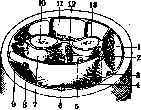
The lateral nerve of the forearm (P.) constitutes a continuation of the n. musculo-cutanei, innervates the skin of the lateral side of the forearm, passes through the fascia of the flexors; 2-a. et n. interosseus volaris; 3-a. et n. radialis; 4- m. flexor carpi radialis; 5- n. medianus; 6-m. palmaris longus; 7 - bed of the deep flexors; 8-a. et n. ulnaris; 9-m. flexor carpi ulnaris; 10-ulna; 11-the bed passes through the fascia of the flexors; 12-membrana interossea; ^-radius. (According to Sog-nelyateral groove of the elbow joint. N. cutaneus antibrachii dorsalis (dorsal cutaneous nerve of the forearm, a branch of the n. radialis) innervates the skin of the dorsum of the forearm, passes to the surface through the fascia of the arm in its lower third. The places where the cutaneous nerves pass through the fascia are the most important points for anesthesia of the entire peripheral part of the nerve. Lymphatic vessels of the forearm-see Lymphatic system, lymphatic system of man. The fasciae of the forearm (fascia superficialis and fascia propria) are a continuation of the fascia of the arm; fascia propria is most strongly developed in the proximal part, from which the muscles of the forearm take their origin; from the anteromedial side, the fascia of the forearm is reinforced by the tendinous expansion of the biceps muscle (lacertus fibrosus). The fascia of the forearm forms a sheath and at the same time a soft, elastic, elastic skeleton for groups and individual muscles of the forearm (fig. 1); it fuses with the periosteum of the ulna along the entire posterior edge, which ensures its firm fixation. In the distal part, above the radiocarpal joint, the fascia of the forearm is additionally significantly strengthened by transverse connective fibers, as a result of which the lig. carpi commune, the common ligament of the wrist, is formed, which in turn is divided into the palmar and dorsal (lig. carpi volare et dorsale); the latter is expressed much better than the former, gives inward partitions going between the tendons of the extensors, and is fixed on the dorsal side of the bones of the forearm.
l. Shangina. Pathology of the forearm. In frequency of traumatic injuries, the forearm occupies one of the first places after the fingers and hand. Impact of the hand on moving parts of machines, gears, drums, transmissions, etc. gives severe injuries to the forearm, accompanied by scalped wounds of the skin, ruptures of muscles, tendons and fractures of bones. According to their nature, injuries can be: a) closed-contusions, fractures, b) open-wounds, open fractures, gunshot wounds, c) burns and frostbite. Closed injuries to the forearm can sometimes be accompanied by detachment of the skin over a large area, subcutaneous ruptures of large vessels with increasing hematoma, which requires surgical intervention-ligation of vessels and suturing of torn tissues. In open wounds, it is necessary no later than 12-24 hours to perform "primary treatment" of the wound, i.e., to excise all crushed, contaminated tissues within the limits of healthy tissues, followed by suturing of nerves, muscles, tendons. With large defects of individual muscles, their tendons are sutured to the bellies or tendons of the remaining functionally similar muscle groups. To avoid necrosis, the skin should be sutured without any tension; in cases with large defects of the skin, the latter are closed by means of primary plastic surgery according to Tirsch or a flap on a pedicle, taken from the abdomen. In the postoperative period, early physio-mechano-therapeutic treatment is applied. Small punctured and incised wounds with a knife, glass, often a broken neck when opening a bottle by drunkards can be accompanied by injury to vessels (more often a. radialis), nerves and tendons. With each injury to the forearm, it is always necessary to carefully examine the skin sensitivity and active movements of the fingers and hand, in order not to miss injuries to nerves or tendons. There are frequent cases when a patient, after suturing an ambulatory skin wound, comes with complaints of disturbance of sensitivity, absence of movement of the fingers and hand due to overlooked and not timely sutured injury of a nerve or tendon. When the n. radialis (deep branch) is injured, paralysis of the extensors occurs-the hand and fingers hang down, movements of dorsal extension are impossible. The thumb is adducted to the palm (m. adductor pollicis-innervation by n. ulnaris), cannot be abducted. When the superficial branch is injured, sensory disturbances occur, less constant due to anastomoses with the branches of ulnaris and medianus. The area of spread of sensory disturbances-the radial half of the dorsum of the hand.-Paralysis of the n. ulnaris manifests as weakening of the movements of flexion and abduction of the hand to the ulnar side. Flexion of the little finger is impossible; flexion of III and IV fingers is incomplete due to partial paralysis of m. flexoris digit, profundi. The thumb cannot approach the wrist bone of the index finger (paralysis of m. adductoris pollicis). The most characteristic feature of paralysis of the n. ulnaris is hyperextension of the main phalanges of the fingers with simultaneous flexion of the middle and nail ones, as a result of which the fingers take the form of claws. With prolonged paralysis of the n. ulnaris, atrophy of the interosseous muscles occurs, and the hand acquires the typical form of "claw hand". Disturbance of skin sensitivity occurs* on the ulnar half of the dorsum of the hand.-In paralysis of the n. mediani, flexion of the hand to the radial side is impaired, movements of flexion of the middle and nail phalanges of I, II, III fingers are impossible, the main phalanges flex due to mm. interossei. The thumb cannot flex and oppose; under the action of the extensors and m. adductoris pollicis, the thumb is applied to the index finger, as a result of which the hand has the appearance of "monkey hand". Skin disorders are noted on the radial side of the palmar surface of the hand (from I to the radial side of IV finger) and on the dorsum of the terminal phalanges of I, II, III fingers. Prolonged paralysis of the n. mediani leads to atrophy of the muscles of the thumb.* If a diagnosis of injury to a nerve or tendon is made, then to find the severed ends, the wound is expanded, having previously excised its edges. The severed ends of the tendons, especially the central one, "recede" due to the contraction of the muscles, for which* it is necessary to make additional incisions along the length. After finding the peripheral and central end of the nerve or tendon, they are refreshed (conveniently with a safety razor blade), after which they are sutured with observance of the corresponding technique of the suture. The nerve is sutured through the perineurium, without capturing its fibers, while the fibers should not be bent, but should be applied end to end. When suturing a tendon, it is necessary to ensure that its ends do not fray and no folds and nodes are formed that hinder free sliding. Convenient is the suture with two needles, in the form of a plait, with punctures on the cut surface; after tying the knots, they do not protrude on the surface of the tendon. The operation should be performed with minimal trauma to the tendon, with strict observance of asepsis, since even a weak inflammatory process causes adhesions of the tendon with surrounding tissues, hindering its movement. With a smooth course, early movements are started (after 7 days, Iselin), mainly active movements, baths, massage. With simultaneous injury to the n. mediani and tendons, erroneous suturing of the tendon to the nerve is possible (see Radiocarpal joint).-Gunshot wounds can be caused by jacket bullets, soft lead bullets and fragments of artillery shells. Wounds from jacket bullets, more often through with small entrance and exit openings and a direct bullet track, usually proceed aseptically. Treatment in the absence of damage to large vessels and nerves is conservative. Injury to vessels can be complicated by the formation of an aneurysm. If the injured nerves are not sutured immediately and the wound is complicated by suppuration, then suturing of the nerve can be started no earlier than 6 months after the cessation of suppuration, if the wound healed per primam, then the best time for suturing is after 2-2th/ months. Incomparably more severe are wounds from shrapnel bullets and especially artillery shells. Wounds are often blind, with the formation of cavities, with crushing of deep tissues; on-, the bottom are fragments of clothing, earth and fragments of the projectile. Such wounds require primary surgical treatment, with large defects: in

tissues the wound is left open. When the wound is contaminated with earth (gunshot wounds, street injuries, agricultural, especially garden), the introduction of anti-tetanus serum is mandatory.
Fractures of the P. constitute, according to Bruns, up to 20% of all fractures. The cause of diaphyseal fractures of both bones, as well as isolated fractures of the radius and ulna diaphysis, is direct force, less often falling on the hand. - Isolated fractures of the ulna occur from direct impact of the poorly protected bone surface against a hard object - falling, protecting with hands from stick blows during a fight or attack. In the latter case, bilateral fracture of the elbow may occur. As a complication of isolated fracture of the ulna, dislocation of the radial head is observed. In fresh cases, dislocation reduction is sometimes possible, in neglected cases bloody reduction surgery is indicated, and even better resection of the radial head, because due to the subsequent scar tissue changes in the ruptured joint capsule and lig. annularis, the radial head cannot always be reduced and held after reduction, and when reduction is successful, recurrences often occur. In fractures of both bones from direct force (direct fractures, impact, lateral compression), the bones break at the same level, more often transversely (fig. 2). Indirect fractures - usually oblique or rotational depending on the mechanism. The radius breaks in the middle, the ulna at the border of the lower and middle thirds (the weakest places). In 3/4 of cases of fractures of both bones, the radius breaks above the ulna (Matti). The fragments are displaced in the direction of the force, secondary displacement - along the length. The radial muscle group is stronger than the ulnar, which is why shortening of the radius is greater. The pronator group is stronger than the supinator group, due to which the peripheral fragment (hand) is positioned in pronation (Bonier). The position of the central radial fragment depends on the height of the fracture: in fracture below the attachment of m. pronatoris teres, the central fragment is in the median position, above the attachment of m. pronatoris teres - the central fragment is in supination position (cross displacement).-With large displacement (fig. 3), especially cross displacement, it is difficult to reduce, and when reduction is successful, it is difficult to hold the fragments in the correct position. The difficulty of reduction is explained by the large number of muscles with different functions and the presence of membranae interosseae. Incorrect position of fragments can lead to healing in cross position, healing of both bones to each other; healing at an angle leads to shortening of the interosseous

^S5?!
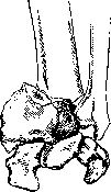
area at a typical location.
and limitation of rotational movement. Reposition of fragments is performed under local anesthesia (10-20 cm3 of 1-2% novocaine between fragments), after reposition the forearm is fixed in a 'semi-physiological' position with a dorsal plaster splint. In this semi-pronated position, the bones are at the greatest distance from each other, the interosseous ligament is stretched, and the danger of the bones fusing together is reduced. The hand is set in a position of dorsal flexion. Further treatment consists of early physio-mechano-therapy. When it is impossible to achieve the correct position of the fragments, surgery to suture the bones is indicated. During the operation, the ulna is first exposed and fixed in the correct position with Lambotte forceps, then the radius is exposed and sutured, after which the suturing of the ulna is completed. Oblique fractures with a large surface area of fracture can be sutured with wire, while transverse fractures are fixed with a metal or preferably a bone plate. The prognosis for fractures of the diaphysis of both forearm bones worsens due to the relative frequency of pseudarthroses (fourth place, according to Matti), explained by the great mobility of the fragments, as well as the ease with which muscles are trapped between them, which are compressed between the diaphyses in extreme pronation (mm. flexor pollicis longus, flexor digitorum profundus) and in extreme supination (mm. extensor carpi ulnaris, extensor digiti minimi) (Wegner). In cases of established pseudarthrosis, resection of the altered ends to normal bone and medullary canal is indicated, followed by bone grafting taken from the tibia. Improperly united at an angle and united together forearm bones also require surgical intervention; in the latter case, after separating the bones, muscles, fascia, or fat are placed between them. Open fractures require 'primary treatment' followed by treatment after suturing, like closed fractures. Particularly unfavorable are open infected fractures: fractures of the forearm from horse bites (coachmen) with crushing of soft tissues and comminution of bone. Treatment of infected fractures consists of immobilization of the forearm as much as possible in the correct position, with main attention paid to combating infection. Purulent extensions are widely opened, counter-incisions are applied for better drainage, etc. In cases of severe infection, amputation may be required. Incorrect position of fragments is corrected secondarily after the inflammatory process has ceased. Fracture of the epiphysis of the radius (fractura radii in loco classico, typico) occurs when falling on an outstretched hand; in this case, the epiphysis is as if knocked out by the carpal bones. The line of fracture when falling on the hand in dorsal flexion is located on the dorsal surface proximally and on the palmar surface distally; in the rarer fracture in palmar flexion, the position of the fracture plane is the opposite. The peripheral fragment is displaced dorsally, due to which the hand acquires the characteristic shape of a bayonet; the deformation is more pronounced on the radial side due to simultaneous supination of the epiphysis. Less constant is the displacement of the fragment to the radial side (Fig. 4). With a longitudinal impact, the dorso-lateral edge of the central fragment is wedged into the spongy part of the epiphysis. The wedging can lead to splitting of the peripheral fragment into pieces and result in T- and V-shaped fractures. The extension form of fracture of the radius is also observed in drivers and tractorists when the starter handle kicks back. In 50-80% of cases, the styloid process of the ulna is simultaneously torn off. The fracture may be accompanied by ligament and joint capsule ruptures and contusions of the metacarpal bones, especially their cartilage covering; in these cases, even after good anatomical union of the fragments, persistent functional disorders remain. Displacement of fragments causes disturbance of finger and hand movement, the protruding central fragment on the palmar surface presses against the flexor tendons, injuring them. Clenching the hand into a fist is impossible. Simple transverse fractures and epiphyseolyses without displacement that occur in children do not require immobilization; treatment with massage, active and passive movements from the first day of fracture is sufficient. When fragments are displaced, complete reposition is necessary, without which satisfactory functional results cannot be achieved. Under local anesthesia, gradually increasing traction is applied to the hand; the arm is bent at the elbow joint at a right angle to relax the two-joint finger and hand flexors; initially, to mobilize the fragments, the angle of displacement is increased dorsally and laterally, after which strong palmar flexion, pronation, and abduction to the ulnar side, with simultaneous pressure on the fragments with the fingers, places them in position. After reposition, a dorsal plaster or wooden splint is applied from the elbow joint to the metacarpophalangeal joint with palmar flexion and abduction of the hand to the ulnar side. The dorsal position of the hand prevents the development of flexor shortening. The splint is kept for 2-2½ weeks until the mobility of the fragments disappears. Immediately after removing the splint, massage, movements, and heat treatment are begun. Movements of the fingers are performed from the first day of fracture. Intra-articular fractures of the upper epiphysis of the radius and elbow joint—see Elbow joint. Burns of the forearm are more common in chemical production (acids), as well as in the dyeing workshop when the hand gets between hot rollers. Treatment of burns by the open method, which eliminates painful dressings, gives faster healing with less coarse scars in third and second degree burns. Deep burns with sloughing of the necrotic area may later cause bleeding from the devitalized vessel wall. Large skin defects are closed by plastic surgery (usually according to Thiersch), which prevents scar contractures. Plastic surgery is undertaken when the wound has cleaned and is covered with healthy granulations. Acute inflammatory diseases of the forearm. Of skin diseases, furuncles are more common (see Furuncle, Carbuncle). Phlegmon of the forearm. Infection penetrates primarily through the skin (furuncles, pyoderma) or as a complication of traumatic injuries, or by extension of purulent processes from the hand, especially in phlegmons of the tendon sheaths of the I and V fingers, which pass under the transverse volar carpal ligament to the forearm. In phlegmons of tendon sheaths, the affected finger is flexed, and light attempts to straighten it are sharply painful; pain is felt along the entire length of the tendon sheath. After destruction of the tendon sheath by pus, the purulent extension spreads upward between the pronator quadratus and flexors, then between both layers of flexors to the shoulder. Depending on location, phlegmon can be superficial, subcutaneous, or deep—subfascial, intermuscular. Subcutaneous phlegmon manifests as rapidly spreading redness, swelling, elevated temperature, and early gives foci of fluctuation. In deep forms, general septic phenomena prevail, and fluctuation may be absent, as the pus is deep under the fasciae and muscles. Treatment consists of early incisions widely opening the purulent extensions; in deep phlegmons, the fascial spaces involved in the process are opened strictly anatomically. The most favorable for pus drainage are incisions in phlegmons of tendon sheaths—along the lateral sides of the palmar surface of the forearm above the wrist joint, giving access to the most sloping parts of the tendon sheath. The incisions are drained with thin rubber strips (cut from old rubber gloves) (Iselin). The prognosis in early detected and properly treated cases is favorable; neglected, severe cases give high disability due to disfiguring scars and subsequent contractures. The prognosis worsens with the addition of putrefactive and especially anaerobic infection (gas gangrene). Osteomyelitic process relatively rarely affects the bones of the forearm (according to Haag's statistics, radius 5.1%, ulna 3.4%). For the clinic of osteomyelitis—see Osteomyelitis. Tuberculous lesion of the diaphyses of the forearm bones is more common in childhood. Often the process spreads from the epiphyseal part to the diaphyseal part through the metaphysis. Characteristic for tuberculosis is the prolonged course with insidious onset: the patient cannot specify the exact time of onset of the disease. The limb quite soon acquires a spindle-shaped form, similar to spina ventosa of the fingers, due to muscle atrophy and simultaneously developing circular ossifying periostitis around the focus of lesion (Oehlecker). The outcome of the process is more often the formation of small sequestra and sinuses with characteristic liquid, crumbly pus. Diagnosis is usually easy due to existing tuberculous lesions of other organs. It is mainly necessary to differentiate from chronic osteomyelitis and syphilitic lesions. Besides the history, the decisive factor is the X-ray picture. In tuberculosis, destructive processes prevail over reparative ones: 1) marked bone atrophy (osteoporosis), 2) absence of reactive bone process around, 3) sequestrum, if present at all, is small, delicate, and has a rounded shape. Conversely, in osteomyelitis: 1) bone atrophy is absent, 2) reactive sclerosing processes prevail, the sequestrum capsule is well expressed, 3) the sequestrum is more massive, elongated in shape, with large toothed edges.
Treatment consists of general strengthening treatment and local treatment of the focus, which consists of immobilization of the extremity, treatment with sunlight, quartz. Sometimes good results are obtained from the application of congestive hyperemia according to Viru. In cases with the formation of sequestra, as well as in long-unhealing fistulas, opening of the cavity with careful curettage of all affected tissue is indicated, otherwise recurrences occur. The resulting cavity is plugged with iodoform paste. The wound is sewn up tightly (Oehlecker). Syphilis of the bones P. is a rather rare disease. More often it is a matter of tertiary period lesions - osteo-periostitis gummosa. Gummatous lesions can be limited or diffuse. Superficial, periosteal gummas represent painful dense nodules with unchanged skin over them. With specific treatment, they can disappear without a trace; when they progress to a necrotizing form, necrotic masses are discharged, the ulcer has a dirty, greasy appearance with a sequestrum at the bottom. After healing, star-shaped scars remain, fused with the bone. Central gummas initially give little external change, later when the process reaches the surface of the bone, a firm diffuse swelling of it forms. Night pains are characteristic when the extremity is warmed in bed; in people working at night, pains appear during the day. The X-ray picture with periosteal proliferations and the presence of parallel processes of bone sclerosis and destruction is quite typical. The greatest difficulty is represented by the differential diagnosis with bone sarcoma. The sclerotic and periosteal changes in syphilis help to exclude it. The Wassermann reaction is also necessary. Treatment is specific - salvarsan, mercury. Tumors of P. In sarcoma of the bones P., the radius is affected three times more often than the elbow. Periosteal and central forms are encountered. Periosteal forms are distinguished by their malignant growth, quickly penetrate into surrounding tissues, which is why treatment usually consists of amputation of the extremity. Central sarcomas with an intact capsule can be resected with subsequent bone grafting. Cancer has the appearance of a cutaneous keratotic cancer; as a professional disease, it occurs in workers of paraffin factories and radiologists. The course is slow with late metastases. Treatment consists in the surgical removal of the tumor with subsequent skin plastic surgery. Among vascular tumors, lymphangiomas and hemangiomas are encountered. Unlike angiectasia, angiomas of embryonic origin develop as a result of abnormal excessive vascular laying. According to structure, simple angiomas (angioma simplex, capillare) and angiomas with cavity formation (angioma cavernosum) are distinguished. With their infiltrating growth and penetration of surrounding tissues, angiomas resemble malignant tumors, differing from them by a much slower growth and absence of metastases. A special type is the so-called racemose angioma (angioma racemosum), representing a tangle of tortuous vessels with arterial blood due to numerous communications between arteries and veins without the intervention of capillaries. These angiomas pulsate, on auscultation a blowing noise is heard, as in an aneurysm. Treatment of small angiomas - excision within healthy tissue; in well-developed inoperable cavernous angiomas, infiltration, galvanocautery can be tried. In angioma racemosum, its excision with ligation of all afferent branches is necessary. In severe cases, with penetration of all tissues, amputation may be required. Besides the described tumors, fibromas, osteomas can be encountered; treatment - surgical. A unique disease of the wrist joint is Madelung's deformation, manus valga. The disease arises in childhood and ends by the period of puberty. Both P. are symmetrically affected, in women more often than in men. The history shows no indication of trauma and inflammatory etiology. The basis of the disease lies in changes in the metaphysis and epiphysis of the radius (meta-epiphysitis radialis inf.) with disruption of the normal ossification process. Under the influence of muscle traction, the carpal bones slip from the articular surface of the radius toward the missing, not yet ossified palmar-ulnar part. Os lunatum as if becomes wedged between the bones of P., due to which the first row of carpal bones forms a triangle instead of the normally present arch. The ulna is dislocated at the same time to the back and outward. P. has the appearance of a bayonet or fork (Gabelhand), the anteroposterior diameter of the joint is sometimes doubled. The function of the joint usually suffers little. Treatment during the period of deformation development consists in wearing a special cuff that to a certain degree delays the development of deformation. In completed cases, surgical assistance may be required with exclusively cosmetic purposes in the form of excision of a wedge from the epiphysis of the radius. Unlike the described 'true' Madelung deformation, a so-called 'pseudomadelung' deformation is distinguished, similar in external joint changes but developing after trauma or inflammatory diseases. Operations on P. Ligation of vessels in bleeding, aneurysm (fig. 5). Ligation of a. radialis does not present difficulties along the entire course of the vessel. In the proximal half, the artery is exposed by a small incision of 5-6 cm between m. brachioradialis and m. flexor carpi ulnaris. The muscular edge of m. brachioradialis is pulled to the radial side, the common belly of the superficial flexors is moved toward the elbow; the artery lies under a fascial sheet through which it shines. It is necessary to remember about the nerve branch running alongside in the middle third of P. - r. superf. n. radialis. When ligating in the lower third of a. radialis, it is exposed in the middle between the radial bone and the tendon of m. flexor carpi radialis, at the pulse counting site. Ligation of a. ulnaris in the upper half is more difficult than ligation of a. radialis, due to its deeper location and abundance of muscular branches. A. ulnaris lies between the superficial and deep flexors (on m. flexor digitorum profundus). A skin incision 6-7 cm long along the line connecting the medial epicondyle of the humerus with the pisiform bone, along the radial edge of m. flexor carpi radialis. After incision of the fascia, the superficial flexor of the fingers is pulled to the radial side, the artery lies under the fascia accompanied by two veins and n. ulnaris coming from the ulnar side. The higher the artery is exposed, the more the superficial flexors have to be pulled to the ulnar side. The incision for exposing a. ulnaris in the distal third is made along the radial edge of m. flexor carpi ulnaris, the tendon of which is closest to the ulna. A. ulnaris lies in the fatty tissue under the fascia between the tendons of m. flexor digitorum sublimis and m. flexor carpi ulnaris. Exposure of the bones of P. is performed in incorrectly united fractures, pseudarthroses, osteomyelitis, tumors. Access to the ulna, due to its superficial location, is simple along its entire course from processus olecranon to processus styloideus. The radius is most easily accessible from the dorsal-lateral side, with P. in a middle position between pronation and supination, with the elbow joint bent at an obtuse angle. The incision is made along the line connecting the posterior-radial edge of the head with its styloid process. After incision of the fascia in the upper half, the bone is exposed between m. extensor digitorum communis and m. extensor carpi radialis, for which it is necessary to cut some fibers of m. supinator, which covers the radius (it is necessary to remember about the deep branch of n. radialis passing through the muscle). In the distal half, it passes between the tendons of mm. abductor pollicis longi et brevis on one side, extensor carpi radialis brevis on the other. In the lower third, the tendons of mm. abductor pollicis longi and extensor pollicis brevis need to be pulled to the back. Amputations of the forearm are performed under an exsanguinating tourniquet. Technically the operation is simple and can be performed by any method (formation of a skin cuff with the formation of dorsal and palmar flaps, method of oblique incision). The periosteum is incised at the same level as the interosseous ligament; tears of the periosteum should be avoided due to the danger of osteophyte formation. The bones are sawed off at the same level. To avoid painful neuromas and fusion with the scar, the nerves are cut off as high as possible.
The aa. radialis, ulnaris, interossea volaris et dorsalis are ligated, and the muscle flaps are sutured over the ends of the bones. The technique of the operation is somewhat complicated in the distal third due to the large number of tendons. It is recommended to prevent complete atrophy of the muscles by suturing antagonists in front of the stump. In amputation of the P., it is necessary to strive to preserve the stump as long as possible (see Prostheses).
Related articles
Mentioned in
Cite this page
“Preoperative Period.” Soviet Medical Encyclopedia. English translation of Bolshaya Meditsinskaya Entsiklopediya, 1st ed. (Moscow, 1928–1936), ed. N. A. Semashko. https://sovietmedicalencyclopedia.pages.dev/article/preoperative-period/