Appendicitis
Historical document, translated for reference. It reflects medical knowledge of the 1920s–30s and is not medical advice.
Summary
This article from the 1928-1936 Soviet Medical Encyclopedia examines the etiology, pathogenesis, pathological anatomy, clinical presentation, treatment, and statistics of appendicitis. It discusses the primary causes of appendicitis as the introduction of pathogenic microorganisms into the appendix wall, with enterogenous transmission being the most common pathway.
Encyclopedia article (1928–1936)
APPENDICITIS. Contents: I. Etiology and pathogenesis............167 II. Pathological anatomy..........170 III. Clinical....................174 Acute A....................176 Chronic A................181 IV. Treatment....................185 V. Statistics...................186 Appendicitis (appendicitis), inflammation of the vermiform appendix of the cecum [syn.: vermiculitis (vermiculitis), epityphlitis (epityphlitis)]. I. Etiology and pathogenesis. The cause of appendicitis is the introduction of pathogenic microorganisms into the wall of the vermiform appendix, among which diplococci, streptococci, and staphylococci have the most significant importance; the intestinal bacillus, anaerobes, and some other microorganisms found in appendicitis probably do not play the role of independent etiological agents, but are introduced sequentially from the lumen into the already inflamed wall of the appendix. Regarding the path of penetration of bacteria into the wall of the vermiform appendix, there are two views. 1. Most modern researchers consider the enterogenous path most likely, i.e., the introduction of microorganisms into the wall of the appendix directly from its lumen; this is evidenced by the presence in the lumen of the vermiform appendix of the same microorganisms that are found in its wall in appendicitis, the penetration of these microorganisms in normal conditions into the wall and the mesentery of the appendix (Yauve, Rubashev), as well as nat.-anat. data on the onset of appendicitis (see below). 2. Some researchers insist on the hematogenous, metastatic path of origin of appendicitis, i.e., they believe that appendicitis occurs as a result of the carriage of microorganisms into the wall of the appendix by the bloodstream. The possibility of this is indicated by the frequent combination of appendicitis with preceding angina, which, according to Kretz (Kretz), should be explained in such a way that the bacteria that caused angina (usually streptococci) penetrate into the bloodstream through the lymphatic pathways and, with its mediation, are carried into the wall of the appendix; in confirmation of his view, Kretz points to his findings in anginas of bacterial emboli in the vessels of the follicles of the appendix with subsequent necrosis of the vascular wall and follicular tissue. The possibility of hematogenous origin of appendicitis is also established by the experimental research of Adrian and Tedesco (Adrian, Tedesco). In general, it must be recognized that appendicitis can arise due to the hematogenous carriage of bacteria into the wall of the appendix, but this is not the usual and most common way of origin of appendicitis; in particular, the combination with angina can be explained by the fact that microorganisms that caused inflammation of the tonsils penetrate further through the digestive pathways and cause appendicitis enterogenously. Thus, the enterogenous introduction of microorganisms into the wall of the appendix is the usual and most common cause of appendicitis. As is known, pathogenic microorganisms can always be present in the lumen of the appendix, but at the same time, not everyone suffers from appendicitis; from this it follows that for the introduction of these microorganisms into the wall of the appendix, facilitating or predisposing conditions are necessary. The main of these is the delay, stagnation of fecal contents in the lumen of the appendix, which serves as a medium favorable for the multiplication and increase in virulence of microorganisms, and which can also cause a decrease in the normal resistance of the mucous membrane of the appendix. It is generally believed that in normal conditions, fecal contents, which can enter from the cecum into the lumen of the appendix, are constantly emptied back into the cecum by the peristaltic contractions of the latter together with the mucous secretion of the mucous membrane. The causes of delay and stagnation of these contents in the appendix can be various factors, either related to the appendix or to the cecum, among which, according to Ashoff (Ashoff), physiological bends and curvatures of the appendix have special importance, which is confirmed by the fact that in most cases of appendicitis, inflammation develops distally to the existing bend in the appendix; in addition, certain variations in the position of the appendix (e.g., position behind the cecum) and its bends on the basis of abnormal embryonic adhesions play a role. Also important is the atonic condition of the musculature of the appendix, due to underdevelopment or atrophy, which sometimes depends on abnormalities in the function of the autonomic nervous system (Abricosov). Furthermore, the emptying of the appendix can be hindered by the accumulation of feces and gases in the cecum, associated with chronic constipation, especially in those cases which are based on a mobile cecum and abnormalities of the ileocecal valve (Rozanov, Yurasov), as well as in those cases where there is atonia of the musculature and significant dilation of the cecum (typhlectasia). In all such cases, in addition to the mechanical factor in the form of overfilling of the cecum with contents, bends that occur in the appendix when the cecum is displaced due to its overfilling with fecal masses, especially when it is mobile, are also of importance. It is also necessary to mention inflammatory processes in the cecum (typhlitis, perityphlitis), in which swelling of the mucous membrane and infiltrates at the site of entry of the vermiform appendix can be the cause of delay of contents in it. In former times, great importance was attached to the formation of fecal stones in its lumen, as well as to the entry of all kinds of foreign bodies into it (fruit stones, insoluble plant residues, pieces of fish or beef bone, enamel plates separated from a carious tooth, pieces of hair, etc.) in the origin of appendicitis. At present, much less importance is attached to this; fecal stones and foreign bodies can contribute to the development of appendicitis only in those cases when, by obstructing the lumen of the appendix, they cause stagnation of fecal contents in it; much more rarely, appendicitis is the result of damage to the mucous membrane caused by its traumatization by a foreign body. On the other hand, in already developed appendicitis, the presence in the appendix of a fecal stone or a large foreign body that prevents the emptying of inflammatory exudate from the lumen and causes anemia of the appendix wall by its pressure can make the course of appendicitis extremely unfavorable and contribute to perforation of the appendix. As for animal parasites of the intestine, worms, their role in the origin of appendicitis is small. Although some researchers, for example, Reindorf, Reinchardt, Lawen, and partly Goldzieher, until recently insisted on the enormous role of intestinal parasites (mainly pinworms) in the etiology of appendicitis, most modern authors deny such a role, believing that these parasites can have significance only in those rare cases when a group of parasites (e.g., pinworms) or a large parasite (e.g., ascaris) closes the lumen of the appendix and causes stagnation of contents in it; in addition, it must be borne in mind that these parasites by their presence in the appendix can cause appendicular pains without appendicitis (see Appendiculopathy). In comparison with the enterogenous and hematogenous origin of appendicitis, much less importance is attached to appendicitis arising as a result of the spread of inflammation to the appendix from neighboring organs, which in general can be assumed only in exceptional cases; even the frequency of the spread of inflammation to the appendix from the right appendages in women, which seemed recently established, is now doubtful. II. Pathological anatomy. Although from a clinical point of view it is very common to speak of acute, subacute, and chronic appendicitis, most pathologists believe that appendicitis always arises as an acute inflammation and that the changes found in clinical subacute and chronic appendicitis are nothing other than various outcomes and consequences of acute appendicitis. As established by Ashoff, the inflammatory process in appendicitis begins in the depths of the crypts of the mucous membrane of the appendix, where in such cases a small area of desquamation of the epithelium is observed with the formation of a small fibrinous-purulent deposit on the mucous membrane; simultaneously, inflammatory edema develops in the mucous membrane
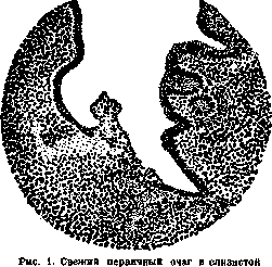
of the appendix.
and infiltration by leukocytes of the submucosal layer, spreading in depth and circumferentially, as well as along the length of the appendix, which gives the process a phlegmonous character [primary infection, A'shof's Primarinfekt (see Figure 1)]. The epithelial covering may remain damaged only at the site of infection penetration, or it may undergo decay over a more or less extensive area, which forms the basis for the formation of an ulcer of the mucous membrane. The serous covering of the appendix at the beginning of the process may only show hyperemia; later, delicate fibrinous deposits appear on it, which have a toxic origin and do not contain microorganisms; only when the phlegmonous infiltration penetrates to the serous surface of the appendix (see Figure 2) is a fibrinopurulent deposit found on it, containing bacteria. In addition, the inflammatory edema and phlegmonous infiltration may spread to the mesentery of the appendix and the cellular tissue around the cecum. The further course of the process may manifest as follows: in the area of phlegmonous infiltration of the appendix wall, purulent melting of tissue occurs and an abscess (or several small abscesses) forms, which later may rupture into the lumen of the appendix or into the peritoneal cavity, or, when the appendix is located behind the cecum, into the surrounding cellular tissue. A severe complication in any of the described periods is necrosis of the appendix wall, transitioning into gangrenous decay and perforation of it, with the outcome being perforative peritonitis; this change is based on the involvement of the appendix's blood vessels in the inflammatory process and their thrombosis (thromboarteritis and thrombophlebitis). Sometimes gangrenous changes of the appendix wall occur at the very beginning of an attack of A.; in such cases, one can assume that the inflammation began in the mesentery.

Figure 2. Ulcerative phlegmonous appendicitis.
In places, the purulent infiltration has penetrated to the serous membrane; perforation threatens. or immediately spread rapidly to it, involving the blood vessels of the appendix passing through it. It should also be added that inflammation from the appendix can spread to the wall and serous covering of the cecum (perityphlitis) and to the cellular tissue around the cecum (paratyphlitis) with the development here of phlegmon or an abscess, which may open into the peritoneum or through the abdominal wall outward, or may spread upward to the kidney (paranephritis) and to the diaphragm (subdiaphragmatic abscess); more rarely, the inflammation descends downward into the cellular tissue of the pelvis (in women, to the right appendages). Purulent inflammation of the veins of the appendix can lead to the transfer of infection through the portal vein to the liver (purulent pylephlebitis and liver abscesses). Through-and-through perforation of the appendix wall when an abscess of the wall ruptures through the serous covering or in the case of gangrene of the latter, as well as rupture into the peritoneum of a paratyphlitic abscess, lead to peritonitis, which may be limited to the area of the appendix and cecum or immediately spread throughout the abdominal cavity (see Peritoneum). As is evident from the above, the patho-anatomical changes in A. can vary depending on the nature and course of the process. In general, the following patho-anatomical classification of these pictures can be given: I. Superficial A., in which inflammation involves only the superficial layers of the mucous membrane. Externally, the appendix is almost unchanged, only congested. The epithelial covering may be intact (superficial catarrhal A.) or destroyed over one or another area (superficial ulcerative A.). II. Phlegmonous A., characterized by purulent infiltration of all layers of the wall. The appendix is thickened, its wall is juicy, congested, the serous covering is inflamed. Subtypes of this form are: 1. Simple phlegmonous A.-with the integrity of the epithelial covering. 2. Phlegmonous-ulcerative A.-with ulceration of the mucous membrane. 3. Apostematous A.-with the formation of abscesses in the wall. 4. Gangrenous appendicitis1-with necrosis of the wall. The last two forms can be: a) without perforation, b) with perforation. The above descriptions and interpretation of the course of patho-anatomical changes in appendicitis are accepted by most modern pathologists; objections are made only by supporters of the hematogenous origin of A. regarding the nature of the changes at the very beginning of A., namely, they point out that the process in the appendix begins with necrosis of the walls of small arteries of the follicles and infarct-like necrosis of the follicular tissue; this is followed by the circular spread of the process, which corresponds to the segmental distribution of blood vessels in the appendix. In recent years, this course of the process and the assumption of the segmental distribution of vessels in the appendix are disputed. In most cases of A., the inflammatory process in the appendix wall, which gave rise to the complex of symptoms of an acute A. attack, stops, and this is followed by recovery processes that can take various patho-anatomical expressions. In superficial catarrhal forms, the condition may end in complete healing, with the appendix returning to normal appearance. In phlegmonous, especially ulcerative, forms, the process of gradual resorption of the purulent infiltrate and granulation of the ulcer surface of the mucous membrane may continue for a long time, eventually leading to the formation of scars in the mucous membrane and sclerosis of the submucosal tissue. In simple phlegmonous forms, the condition may be limited to sclerosis of the wall and atrophy of the mucous membrane with subsequent expansion of the lumen. Scar formation causes narrowing, and sometimes local or complete obliteration of the lumen of the appendix. The result of partial obliteration of the lumen may be the overfilling of the distal part of the appendix with continuing to be exuded exudate or secretion of the mucous membrane, which leads to the transformation of part of the appendix into a cyst, filled with serous fluid or mucus (hydrops and mucous cyst--A cyst of the appendix) or with pus (empyema of the appendix, see Figure 3). At the sites of former abscesses and areas of partial destruction of the appendix wall, defects and scars may remain, which have a tendency to bulge with the formation of diverticula, usually filled with mucus. In some cases, after perforation of the appendix wall toward the mesentery or into previously formed adhesions, the process may subside, and the perforation channel may scar over, or, remaining open, it may be filled with mucus coming from the lumen of the appendix; such m masses become encapsulated at one or another point along the perforation channel, while in rare cases they spread throughout the peritoneum, giving a condition called pseudomyxoma peritonei. Finally, inflammatory processes of the serous covering of the appendix and cecum leave connective tissue thickenings of it and adhesions with surrounding parts. All the above-mentioned changes in the appendix, following the acute period of A. and based on recovery processes after acute inflammation, can be conditionally designated as subacute and chronic appendicitis, dividing them, depending on the nature of the patho-anatomical changes, into three basic forms--granulating, sclerosing, and obliterating. The changes underlying these forms can be a source of appendiceal pains (see below), even in the absence of an inflammatory process (see Appendicopexy). On the other hand, many of these chronic changes in the appendix, which developed after acute A., create very favorable conditions for reinfection and a new attack of acute A. A feature of the patho-anatomical picture of such an attack of A. is that one of the above forms of acute A. is superimposed on the changes characteristic of the so-called chronic appendicitis; in particular, during perforation of the appendix during a recurrent attack, the rupture may occur into the area of previously formed connective tissue adhesions around the appendix, which may prevent the development of general peritonitis. The question of the existence of an independent chronic appendicitis, developing gradually and without preceding acute inflammation, is controversial. While some researchers (Gol'dshter and others) admit such a possibility and speak of catarrhal, lymphoid (follicular), and atrophic forms of independent chronic A., most modern pathologists deny such A.,

Figure 3. Empyema (A) of the vermiform appendix.
assuming that in these cases we are dealing with processes that are a continuation of acute inflammation, or with non-inflammatory changes; the latter, for example, include lymphoid or follicular appendicitis, in which hyperplasia of lymphoid tissue is simply a manifestation of lymphatic status, as well as atrophic and sclerotic changes of the appendix, which may be an expression of appendiceal involution (see Appendix). The existing disagreement until recently between pathologists in the classification of certain changes in the appendix is reflected both in the question of recognizing or not recognizing a chronic appendicitis as a separate entity, and in the percentage of appendices removed by appendectomy, in which microscopic examination does not reveal signs of appendicitis. In various researchers, this percentage varies between 5 and 50. In some of these cases, the appendix is found to be completely normal, and then one can think that the appendicular pains were not associated with the appendix (see Appendicopathy); the persistence of pains after the operation often confirms this; one can also assume (especially if the pains disappear after the operation) that the pains were caused by a violation of the function of the appendix in the form of spasm of the musculature (Rossle) or, conversely, in the form of weakness of its peristalsis, leading to the accumulation of feces in it (Abrikosov). Some of these cases are classified by some authors as appendicitis under the name of rudimentary appendicitis (Ashoff) or chronic appendicitis, while others consider them as a manifestation not of inflammation, but of involution (see above); the mentioned difference in interpretation of such cases gives the above-mentioned difference in the percentage of appendectomized appendices without appendicitis. A. Abrikosov. III. Clinic. The concept of inflammatory processes in the right iliac fossa and the names of these inflammatory processes changed according to the understanding of them by individual authors. The name 'appendicitis' was introduced by the American Fitz in 1886; a commission of American physicians, elected to study the question of perityphlitis, retained this name as corresponding to the essence of the disease, and since then it has come into general use as a definite nosological unit. This affliction, with its prevalence, its typical clinical course, and its often fatal outcomes, was known to physicians of ancient times. There is mention of this by Celsus and Areteus; Lannelongue, who studied an Egyptian mummy with a typical appendicular incision on the abdomen, expresses the assumption that already 5,000 years before the Christian era, Egyptians intervened surgically in this disease. Without delving into distant centuries, it is necessary to recall the appearance in the 1820s of Dupuytren's 'Theory of iliac abscesses' and Albers' classification of inflammations of the cecum: 1) typhlitis ac, 2) typhlitis chr. and 3) perityphlitis (1838). The views of these authors formed the basis of all subsequent works on the question of 'inflammation of the cecum' and made it possible to accumulate extensive material concerning the etiology, pathological anatomy, clinical course, and treatment of these inflammations. Later, Pirogov distinguished five types of abscesses of the iliac fossa: 1) acute-phlegmonous abscesses with sharply expressed local phenomena-in people of robust constitution; 2) torpid-phlegmonous abscesses-in people flaccid, lymphatic, already suffering from some severe diseases; weakly expressed local phenomena, often with exhausting fever and with frequent purulent infection; 3) cold abscesses; feverish phenomena, if they occur, are very weak; local phenomena are clearly expressed, but without tension of the abdominal walls; abscesses develop in people cachectic; 4) acute infiltrates-general phenomena are similar to those in acute-phlegmonous abscesses; 5) torpid infiltrates-with general phenomena similar to those in torpid-phlegmonous abscesses. Local phenomena in both of the latter types have the peculiarity that the suppurations spread to large spaces in the retroperitoneal tissue, for example, to the linea alba, to the diaphragm, and into the cavity of the small pelvis. - All these works supported the theories of Dupuytren and Albers, but the accumulating pathological-anatomical material and more profound clinical observations eventually led to emphasize in all these diseases in the right iliac fossa the changes found in the vermiform appendix. At present, in the differential diagnosis of a patient who complains to the physician of pains in the right iliac fossa, it is correct for the physician to first direct his attention to appendicitis and, only after excluding it, to proceed further with the differential diagnosis. As for the history of the doctrine of inflammation of the vermiform appendix and methods of its treatment, it is necessary to note that surgeons and therapists in the last 10-15 years have come to the conclusion that appendicitis is a condition that should be under the observation of a surgeon, even if only consultatively. The fact is that large statistical data have clearly shown that unrecognized or late-diagnosed appendicitis, not operated on in a timely manner, often threatens the life of the patient. Clinically, it is necessary to distinguish two large groups of inflammations of the vermiform appendix: acute and chronic appendicitis. If in the most strict analysis of each individual case of acute appendicitis, in approximately 25%, it is not possible to find the etiological moment that served as the impulse for this attack, then a fair explanation for this can be the opinion of Sonnenburg that in acute appendicitis the attack does not coincide with the beginning of the disease. This attack is, as a rule, preceded by a hidden inflammation, which leads to various essential changes in the vermiform appendix and thereby prepares the ground for the acute attack. When the attack subsides, when it first passes into the subacute and then into the cold form, changes remain in the vermiform appendix from the experienced acute attack, and chronic appendicitis always contains the danger of the possibility of a new outbreak of acute appendicitis, which can be fatal for the patient. There are sometimes cases when the chronic course of the disease is not interrupted by exacerbation; such cases are designated as A. larvata, hidden appendicitis. Acute A. Clinically, acute appendicitis can be subdivided into three groups: 1) acute catarrhal A. (a. catarrhalis; a. simplex); the peritoneum in this form may not participate, but it may also participate; in the latter case, at laparotomy, one can find serous or even seropurulent exudate; 2) purulent A., perforating (a. purulenta, perforativa), caused by ulceration of the appendix and its perforation (often by a fecalith), with its consequence-purulent peritonitis, which either quickly localizes, becomes encapsulated, or passes into general peritonitis; 3) gangrenous A. (a. gangraenosa), the most acute form, sometimes with a lightning onset; rapidly increasing picture of septic peritonitis, which soon becomes general and very rarely has a tendency to become delimited. This subdivision of acute appendicitis for the purposes of clinical diagnosis, as a division based on the pathological-anatomical changes in the appendix, has more grounds than the division encountered in hospital practice of appendicitis into mild, moderate, and severe cases. To the group of acute appendicitis should also be included those cases that go under the diagnosis of paratyphlitis (inflammation of the tissue lying behind and around the cecum) and have a tendency to purulent melting of the tissue and to the formation of a retrocecal abscess. These paratyphlites, although they may originate from various pathological changes in the cecum itself, most often have as their source an inflammatory process in the vermiform appendix lying retrocecally. The attack of acute appendicitis in most cases begins suddenly in the midst of complete health, often at night, during sleep. Pain in the abdomen appears either immediately acute or gradually increasing. These pains are first felt by the patient in the umbilical region, then spread and localize, mainly in the right iliac region, and radiate to the epigastric region; nausea and vomiting appear. The patient complains of a sensation of heat throughout the body and in the abdomen, a feeling of chill, sometimes a shaking chill. On palpation of the abdomen, it is painful throughout, but even slight pressure on the right side causes severe pain, radiating to the epigastric region. An enema given to the patient is often ineffective in terms of good emptying of the intestine and passage of gases; usually it brings little relief, the painful sensations remain the same. Measurements show a rise above 38°, often up to 39°, but sometimes the temperature rises to 40°; a thermometer placed in the rectum gives a difference of half a degree and even a little more. Heat on the abdomen brings relief only in rare cases; the patient feels better if ice is applied. The pulse is rapid. All these phenomena sometimes can subside quite quickly, within a few hours, sometimes after 1-2 days: the pulse becomes even, the temperature drops to normal, the painful sensations in the abdomen pass, only tenderness to pressure in the right iliac fossa, with radiation to the epigastric region, and rigidity of the abdominal muscles on the right below remain; ice becomes unpleasant to the patient, a hot water bottle, a bottle with hot water, a thermophore or a heating compress give a more pleasant sensation.
The tongue, which was dry and coated, clears and becomes moist; vomiting does not recur, nausea passes, a certain appetite appears, gases begin to pass on their own and with an enema. During the attack, the most comfortable position for the patient is lying on the back, sometimes with legs bent at the knees; when the attack subsides, the patient willingly stretches out his legs and expresses a desire to turn on his side, and the position on the right side does not cause painful sensations, but if the patient turns on the left side, it causes pain in the area of the cecum. The described picture of the disease gives grounds for the diagnosis-acute catarrhal appendicitis. Usually several more days pass, and the patient, having sufficiently recovered from the attack, switches to a normal diet and resumes his usual activities. Such a mild course of the acute attack is observed far from always, the inflammatory process of the vermiform appendix can be deeper, and the reaction of the peritoneum to this inflammation more widespread. The general condition of the patient does not improve, temperature does not fall, pulse becomes more frequent, tongue is bad, covered with a dirty coating; usually there is constipation; abdomen is slightly swollen, gases pass poorly; rigidity of the abdominal wall on the right becomes very significant, and with light percussion, dullness is noted over a greater or lesser extent, giving reason to assume effusion in the abdominal cavity, which can be serous, serous-purulent or even purulent. If due to weakening of the process in the first two cases the matter can end favorably and the infiltration in the abdomen will begin to quickly resolve, then the general condition of the patient will improve, but if the patient's condition continues to remain poor, painful sensations appear in the left side of the abdomen and even throughout the abdomen, repeated vomiting appear, increased pulse, then there is reason to assume perforation of the appendix and purulent effusion in the abdominal cavity. When localizing the purulent effusion, the matter ends either with the formation of an abscess, or prolonged resolution of this pus may occur, and a dense infiltrate, of greater or lesser size, is not only well percussed but also distinctly palpable. Besides the general condition of the patient and characteristic phenomena from the abdomen, in this severe reaction from the side of the peritoneum and the entire organism to perforation of the vermiform appendix-for clinical conclusions about the necessity and time of surgical intervention, as well as for prognosis, there is great assistance in the study of the blood: increase in leukocytosis speaks for the spread of the purulent process, increase in neutrophilosis aggravates the prognosis. When a localized abscess forms, as said, it is possible to palpate a tumor, which is formed due to the accumulated pus and infiltrated neighboring organs-loops of intestine, inflamed omentum, as well as inflamed parietal peritoneum. On the basis of clinical experience it can be stated that in the process saving the patient from the localization of the peritoneum from the spread of pus, the omentum plays a primary role; and in operations one can often see how it envelops the vermiform appendix and together with the adherent loops of intestine and peritoneal sheet forms a barrier through which pus cannot penetrate further. With low resistance of the body, the tendency to form adhesions may be expressed very insignificantly; therefore, although the patient may not develop general peritonitis, pus can spread very widely in the abdominal cavity, which presents great danger to life; accumulations of pus can be found in the pelvis and in the left iliac fossa. The same will be the case with very severe infection. The most severe picture of the disease of the vermiform appendix is obtained in gangrene of the appendix, in gangrenous A.-a stormy onset with shaking chills and rapidly increasing picture of septic peritonitis; small, frequent pulse, abdomen is not swollen but board-like, touching the abdomen is painful everywhere, breathing is shallow, frequent; deeper breathing is impossible for the patient, as, causing movements of the abdominal muscles, it immediately causes sharp pain in the abdomen. The face of the patient already after a few hours from the beginning of the attack gives the characteristic picture of the so-called facies hippocratica-sharply sunken facial features, sunken eyes, grayish-earthly color of the facial skin; extremities are cold; sticky, clammy sweat appears. It goes without saying that between these most typical pictures of acute appendicitis there can be various transitional forms, which do not fit into the framework of typical descriptions and symptoms. It is necessary to also indicate such signs that enable the physician to make the correct diagnosis in gangrenous A. and, what is most important, to intervene surgically in a timely manner. These are the two signs noted by Sonnenburg: 1) in gangrenous appendicitis, slight edema of the skin can often be found in the right iliac region, spreading also to the lumbar region: 2) sometimes a short time after the onset of the attack, the disappearance of hepatic dullness can be ascertained. Regarding surgical intervention, it can be said that, since in all peritonitis their source most often is inflammation of the vermiform appendix, then in all laparotomies performed for acute inflammation of the peritoneum where the source of this inflammation is not clearly clarified to the surgeon before the operation, every surgeon, upon opening the abdominal cavity, should first of all pay attention to the area of the cecum and to the vermiform appendix. Prognosis. In gangrenous, most acute, septic A., i.e.; in diffuse septic peritonitis, if the operation is not performed in the first day, and more not in the first hours from the onset of the disease, the prognosis is unquestionably bad. The prognosis in the catarrhal form of A. is usually good, if there are no grounds to expect complications from other organs. Great difficulties are presented by the prognosis in the purulent form of A., with perforation of the appendix, when there is a picture of spreading or already localized peritonitis and a dense infiltrate or encapsulated abscess is found. The prognosis must be made very cautiously with the most careful and constant observation of the patient, of his general condition and of those objective data which the examination of the abdomen gives. The correct ratio between temperature and pulse gives much in the sense of improvement or worsening of the prognosis. The picture of the blood, leukocytosis, is of great importance for prognostic conclusions; therefore, frequent blood studies, sometimes even daily, are extremely desirable, since an increase in leukocytosis, especially rapid, gives reason to think of the formation of an abscess, and an increase in neutrophilosis, indicating a decrease in the body's resistance, will force one to proceed to active intervention even in cases where there were grounds for expectant treatment. With favorable course, pulse and temperature quickly equalize and approach normal, painful sensations in the abdomen become localized and cease to be so acute, leukocytosis falls, general condition of the patient improves, infiltrate decreases both percussively and on palpation; in this case the prognosis is good, and one can think of the speedy elimination of the process. With unfavorable course of the inflammatory process, no improvement is observed either from the side of the pulse or from the side of temperature; the latter may remain high or be hectic, which will indicate a purulent accumulation, not sufficiently firmly localized by adhesions, and therefore easily absorbable; leukocytosis is high and increases-prognosis worsens and makes one be on guard, in terms of timeliness of surgical intervention. When observing the patient, one must be very attentive to various possible complications. One must always keep in mind the possibility of phlebitis and thrombosis of the mesenteric veins, carrying blood to the portal vein of the liver, and consequently, the possibility of spread of septic infection. Transfers to the lungs of septic emboli are not uncommon, giving impetus to the development of severe pneumonias. A very severe complication of acute A. are pararenal and, especially, subdiaphragmal abscesses. Sometimes observed in appendicitis, gastric and intestinal hemorrhages can be considered as septic, of embolic origin. In A., pains in the area of the bladder can be observed. It is not uncommon for abscesses of appendicular origin to spontaneously rupture into the rectum, bladder, and vagina and subsequently sometimes form difficult-to-close fistulas. It is necessary to point out the severe course of A. during pregnancy, which often ends in spontaneous abortion.
In differential diagnosis, one must consider renal colic, which can give rise to intense painful phenomena when a stone is impacted, especially during its passage through the ureter; diseases of the liver and gallbladder (acute suppurative cholecystitis), diseases of the pancreas (acute pancreatitis); intestinal obstruction of various origins, which can also lead to an incorrect diagnosis (one must always remember intestinal obstruction when observing a patient in whom the diagnosis of A. has already been made, as this obstruction can be a complication of A.—'strangulation ileus' forming on the basis of existing inflammatory adhesions). If perforation of the vermiform appendix is indeed the most frequent cause of acute peritonitis, one must not forget that peritonitis can also result from perforations of gastric ulcers, duodenal ulcers, perforation of the intestine during typhoid fever, especially in the so-called 'ambulatory typhoid.' In women, a careful differential diagnosis must be made with acute diseases of the appendages; right-sided ectopic pregnancy has also been a cause of errors. Due to the topographical proximity of the right female appendages and the vermiform appendix, and the possibility of inflammation spreading from the appendix to the appendages (lig. appendico-ovariale), differential diagnosis in women must be carried out with particular care. Examinations through the rectum and through the vagina are necessary. Such examinations are necessary not only for the correct diagnosis but also in the further observation of the patient, as abscesses may form in the patient which can rupture into the rectum, vagina, and urinary bladder. Long-drawn-out, slowly, poorly resolving infiltrates in the region of the cecum, especially in middle-aged patients, suggest the possibility of the existence of a malignant tumor (as well as actinomycosis) in the patient. Examination for leukocytosis in many cases makes it possible to put the diagnosis on the right track: typhoid fever, acute intussusception of the intestines, impaction of a stone in the right ureter, at least in fresh cases, do not give leukocytosis. Differential diagnosis between acute A. and acute cholecystitis in cases where the vermiform appendix is in an abnormal position and lies high up near the liver can present the greatest difficulties: the correct diagnosis is made only tentatively; in some cases, a carefully taken history can help. In differential diagnosis with acute diseases of the female genital sphere, para- and perimetrites, salpingites and oophorites, and abscesses forming on the basis of these diseases, the starting point for a correct conclusion can be the indication that abscesses of appendicular origin spread from the cavity of the large pelvis toward the small pelvis, whereas abscesses of gynecological origin are concentrated, for the most part, in the small pelvis and only when the process spreads do they pass into the large pelvis. This is the case in the majority of instances, but diagnosis is difficult if there is a case in which the position of the appendix is downward, inward, and its end lies in the small pelvis. Sometimes Rovsing's symptom (Rovsing), pathognomonic for A., helps: jerky pressure on the descending colon is painfully reflected in the cecum, involved in the inflammatory process; these painful sensations will not be present in disease of the appendages. If one percusses the iliac bones, striking with a finger or hammer on spina anterior sup., normally a clear high tympanite is obtained, the same on both sides; in A., however, on the affected side the sound will be lower and shorter (Ferretti's symptom); in disease of the appendages the sound remains the same on both sides. In pediatric practice, when examining a sick child, one must keep in mind that there have been gross diagnostic errors when lobar pneumonia of the right lung was confused with acute A., and a completely unnecessary operation was performed, aggravating the already serious condition of the sick child. If no operation was performed during the acute attack of A. (see below), but reverse development has begun and the inflammatory process is resolving, then acute A. gradually, over a greater or lesser period of time, passes into the so-called cold period, into the stage of chronic appendicitis. Chronic A. On the basis of numerous studies by various pathologists, it can be asserted with sufficient conviction that chronic A. is a continuation of an acute attack once suffered (see above). Chronic A. in general is characterized by the following symptoms. The patient complains of painful sensations, sometimes exacerbating, in the right half of the abdomen, predominantly in the region of the cecum. These pains often radiate to the right leg or to the urinary bladder (less frequently). In addition, there are complaints of painful sensations in the epigastric region and constipation, sometimes alternating with diarrhea (symptoms of colitis). These constipations are found in the patient's history in the vast majority of cases of chronic A. One often encounters indications of constipation beginning in the earliest childhood of the patient. There may or may not be an exact indication of an attack of acute A. once suffered, even if very mild, despite the most careful taking of the history. There are frequent complaints of a constant struggle with constipation, of constant laxatives and enemas, which sometimes have no effect at all, but when administered are accompanied by pain, often in the region of the cecum. Diagnosis is facilitated if the patient definitely indicates an attack of A. that he has suffered. In cases where there is no acute attack of A. in the history, the temperature may be completely normal or show only insignificant increases of 0.2-0.3 degrees; in chronic A., where its transition from acute is obvious, the temperature may show somewhat greater increases. The patient complains that neither dietary regimen nor the strictest diet nor all therapeutic instructions—heat, rest, belladonna, etc.—bring relief, and the painful sensations, now weakening, now intensifying, remain as before. Objective examination. If the patient comes to the physician soon after an attack of acute A., remnants of an infiltrate can often be palpated in the region of the cecum. These infiltrates sometimes persist for a very long time. It has been observed that infiltrates completely resolve only after 4-6 months, but, on average, 6 weeks after the temperature drop in the acute attack, a lull in the process sets in, the cold period. Palpation of the region of the cecum is painful; it must be noted that direct palpation of the appendix, in the opinion of most surgeons, is very rarely successful. For the diagnosis of chronic A., a number of symptoms, more or less characteristic of this condition, have been indicated. But it must be kept in mind that one cannot rely on each of them separately for the exact establishment of the diagnosis, and only the general symptom complex of phenomena and a careful analysis of all clinical data will allow the correct diagnosis to be made and the correct surgical treatment to be proposed, and perhaps in some cases also therapeutic treatment.—Mac Burney's point. Pressure on the point corresponding to the midpoint between the right anterior superior iliac spine and the navel gives a painful sensation. Lanz's point. If one connects the right and left anterior superior iliac spines and divides them into 3 parts, then pressure on the border of the right third with the middle gives a painful sensation, as this point, according to Lanz, corresponds to the most frequent anatomical position of the appendix. Kummel's point, one finger below and to the right of the navel, corresponds to the root of the mesentery of the ileal segment entering the large intestine. Kocher's symptom, especially in acute A.—painfulness in the epigastric region. Often the attack itself begins with pain precisely in this location. This pain can be interpreted as radiating from the appendix and as independent—from spasm of the pylorus due to reflex. Rovsing's symptom—see above. Sitkovsky's symptom—painfulness in the right iliac fossa when the patient lies on the left side. In addition to many others, it is necessary to mention also Petitov's triangle symptom, or the posterior symptom: pressure 18! immediately above the iliac crest causes pain, which usually does not occur in either paranephritis or in stone of the kidney or ureter. The prognosis follows from the fact that every chronically changed appendix and its mesentery harbor the possibility of a new attack of acute appendicitis, the outcome of which, despite all timely measures taken, can be fatal for the patient. A guarantee against an acute attack can only be the surgical removal of the appendix. If in the diagnosis of chronic A. all possibilities that create contradictions between clinical and pathological anatomy (mobile cecum, typhlotonia, typhllectasia, spasm and insufficiency of the Bauhinian valve, etc.) have been taken into account, and these possibilities have been considered during the operation, then in the vast majority of cases, together with the removal of the appendix, the operation will free the patient from painful sensations and will correct (or to a large extent correct) his intestine. On the differential diagnosis of chronic.
There is no need to dwell on A., since when examining the patient, the same conditions must be taken into account as indicated in the differential diagnosis of acute appendicitis. IV. Treatment. At present, it must be considered established that A. is a surgical disease, and its treatment should be only operative. In an acute attack, immediate surgical intervention is necessary within the first 48 hours from the onset of the disease; the wound can be closed tightly. If the patient is brought in after 48 hours, then according to some authors, immediate surgery is also indicated, but most surgeons prefer expectant treatment (of course, if there are no indications for immediate surgical intervention: formation of an abscess, increasing peritoneal phenomena, etc.), hoping to transfer the patient to the cold period. The treatment therefore comes down to the following: absolute rest, cold on the abdomen, broth, tea, coffee; enemas, by no means laxatives; of narcotics, only belladonna, since opium, morphine, pantopon can mask the picture of the spread of peritonitis. In case of operation - general anesthesia, wide incision; great care when separating adhesions to find the appendix; sometimes the entire operation comes down only to an incision, opening the abscess and tamponade, while the appendix walled up in adhesions remains unremoved. Operation for chronic A. Anesthesia is local or spinal, or general anesthesia. The incision should be such that it allows free examination of the cecum and ascending colon, and the ileum at its junction with the large intestine. The most commonly used incisions are the oblique one with muscle separation, or the Lennander's - along the edge of m. recti abdom. dext. During the operation, the greatest care must be taken to the mesentery of the appendix being severed, as it may contain pathogenic microbes in a latent state (Jaure, Rubashev). The mesentery is cauterized with a thermocautery or treated with iodine, carbolic acid. The treatment of the stump by various surgeons is done differently: some, before cutting off the appendix, ligate it, having separated the serous membrane at the site of the incision, others do not separate this membrane; many crush the appendix at its base with some strong instrument, but all surgeons agree that before immersing the stump into the cecum, before invagination of the stump, it is cauterized with a thermocautery or, like the mesentery, treated with iodine or carbolic acid. -As for incisions, the Lennander's incision should be considered more convenient if examination of the appendages is anticipated; besides a number of other examinations, every incision must provide quite sufficient access for free manipulation with the appendix to be removed, so as not to tear it or for it not to burst under the surgeon's fingers, for example, when there is fluid accumulation in it (hydrops of the appendix, empyema of it). When performing the appendectomy adopted for the operation (as well as for any other operation), it is always necessary to remember the anatomical location of the nerve fibers innervating the abdominal muscles, in order not to get muscle atrophy and postoperative hernia in the future, which are often encountered when performing the operation for acute A., where one cannot consider the width of the incision and where the wound is not closed in order to be able to fight the pus (see Figure 4). V. Statistics. The vast literature devoted to appendicitis makes it possible to draw some interesting statistical conclusions: in years of famine, the number of A. diseases decreased; the explanation for this is sought in the fact that in years of famine the population mainly ate plant food; the urban population, where meat consumption predominates, gets sick with A. more often than the rural population, which eats almost exclusively plant and dairy food. On the basis of studying statistical materials, no exact indications of the dependence of A. on the patient's sex can be given, although recent statistics show the predominance of women over men; Neumann's statistics - out of 3,258 chronic A., men - 1,062 and women - 2,196. Regarding the significance of profession as a predisposing factor

Figure 4. Innervation of the abdominal musculature.
for disease of the vermiform appendix, on the basis of studying the statistical material, no definite conclusions can yet be drawn. On the basis of material from the Botkin Hospital in Moscow, a comparison can be made between the morbidity rate among people of intellectual professions and people engaged in physical labor - 1 : 1.8. As for age predisposition, the age of the second and especially the third decade is most predisposed to A.; after 40 years, the disease is relatively rare. -The mortality rate for A. is as follows: Medical Institutions Göttingen Clinic Swiss collective clinic Würzburg Clinic Moscow Botkin Hospital Years 1896-1910 1908-1912 1916-1921 1911-1927 Number of cases % mortality 9.0 8.1 6.8 1.8
Related articles
Mentioned in
Cite this page
“Appendicitis.” Soviet Medical Encyclopedia. English translation of Bolshaya Meditsinskaya Entsiklopediya, 1st ed. (Moscow, 1928–1936), ed. N. A. Semashko. https://sovietmedicalencyclopedia.pages.dev/article/appendicitis/