Intubation
Historical document, translated for reference. It reflects medical knowledge of the 1920s–30s and is not medical advice.
Summary
Intubation is the surgical procedure of inserting a special tube into the larynx through the mouth in cases of laryngeal stenosis threatening suffocation. The article details the historical development of the technique from its accidental discovery in 1803 to its widespread adoption by 1890.
Encyclopedia article (1928–1936)
INTUBATION, the operation of introducing into the larynx through the mouth a special tube in cases of laryngeal stenosis threatening the patient with suffocation. At present, I. is used almost exclusively in diphtheria of the larynx (croup). Until 1803, it was thought that the larynx was so sensitive that it was impossible to enter it with any instrument, but one day Desault, while inserting a stomach tube for artificial feeding, accidentally entered the larynx and instead of the esophagus, inserted it into the trachea. To his surprise, he convinced himself that the respiratory tract could tolerate the presence of this "foreign body" without consequences. In 1815, he, together with Thuillier, applied the insertion of a tube into the trachea for therapeutic purposes in laryngeal edema. The experiment was crowned with success. This was undoubtedly the first I. After this, many doctors introduced instruments into the larynx and trachea for various purposes (insufflation of medicinal powders, pouring of liquids, facilitation of breathing, etc.). In 1857, Bouchut presented to the Paris Academy of Medicine a report on 28 cases of stenosis treated by him by means of "tubage," which consisted of introducing into the larynx tubes invented by him (d'une nouvelle methode chirurgicale du traitement de croup par le tubage). After stormy debates, the academy rejected the new operation as unnecessary and harmful, recognizing the right of citizenship only for tracheotomy. By this vote, I. was buried for a long time. But in 1885, the American O'Dwyer published two cases of croup treatment by a new method, which he called "intubation." His instruments were at first very imperfect: the apparatus introduced into the larynx could not actually be called a "tube." It consisted of two metal plates, connected by a spring and diverging after insertion into the larynx. The mucous membrane and membranes, protruding into the slit between both halves of the instrument, soon narrowed its lumen again. But O'Dwyer persistently worked on improving his instrument, studied casts of the larynx of children of different ages, and gradually came to the type of tubes used
at present. As early as 1887, Dillon Brown could present to the New York Academy of Medicine a report on the fate of 806 intubated patients by various authors on the American continent. By the time of the X International Congress of Physicians in Berlin in 1890, the number of intubated patients was already counted in the thousands. At this congress, the 70-year-old Bouchuy was present, who lived to see the triumph of his idea. (O'Dwyer, developing his method, knew nothing about the works of Bouchuy.) The first I. in Europe were performed in 1886 in Spain, and then the method quickly spread throughout the continent. In Russia, the first I. were performed by K. Rauchfus (reported in 1890). Then I. began to be used in the hospitals of Voinov and Finkelstein in St. Petersburg. In Moscow, the first I. was performed by Polyektov in Filatov's clinic.
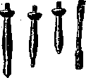
Instruments. The intubation instrumentation consists of 1) a set of tubes, 2) an introducer, 3) a laryngoscope, and 4) an extractor. - Tubes. In form, the tube is divided into a head, neck, and body. The front part of the head is cut off, the back protrudes and is placed in the inter-arytenoid incision. On the left side of the head (the sides of the tube are designated everywhere in relation to the patient, not to the operator), there is a small hole for passing a silk thread. The head itself lies at the entrance to the larynx on the vocal cords. In front of it are the root of the epiglottis and the upper part of the thyroid cartilage, on the sides are the ary-epiglottic ligaments (lig. ary-epiglottica), behind are the arytenoid cartilages. Behind the head follows the narrowest part - the neck, lying between the vocal cords;1 below the neck the tube is swollen in the form of a body (Fig. 1a). At present, there are 2 main types of instruments, differing in that in one (in the so-called "closed" I.) the tube is equipped with an obturator - a mandrel, completely closing the lumen of the tube and removed only when the tube is inserted into the larynx. Tubes of the second type (in the so-called "open" I.) do not have a mandrel. The lower opening of the tube when inserted into the larynx can easily injure the loosened mucous membrane. Therefore, a mandrel is inserted into the tube, the rounded end of which should tightly close the lower opening and protrude more or less from it. Thus, on the one hand, the mucous membrane is protected from injury, and on the other hand, the lower end of the tube, facing a narrow rounded tip, more easily slips into the glottis. In open I., the lower end of the tube itself must be changed accordingly. With the help of a mandrel, the tube is connected to the introducer; which is a long metal Fig. 1. a - O'Dwyer's tube; b and c - Sevestre and Bayeux; d - mandrel. rod with a handle, bent at the distal end at a right or slightly acute angle. If there is no mandrel, then the end of the introducer is equipped with a device for holding the tube. The instrumentation for closed I. In the original model of O'Dwyer, the tubes, made of hard rubber (ebonite), are of considerable length: from 2.5 to 7 cm, depending on age. The mandrel is a thin steel bent rod consisting of two halves connected by a hinge and connecting with the introducer by means of


a screw. At present, in Europe, the tubes of Sevestre and Bayeux are most often used. Being approximately */3 shorter than O'Dwyer's (Fig. 1 b and c), they are made of fusible metal on a thin silver sleeve and have a bent mandrel. In the set made by Collin, there are 6 sizes of tubes. Simple and ingenious is the method of connecting the mandrel with the introducer; the latter consists of three parts: the middle - the immovable part - ends in a small sleeve with a quadrangular hole and a horizontal slot on the back side. A correspondingly shaped tooth on the upper end of the mandrel tightly fits into this hole, having the same slot on the back (Fig. 1 d). On the left side of the rod is a slide, entering with its front end into the slots of the sleeve and the mandrel tooth and securing them. On the right side of the rod is the apparatus for ejecting the tube from the mandrel ("propulseur", or "declancheur"). It consists of a lever with a short back and a long front arm and ends in front with a small horizontal hook lying on the head of the tube. The tip of the thumb of the hand holding the instrument is placed under the back arm (Fig. 2); at the right moment it slightly lifts this arm, the front end of the lever descends and pushes the tube off the mandrel (Fig. 3 v and b). The entire introducer is extremely easy to disassemble, clean, and boil.-Instruments for open I. In 1892, From proposed his model of intubation set without a mandrel. The tubes are made of hard metal, gold-plated, with thin walls and correspondingly wide canal. The body of the tube expands conically downward, without a separate body, and ends in a narrow bridge lying in the sagittal direction. On both sides of the bridge are two large oval windows, as a result of which the lower end takes the form of a wedge and the appearance of a stirrup. The head of the tube has the appearance of a small cup and is polished flat on top, thanks to which it sits very low, is easily covered by the epiglottis, and does not interfere with swallowing. On its upper surface is engraved the age number for which this tube is intended. On the head two holes: one - small (on the left) for a thread, the other - large size (on the back edge) for the hook of the extractor (Fig. 3c). In size, the tube
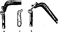
Fig. 3.
The tubes belong to the long type, but they are much lighter than the Collin tubes. The set contains 6 numbers. The introducer has the appearance of a curved forceps with sliding branches; it can also serve as an extractor. In the latter respect, it proved so convenient that it can easily be used to extubate tubes of other types when necessary. A special extubator is included with the set, which is worn on the finger, but in practice it is inconvenient and can easily injure the mucous membrane. There is no declancher; but when the end of the tube has passed through the glottis, the branches of the introducer are shifted and the tube is easily lowered into the larynx by the finger. The instrument is generally very convenient, easy to intubate with, the tubes fit tightly in the larynx, are rarely coughed up, and due to their wide channel, they freely allow bulky films to pass through. The great advantage of the 'open' intubation is the absence of the moment of suffocation (apnea): as soon as the end of the tube passes through the glottis, air freely passes through its channel, and the operator can, without worry, complete the lowering of the tube and the release of the beak of the introducer. With the 'closed' method, on the contrary, the most painful moment, especially for a beginner, is the removal of the mandrel. Technique of intubation. Before proceeding with the operation, it is necessary to carefully inspect and check the instruments. Of course, all instruments must be cleaned and boiled. Having selected the required size of the tube, a strong silk thread (preferably braided) is pulled through the hole on the left side of its head. If it is planned to leave the thread after intubation, it is tied in a knot at a distance of 1-2 cm from the head; the ends of the thread are also tied in a knot so that the length of the entire loop slightly exceeds the length of the introducer. Special attention should be paid to the hinges of the curved mandrels: whether there are cracks, breakages or lateral bends in them. Then, after assembling the introducer (disassembled for boiling), the mandrel is attached and the tube is put on. It is necessary to make sure that all parts of the instrument work well and the tube easily slides off the mandrel. The latter can be lubricated with borated vaseline or a solution of menthol in oil (1%). The instruments in a tray with boric water are placed on the table to the left of the operator. In addition, one must always remember the rule: if proceeding with intubation, a tracheotomy set must always be at hand, because in case of collapse, sudden spasm of the larynx or the film getting caught under the tube, every lost moment can cost the patient their life. Intubation can be performed with the patient sitting or lying down. In the first case, two assistants are needed. The patient with arms extended along the body is tightly wrapped in a sheet up to the shoulders; one of the assistants sits on a chair and takes the patient in their arms, the patient's legs are firmly held between the knees, the torso is grasped with hands and pressed against the assistant's chest. The second assistant stands behind the first assistant's chair. The operator (or the second assistant) inserts a closed mouth gag between the patient's teeth on the left side. The instrument must be pushed as far back as possible so that the labial frenum is slightly stretched. Having made sure that the branches of the gag are on the teeth, they are spread as wide as possible and pressed against the patient's cheek. The second assistant, standing behind the first assistant's chair, holds the patient's head with both hands. The fingers of the left hand press against the branches of the gag, with the tip of the middle finger placed between the anterior branches of the instrument and slightly pulling it backward. It happens that during intubation, a poorly locked or worn-out toothed gag slips off, and if there is no finger between the anterior branches, the patient's teeth close tightly, trapping the operator's finger and sometimes causing serious injuries. If the patient's teeth do not enter the depressions of the cheeks but only press against their edges, the instrument will inevitably slip and can not only injure the gum and cheek mucosa but even dislocate a tooth. When there are no molars yet, the mouth gag holds poorly, and in very small children when there are not even front teeth, it is advantageous to operate without a gag at all; their mouth is so small that the introduction of a pacifier opens it almost as much as the instrument. The main condition for success of the operation is the correct and completely immobile position of the patient. Their face should be facing forward, the head must be held straight, slightly tilted. The most dangerous thing is to tilt the head back, which inexperienced assistants often do. In this case, the entrance to the larynx, open upward and backward, deviates even further back, and the bodies of the cervical vertebrae move forward. In this position, the introduction of the tube into the larynx becomes very difficult, and sometimes impossible. The operator's chair is placed opposite the patient. It is more convenient if it is somewhat lower than the patient's chair. This is especially important for beginners: such a position forces
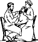
Figure 1. Position of the patient and personnel during intubation (introduction of finger).
one to act only with the wrist, hindering the application of force. The handle of the instrument is held in the whole hand, with the thumb on top; the thread of the tube, loosely tightened, is pinched between the index and middle fingers. The thread should not be pulled tightly, as it, forming a kind of bowstring between the head of the tube and the operator's fingers, presses against the patient's tongue and hinders the immersion of the tube into the larynx. If one wishes to remove the thread after the operation (as is done in France), one must first pass the tip of the little finger through the loop, making sure that the loop does not twist. With this method, after completing the intubation, it is easy to determine which part of the thread is on top, and by cutting it above the knot, pull out the lower part from the tube opening. The operator's left index finger is inserted into the patient's mouth, and upon reaching the entrance to the larynx, palpates the identifying points (Fig. 4). The first to be encountered is the epiglottis, and behind and below it are the arytenoid cartilages, like two dense buttons with a notch between them; even deeper, the posterior wall of the larynx is felt under the pulp of the finger in the form of a hemispherical protrusion of cartilaginous consistency. The soft, thin epiglottis often bends backward, sometimes even rolling into the tube under the palpating finger: it must be unfolded, which is not always easy for an inexperienced person. When the tip of the finger is over the entrance to the larynx, the epiglottis is pressed against the root of the tongue (Fig. 5), the aryepiglottic ligaments (lig. ary-epiglottica) and the arytenoid cartilages should be clearly felt at this moment. At this moment, the tube is inserted into the patient's mouth and advanced to the entrance to the larynx, following all the time along the radial side of the left index finger (Fig. 6). The instrument must be guided along the midline, holding it strictly in the sagittal plane. Sometimes the size of a child's mouth is so small that the knee of the introducer presses against the hard palate and hinders the advancement of the tube; in this case, the handle of the instrument can be deviated to the right, without changing the position of the tube, but only by rotating it around its longitudinal axis. But as soon as the end of the tube reaches the epiglottis, the instrument is turned again, placing it in the sagittal plane. Only in this last position of the introducer can one proceed with inserting the tube into the larynx. Now the end of the index finger is moved away and the end of the tube is placed in its position, and the finger is placed behind it so as to again feel the ends of the arytenoid cartilages (Fig. 7), and slightly pull the handle of the introduator toward oneself, pressing the tube against the anterior wall of the laryngeal entrance. At this moment, the tube is directly over the entrance; but since the laryngeal opening is directed downward and forward, if the instrument is simply lowered, the tube will almost inevitably slip into the esophagus. It is necessary to slightly raise the handle of the introducer (it is especially important here to strictly maintain the sagittal plane), then the end of the mandrel will turn forward, and the tube will enter the laryngeal entrance without any effort and will press against the vocal cords, which at this moment reflexively narrow the glottis. To pass through it, a slight effort must be applied. Under no circumstances should force be used, otherwise a false passage may be made. Sometimes the reflex is so pronounced that the glottis closes tightly and does not allow the tube to pass; in this case, one must, without worrying, wait a few seconds, slightly pressing on the instrument: the child will attempt to take a breath, the glottis will open, and the tube will slip into the larynx. At this time, it should be remembered that during inspiration the larynx descends, and the identifying points should not be lost. After passing through the glottis, the tube is immersed as far as the instrument allows; only then is the mandrel slightly shifted with a light movement of the declancher; the edge of the head is found with the left finger, and by pressing against it, the tube is lowered; at the same time, not
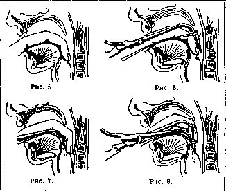
they quickly raise the mandrel (fig. 8) in a straight line upward and slightly backward; when its end emerges from the tube, the introducer is removed by lowering its handle from the child's mouth; the tube is then inserted with a finger into the larynx as deeply as possible. That the operation was successful is concluded first by a special metallic, so-called "tubular" breathing sound, and second by palpation: the head of the tube should be distinctly felt at the entrance to the larynx, but should not protrude from it. One should never forget to check with a finger the position of the tube. If the thread cannot be removed, it is, to avoid being bitten off, tried to be placed in some gap between the teeth, and the end is wound from top to bottom around the child's left ear and attached to the cheek with a piece of adhesive tape.-For I. in the recumbent position, the patient, wrapped in a sheet, is placed on a table or bed without a pillow. One assistant stands at the feet on the left side of the patient and, firmly grasping him by the elbows, presses his legs between his left shoulder and chest. The second assistant stands at the head and grasps the head, holding the mouth gag. The operator* stands on the right side of the patient. The techniques for I. are the same as those described above. One can manage with only one assistant; in this case, the ends of the sheet in which the patient is wrapped are tied under the bed or table, and the assistant holds the head. Main rules of I. The entire operation must be performed gently, without any violence. Trump (Truмpр) advises handling the instrument during I. as if it were made of fragile glass. The most important and dangerous moment is, of course, when the end of the tube is positioned at the entrance to the larynx. Here one must not forget the following. 1. The end of the left index finger, without pressing on it at all, should be slowly raised slightly above the horizontal line. If the handle is raised quickly and too high, the end of the tube will press against the incisure of the thyroid cartilage or the anterior commissure of the vocal cords and easily make a false passage. 3. During the previous maneuver, one must remember that the instrument should be in the strictly sagittal plane. The quite natural slight supination of the operator's right hand tends to direct the end of the tube into the right Morgagni's recess of the patient. Indeed, the largest number of injuries and false passages occur in the right half of the larynx.
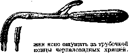
Care of the intubated child. To prevent the patient from pulling out the tube by grasping the thread, he must be swaddled. But it is much more rational to use Everbushev's elbow bandages, which do not allow the patient to bend his arms at the elbows. These bandages, leaving freedom of movement in the shoulder and wrist joints, do not disturb the patient. In the absence of bandages, one can simply bind to the patient's arms thin and narrow, cotton-wrapped splints or cardboard splints bent into a trough.-Extubation. Removal of the tube with the help of an extubator (fig. 9) differs little in technique from I. It is always done with the patient in a sitting position. The head is placed straight or slightly tilted back. The left index finger locates the head of the tube and slightly pushes the epiglottis forward; the beak of the extractor is carefully introduced along the finger into the opening of the tube. Getting into this opening is by no means simple! The beak of the instrument slides along the head of the tube and easily gets between it and the wall of the larynx. If at this moment the branches are spread and an attempt is made at extraction, one can seriously injure the mucous membrane. Therefore, one must be very sure with the tip of the finger that the instrument is indeed introduced into the channel of the tube. It should be inserted as deeply as possible and then, by spreading the beak with strong pressure on the posterior end of the movable branch, carefully extract the tube. The left finger should be withdrawn together with the tube, constantly touching its head, so that in case the instrument slips, the extraction can be completed with a finger. In 1895, Baye proposed an excellent method for pushing out tubes-"enucleation"; unfortunately this method is applicable only to short tubes for "closed" I. It is clear that if the thread has not been removed and the patient has not bitten through it, extraction is performed simply by pulling on this thread. Indications for I. The question of when to proceed with I. is extraordinarily important. On the one hand, it is desirable to introduce the tube as late as possible, in the hope that the serum will have time to take effect and it will be possible to avoid the operation; on the other hand, by waiting too long, one may miss the moment, and the patient cannot be saved. One must catch the moment when persistent stenosis begins to turn into asphyxia. Many methods have been proposed for determining this moment; the simplest and most reliable of them is the appearance of the so-called "paradoxical pulse" (the pulse wave begins to fall on inspiration and rise again on expiration). It is especially important not to miss this moment in small children (under 1 year), in whom asphyxia can easily and suddenly occur.
Time for extubation. According to the opinion of most authors, the tube should be removed as soon as the larynx is cleared of membranes, since the latter protect the mucous membrane from pressure from the tube and consequently from the formation of bedsores. The first attempt at extubation should be made at the end of the second or beginning of the third day, if the membranes in the pharynx are coming off well and the temperature has fallen below 38°. Practice shows that with this method of action, it is usually possible to manage with a single I.-Injuries can be inflicted both during intubation and during extubation. Small erosions and scratches of the mucous membrane are not serious. This cannot be said about false passages, which usually lead to the formation of abscesses and necrosis of tissue. False passages most often occur when the end of the tube enters the Morgagni's recess; the swollen, softened mucous membrane is very easily injured, and the tube makes its way between the mucous membrane and the muscles of the larynx. The secretion from the mucous membrane flows into the resulting pocket, and suppuration begins. Injuries during extubation are very rare; in the literature, cases of injury to the nil vocal cords by the extubator and even fractures of the cricoid cartilage are described. Much more often one has to observe bedsores formed from pressure of the tube. Bedsores sometimes form extremely quickly (especially if there are no membranes, e.g., in measles stenosis and false croup); on the other hand, sometimes even several days' presence of the tube leaves only small superficial ulcers. Most often, bedsores form on the vocal cords and in the area of the cricoid cartilage. All bedsores can be divided into 3 groups: 1) only the mucous membrane is necrotized; 2) the ulcer reaches the cartilage, but the latter is not exposed; 3) the perichondrium has died, and the cartilage itself is more or less damaged, and often partially necrotized. These groups differ sharply in their consequences. The first degree usually does not hinder extubation, and the ulcers heal without a trace; with the second, extubation may be difficult, and the ulcers leave scars; the third already represents a severe lesion, often accompanied by the shedding of necrotized cartilage with subsequent collapse of the larynx, which forces one to resort to tracheotomy; subsequently, complete atresia of the larynx may occur. Mucus falling on the epithelium-denuded surface of the bedsore decomposes, and the hydrogen sulfide formed thereby causes black spots to appear on the metal tube. These spots are an indication of the beginning necrosis of the mucous membrane. In severe bedsores of the third degree, when cartilage necrosis occurs, the tube no longer holds firmly in the larynx, and constant expectoration of it begins with rapidly developing asphyxia. In these cases, O'Dwyer still used tubes coated with a layer of gelatin in which alum is dissolved. Bokay is also a hot supporter of this method.
Related articles
Mentioned in
Cite this page
“Intubation.” Soviet Medical Encyclopedia. English translation of Bolshaya Meditsinskaya Entsiklopediya, 1st ed. (Moscow, 1928–1936), ed. N. A. Semashko. https://sovietmedicalencyclopedia.pages.dev/article/intubation/