Parathyroid Glands
Historical document, translated for reference. It reflects medical knowledge of the 1920s–30s and is not medical advice.
Summary
The parathyroid glands are special endocrine glands located near the thyroid gland. This article describes their anatomical structure, location, and historical discovery, noting that they are distinct organs with specific functions separate from the thyroid gland.
Encyclopedia article (1928–1936)
PARATHYROID GLANDS (glandulae parathyreoideae) (syn.: parathyroid glands, accessory thyroid glands, French glandules thyroïdiennes, glandes satellites de la thyroïde, German akzessorische Schilddrüsen, Beischilddrüsen, Nebendrüsen der Schilddrüse, Epithelkörperchen - epithelial bodies), special glands of internal secretion, located near the thyroid gland in the form of two pairs of small glandular organs. The existence of small glandular formations occurring more or less regularly near the thyroid gland became known to researchers comparatively recently. Certain indications of them can be found in Remak in 1858 and in Virchow in 1863, however most authors did not consider them independent organs, but regarded them as accessory parts of either the thyroid or goiter glands. Even Sandstrom, who in 1880 first gave an exact description of these small glands and proposed for them the name 'glandulae parathyreoideae', considered them to be built from tissue of the thyroid gland remaining in a state of embryonic underdevelopment. This opinion was predominant until the experimental development of the question of the function of these small glands. Gley and Hofmeister in 1891 first began to speak of the independence of the parathyroid glands, but this was finally established by the Prague anatomist Kohn, who in 1896 pointed to the constancy of presence and position of the parathyroid glands and to their functional specificity. In order to emphasize the complete lack of relation of the parathyroid glands to the thyroid gland, Kohn proposed to call them 'epithelial bodies' (German Epithelkörperchen), that is, a term put forward in 1888 by Maurer to denote similar formations discovered by him in amphibians. A whole series of subsequent experimental researches (see below - physiological part) and observations from the field of pathology (see below - patho-anatomical part) further contributed to the final establishment of the position that the parathyroid glands represent quite special endocrine organs with special functions.
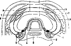
Anatomy. Normally in humans there are four parathyroid glands, located two on each side. On the corpse they are found, having separated from the thyroid gland the muscles and having drawn forward one or the other lobe of the thyroid gland; one can also extract all organs of the neck and discover the parathyroid glands on the posterior surfaces of the lateral lobes of the thyroid gland, somewhat moving aside the unopened esophagus. On both sides the upper parathyroid gland, which Welsh calls glandula parathyreoidea superior posterior, lies on the postero-medial edge of the lateral lobe of the thyroid gland, namely - at the level of the middle third of this edge, directly behind the ascending branch of a. thyreoideae inf. and n. recurrentis, in the loose connective tissue surrounding the esophagus (fig. 1). Verebely in 76% found this parathyroid gland in the middle of the extent of the indicated edge of the lateral lobe of the thyroid gland, in 15% above the middle, in 9% below it. Usually the upper parathyroid gland is 2-3 mm distant from the posterior surface of the thyroid gland, but may be found closely connected with its capsule and even located under the capsule. The lower parathyroid gland, according to Welsh - glandula parathyreoidea inferior anterior, is usually somewhat larger in size than the upper, lies in the loose connective tissue near the capsule of the posterior surface of the lower part of the lateral lobe of the thyroid gland (fig. 2), usually where in this part there is a depression of the surface; with relation to a. thyreoideae inf. the lower parathyroid gland lies at the level of its entry into the tissue of the thyroid gland, but forward from both it and n. recurrens. In general topographically the lower parathyroid glands are closely connected both with the lower poles of the lateral lobes of the thyroid gland and with the upper parts of the goiter gland.
From the above-mentioned normal position of the parathyroid glands, frequent deviations are observed. Thus, the upper parathyroid glands, especially the left one, may be found located considerably higher than their usual place, for example at the upper edge of processus pyramidalis, or else below - near the lower parathyroid glands; on the other hand, the lower parathyroid glands may also be found lying
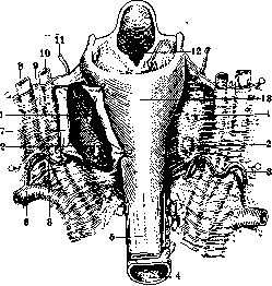
Figure 2. Bed of the thyroid gland and relation of the parathyroid glands within this bed (from behind): 1 and 2 - upper and lower parathyroid glands; 3 - inferior thyroid arteries; 4 - trachea; 5 - esophagus; 6 - subclavian artery; 7 - posterior wall of the sheath around the thyroid gland; 8 - internal jugular vein; 9 - vagus nerve; 10 - common carotid artery; 11 - superior thyroid artery; 12 - larynx; 13 - pharynx.
near the upper parathyroid glands or much lower than their usual position, for example, 4-5 cm below the thyroid gland, at the level of the 10th-12th tracheal cartilage rings. Sometimes one or another of the parathyroid glands is located within the tissue of the thyroid gland or, more rarely, within the goiter. In addition to irregularities in the position of the parathyroid glands, various deviations from the usual number of parathyroid glands may occur. Findings of fewer than four parathyroid glands, often mentioned by previous authors, hardly deserve attention in view of the frequent difficulty in finding these formations, especially since recent studies establish that with careful research, cases where fewer than four parathyroid glands are found are extremely rare. It happens that on each side both parathyroid glands are fused together (Erdheim). Cases have been described where on one side there were three parathyroid glands and on the other only one. Significantly more often, a greater number of parathyroid glands than four is observed. Among such cases, it is relatively common that one of the parathyroid glands is divided into two unequal parts, which is sometimes visible to the naked eye and sometimes (with a common capsule) is revealed only under the microscope. Pfeiffer and Mayer saw instead of one parathyroid gland a whole series of small glands arranged in the form of a chain. Finally, cases of additional parathyroid glands located outside the connection with the four normal parathyroid glands are also not particularly rare; thus, various researchers found 5, 6, 8, 12 parathyroid glands located in the area of the thyroid gland, above and especially below it; cases of numerous parathyroid glands scattered in the fatty tissue of the mediastinum have been described. Excess parathyroid glands or groups of their cells are often found inside the thyroid gland, in the goiter gland. Askanazy once found an additional parathyroid gland in the thickness of the phrenicus nerve. The shape of the parathyroid glands is usually oval and slightly flattened, more rarely round, bean-shaped or kidney-shaped; the greatest diameter, i.e., the length of each parathyroid gland, is always located from top to bottom, i.e., parallel to the course of the esophagus and trachea. The color of the parathyroid glands is slightly brownish or brownish-yellow. The surface is smooth. The size of the parathyroid glands varies within relatively small limits: for adult parathyroid glands, Welsh determines the length as 6-7 mm, Erdheim as 8 mm, other researchers, for example, Verbeely, Moller, as 3-18 mm. The thickness is determined by Welsh as 4 mm, by Erdheim as 5 mm, by Verbeely, Moller as 2-8 mm. At the same time, the lower parathyroid glands are always somewhat larger than the upper ones. The average weight of each parathyroid gland in an adult, according to Welsh, is 0.035 g. The blood supply to the parathyroid glands goes through special branches of the inferior thyroid artery, with each parathyroid gland having its own arterial branch (superior and inferior parathyroid arteries). Ginsburg observed that with a high position of the upper parathyroid gland, its artery may branch off from the superior thyroid artery. The arteries, plunging into the tissue of the parathyroid gland, branch within it, while the veins form a network on the surface and collect into trunks going along the course of the parathyroid arteries. Nerve trunks also go along with these. Sometimes at the point where the vascular-nervous bundle enters the gland, there is a depression which some authors designate as the hilus of the parathyroid gland. Despite the fact that the parathyroid glands have more or less constant size, shape and position, finding them both on the dissecting table and during the appropriate operation or experiment is not always easy; the possibility of irregularities in the position and number of parathyroid glands further complicates the matter. Most often, inexperienced people mistake small lymph glands, lobules of fatty tissue, and nodules of thyroid tissue, sometimes bulging from under its capsule, for parathyroid glands. In difficult cases, one should be guided by the presence in the parathyroid glands of their own arteries (parathyroid arteries), coming from the inferior thyroid artery, and by the brownish tint characteristic of the parathyroid glands. Histology. Schematically, the structure of the parathyroid glands can be represented as follows: they have a connective tissue capsule, from which connective tissue strands extend into the gland; in the spaces between these branching connective tissue strands is the parenchyma, consisting of epithelial cells. The ratio between the connective tissue stroma and the epithelial secretory parenchyma, i.e., the type of glandular structure of the parathyroid gland tissue, varies, which is partly related to age. Kon distinguishes three types of parathyroid gland tissue structure: 1) The compact type, when the connective tissue stroma is represented only by individual strands, and the parenchyma is arranged in the form of a solid mass of epithelial cells without any division of them into lobules or cells. This type of structure is characteristic of the parathyroid glands in the embryonic period and in childhood; it is generally common in young age (up to 20-30 years), but sometimes it is also found in advanced age. 2) The reticular type, when the parenchyma is divided by a network of connective tissue strands into groups of epithelial cells of equal size and without signs of any proper glandular structure; this type of parathyroid gland structure is the most common. 3) The alveolar type, when the parenchyma seems to be divided by connective tissue into more or less regular lobules and glandular alveoli. Various transitions are observed between these three types. In addition, very often among tissue built according to one type, areas having another type are encountered: thus, in children with compact type parathyroid glands, areas with alveolar type structure can be observed, on the other hand, in adults and elderly people in parathyroid glands of reticular and alveolar type, areas of compact type are sometimes found. With all the above-mentioned types of structure, the epithelial cells forming the secretory parenchyma of the parathyroid glands are not uniform in appearance. Initially, two types of epithelial cells of the parathyroid glands were distinguished: 1) chief cells, which constitute the main mass of the parathyroid gland tissue, and 2) oxyphilic cells, located in groups among the chief cells. Later, however, a larger number of cell forms were distinguished. The most common at present is the division of the epithelial cells of the parathyroid glands into 4 types, proposed by Getzowa: 1) Light chief cells, the distinctive feature of which is their light, completely non-staining protoplasm and sharp contours, which makes them resemble plant cells. Their size is medium, but sometimes they are very large; occasionally (in children) they locally take on a cylindrical shape and are arranged like a palisade or even form what are like glandular tubes with lumens. These light cells constitute the main mass of the parathyroid glands in the embryonic period and in early childhood. 2) Dark chief or pink-red cells, whose protoplasm is finely granular and stains with background dyes. These cells originate from the light ones and constitute the main mass of the parathyroid gland parenchyma in adults. The two types of cells mentioned by Getzowa belong to what was previously united under the concept of chief cells. 3) Oxyphilic cells, discovered by Welsh, have coarsely granular, strongly eosin-staining protoplasm and larger dimensions than chief cells. These cells are usually located among the chief cells only in small groups. In children, they are usually not found, as they appear in the parathyroid glands only after 10 years, and their quantity increases with age; they are most easily detected after 20-30 years. The number of oxyphilic cells in the parathyroid glands in comparison with chief cells is small: very rarely, mainly in old age, it exceeds 1/10 of the latter (Getzowa). 4) Syncytium-like cell groups, having the appearance of closely located nuclei with well-staining protoplasm without clear boundaries between them, and apparently represent closely adjacent and as if fused into a solid mass dark chief cells. Syncytium-like groups are found almost exclusively after 40 years of age. All cells of the parathyroid glands, except oxyphilic cells, contain many fat droplets (phosphatides, neutral fat), which appears in the epithelium of the parathyroid glands after 4 months of extrauterine life. In oxyphilic cells, fat is not constantly present and in small quantities. The chief cells of the parathyroid glands also contain glycogen, found in particularly large quantities in childhood in light chief cells. Sometimes glycogen in large quantities fills perivascular lymphatic spaces. Often among the cells of the parathyroid glands, especially in the peripheral parts, there is an accumulation of a substance similar to colloid or having the appearance of small or larger spherical masses; where larger spheres lie, epithelial cells are arranged around them in the form of a row of cubic cells, which gives great similarity to a follicle of the thyroid gland. Sometimes there are so many such formations that the entire tissue resembles the structure of the thyroid gland. These pictures of colloid in childhood are found in the parathyroid glands extremely rarely; usually they are found in adults and especially in the elderly. The 'colloid' of the parathyroid glands, as recent studies have shown, has nothing in common with the colloid of the thyroid gland; its chemical structure is different; in particular, it does not contain iodine or contains it only in traces.
Apparently, the 'colloid' of the parathyroid glands represents a coagulated protein mass, possibly a product of secretion, only morphologically similar to the colloid of the thyroid gland. On this basis, the assumption of previous authors about the possibility of parathyroid tissue transforming into thyroid tissue is now decisively rejected. The stroma of the parathyroid glands consists of loose connective tissue, which also forms the capsule; inside the glands, the stroma is distributed either in the form of separate strands or in the form of a network, depending on the particular type of parathyroid gland structure (see above). In addition, according to the method of Bielschowsky, a network of reticular (mesh-like) fibers is found between the cells of the parenchyma. The connective tissue stroma is very rich in blood vessels, especially peculiar wide 'sinusoidal' capillaries, which are in very close contact with the epithelial cells of the parenchyma. Lymphatic vessels, sometimes very wide, accompany the blood vessels. Nerve trunks also pass through here. In the connective tissue, fat cells are often found, and near the vessels, sometimes stellate cells with fine-grained brown pigment that does not give an iron reaction can be seen. Very often with age (according to Erdheim after 5 years, according to Arndt after 7 years), groups of fat cells appear in the connective tissue, gradually increasing in number; in adults, the replacement of the parathyroid stroma to some degree by fatty tissue is almost constant; thus, Herxheimer found that in only 4% of adult parathyroid glands is fatty tissue not found at all, in 42% it is present in moderate amount, and in 54% in large amount. Embryology. Information on the embryonic development of the parathyroid glands became more or less accurate after Kon established the functional independence of these glands (see above). Until that time, although it was known that the parathyroid glands are formed from the III and IV branchial clefts, their formation and further evolution were not clearly distinguished from the development of the thymus and thyroid glands. At present, it can be considered established that the primordium of the parathyroid glands in human embryos and all animals occurs in the III and IV branchial clefts separately from the primordia of the thyroid and thymus glands, in the form of a group of epithelial cells, which then in each cleft form a kind of diverticulum and separate as independent organs. This development of the parathyroid glands, however, proceeds parallel to the development from these same branchial clefts of the primordia of the thymus and thyroid glands, so that the connection between the developing parathyroid glands and the metameres of the thymus and thyroid glands can be very close; among other things, this prompted Groschuff to denote the parathyroid glands developing from the III branchial clefts together with the thymus gland as 'parathymus,' and those developing from the IV branchial clefts in close connection with the lateral lobes of the thyroid gland as 'parathyreoidea.' The nomenclature of Verdun is more widely accepted, who designates the parathyroid glands developing from the III branchial clefts and subsequently located at the lower poles of the thyroid gland with the number III, and those originating from the IV branchial clefts and later found to be located behind the middle third of the lateral lobes of the thyroid gland with the number IV. In humans, as in all higher vertebrate animals, the primordium of the parathyroid glands occurs in the dorso-cranial walls of the branchial clefts; in contrast to this, in lower vertebrates they develop much more ventrally. As an ontogenetic variant, some have found development of the parathyroid glands from the II and V branchial clefts. Comparative anatomy shows that the presence of parathyroid glands with the developmental course described above and the histological structure given above is characteristic of all vertebrate animals, both lower (amphibians, reptiles, fish, etc.) and higher (birds, all mammals); the difference from the human parathyroid glands lies in their different number in different animals and in the difference in their anatomical position. The greatest constancy in the series of animals is shown by the parathyroid glands III (according to Verdun's designation), i.e., those that develop from the III branchial clefts; this pair of parathyroid glands is always found and is located in all animals more or less identically. In contrast to this, the parathyroid glands IV in some animals (e.g., in pigs, hedgehogs, moles, seals, guinea pigs, mice, rats) are either absent altogether or occur extremely rarely. In many animals, the parathyroid glands IV are present, but lie inside the thyroid gland, which with the parathyroid glands III happens very rarely (sometimes in mice and rats); such a position of the parathyroid glands IV inside the thyroid gland is characteristic of the cat, dog, wolf, badger, fox, rabbit, horse, macaque monkey. The constancy of the position of these parathyroid glands in these animals inside the thyroid glands prompted Kon to distinguish in them 'external' and 'internal' parathyroid glands. An increase in the number of parathyroid glands in animals is very common; thus, in cows, goats, cats, dogs, rabbits, rats, additional parathyroid glands, sometimes numerous, have been found very often inside the thymus gland, in the mediastinal fat, at the root of the aorta, etc. The fact that many animals normally have 'internal' parathyroid glands, i.e., those located inside the thyroid gland, and on the other hand, the frequent presence of additional parathyroid glands lying in the mediastinum, is of great importance for evaluating various experiments with parathyroidectomy. Pathological anatomy. The parathyroid glands are often subject to various pathological-anatomical changes; however, the significance of these changes in terms of their effect on the function of the parathyroid glands has been very little studied. It can be noted that among pathological processes, only hemorrhages into the parathyroid tissue have the most definite consequences for the function of the parathyroid glands, which often underlie tetany, and hyperplastic processes of the parathyroid glands, which are often associated with a disturbance of calcium metabolism and in particular with some general lesions of the skeletal system. Malformations of the parathyroid glands are rare, and cases of complete absence of formation, aplasia of the parathyroid glands, have not been described at all. It is important to note that in cases of complete aplasia of the thyroid gland, the parathyroid glands are developed normally and generally show no special deviations from the norm; this is one of the proofs of the absence of any genetic dependence between the parathyroid glands and the thyroid gland. Cases of congenital hypoplasia of the parathyroid glands are described extremely rarely; more often one can speak of underdevelopment of the parathyroid glands as a result of their damage in early childhood (hemorrhages, syphilis).-As for anomalies in position, number, and size of the parathyroid glands, all such cases are considered anatomical variations (see above), not malformations.-Some cysts of the parathyroid glands are the result of improper development, especially those lined with ciliated epithelium; since they represent remnants of branchial clefts, they should be considered close to branchiogenic cysts. In addition to such congenital cysts, which are the result of improper development, acquired cysts can also occur in the parathyroid glands; some of the latter, containing either liquid or colloid-like mass, belong to retention cysts, while others apparently result from the breakdown of tissue of one origin or another. Atrophy of the parathyroid glands, manifested by their decrease in size and hardening due to atrophic changes in the parenchymatous elements and proliferation of the interstitial connective tissue, mainly along the course of blood vessels, is often observed in old age, as well as in some wasting diseases (e.g., tuberculosis). The same changes in the parathyroid glands of the type of sclerotic atrophy were found by Melnikov and others in goiter, Marinesco in pellagra, Thomson in pedatrophy, Erdheim in epilepsy, Kraus in diabetes. The qualification, and hence the designations, of these changes in the parathyroid glands by various authors are different; in addition to classifying them simply as atrophies and sclerotic atrophies, they are also designated as 'sclerosis' of the parathyroid glands and as 'chronic fibrous parathyroiditis.' Sometimes sclerotic atrophy of the parathyroid glands is observed simultaneously with similar changes in other endocrine glands (thyroid gland, pancreas, testes, etc.), which underlies the so-called 'polyglandular insufficiency.' It should be noted, however, that in some of such cases, simple atrophy of many endocrine glands, including the parathyroid glands, without any sclerotic changes was observed (Lindemann). Degenerations in the parathyroid glands in acute infections show protein granular degeneration of the chief cells. Fairly often (according to Petersen in 25%) vacuolar degeneration of the chief cells is observed; as a result of the latter, the cells may melt and form cavities. Fatty degeneration of the parathyroid glands in the sense of proliferation of fatty tissue in their stroma, as already indicated above, is not a pathological phenomenon: only those cases in which fatty tissue appears in large quantity in the parathyroid glands at a young age can be regarded as manifestations of pathological lipomatosis. It is even more difficult to speak of degenerative fatty degeneration (fatty degeneration) of the parathyroid glands, since their cells constantly contain fat; again, only with too great an accumulation of fat in the cell protoplasm and in the presence of necrobiotic changes in the nuclei can fatty degeneration be assumed, as some have noted in diphtheria, in septicemias.
The same can be said about pathological manifestations in the glycogen content of the parathyroid glands; indications of an increase in its quantity in the chief cells in some cases of tetany are doubtful. Much more convincing are observations about the decrease in quantity and disappearance of glycogen from the cells of the parathyroid glands in some cases of pneumonia, nephritis, cirrhosis of the liver, acute yellow atrophy of the liver, marasmus, etc. A very frequent type of degeneration of the parathyroid glands is amyloidosis, which almost always accompanies general amyloidosis. The deposition of amyloid occurs in the walls of small vessels (arteries, veins, capillaries); in a severe degree of amyloidosis, the entire interstitial tissue of the parathyroid glands may be involved. The parenchyma of the parathyroid glands in amyloidosis is successively subjected to atrophy and degenerations. Of the salt deposits in the parathyroid glands, the deposition of lime can often be observed, for example, in connective tissue in a severe degree of sclerosis of the parathyroid glands or in the walls of arteries in general lime metastases. Pathological pigmentations in the parathyroid glands manifest only as the deposition of hemosiderin as a result of previous hemorrhages (see below); the appearance of lipofuscin in the parathyroid glands of elderly people, mentioned by Hueck, is currently rejected. All the above-mentioned atrophic-degenerative changes in the parathyroid glands in most cases are not accompanied by any disturbances of their functions; only in individual cases, for example, sclerotic atrophy or a pronounced amyloidosis, were there signs of loss of parathyroid gland function in the form of tetany. Of circulatory disorders, anemia and hyperemia, as well as the manifestation of edema, which may occur in the parathyroid glands as a particular manifestation of these changes throughout the body or in the neck, are not of great importance. Much more attention is attracted by hemorrhages into the parathyroid glands, which many researchers qualify as the main pathological-anatomical basis of tetany in children. Hemorrhages into the parathyroid glands are most often found in early childhood and are associated with the act of birth (injury to the neck or asphyxia during birth); there are observations indicating that hemorrhages into the parathyroid glands in children can occur as a result of attacks of severe coughing. In adults, hemorrhages into the parathyroid glands are rare and their origin is not always clear. Examination of the parathyroid glands in hemorrhages into them reveals areas of blood impregnation in their tissue, and often as a result of tissue destruction, the formation of blood false cysts; after the resorption of blood in the interstitial tissue of the parathyroid glands, the deposition of hemosiderin remains for a long time, and sometimes there is a significant proliferation of interstitial connective tissue. One can think that as a result of hemorrhages into the parathyroid glands of children, a delay in their development may occur, leaving permanently hypoplastic parathyroid glands. All such findings, i.e., more or less fresh hemorrhages, deposition of hemosiderin in the stroma, sclerosis with the presence of hemosiderin, hypoplasia (in adults) with hemosiderin in the capsule, were often found in cases of tetany, which served as the reason to consider hemorrhages into the parathyroid glands as the main cause of tetany in children and one of the causes of tetany in adults. However, at present many object to this point of view, mainly on the grounds that hemorrhages into the parathyroid glands also occur without any tetany (according to Auerbach in 76% of all children) and also sometimes in cases of more or less prolonged tetany, completely fresh hemorrhages are found in the parathyroid glands; the latter circumstance makes one think that hemorrhages into the parathyroid glands may be a consequence of tetanic convulsions. (For more about the pathogenesis and pathological anatomy of tetany-see Tetany.) Inflammations of the parathyroid glands are rare and have no independent significance. Erdheim saw embolic abscesses in the parathyroid glands in pyemia of ear origin; Koopmann in cases of sepsis sometimes found lymphoid infiltrates around the vessels of the parathyroid glands. Kraus discovered chronic interstitial parathyroiditis with infiltration of the stroma by lymphoid and plasma cells in a case of scleroderma. In addition, in inflammation of the neck fascia or thyroid gland, the inflammatory process can also involve the parathyroid glands. Such inflammations usually did not cause any functional disorders of the parathyroid glands, although, on the other hand, Dieterich, by experimentally causing inflammation of the parathyroid glands in dogs and rats, obtained tetany in them. The phenomena of sclerosis often encountered in the parathyroid glands, especially in elderly persons, hardly often belong to the consequences of an inflammatory process and understandably do not deserve the often applied designation 'chronic fibrous parathyroiditis' in relation to them. Tuberculosis of the parathyroid glands occurs in general miliary tuberculosis in the form of the presence of individual tubercles in the tissue of the parathyroid glands. Möller found tubercles in the parathyroid glands in half of the cases of general miliary tuberculosis he examined, and in one case concerning a child with a particularly large eruption of tubercles in two parathyroid glands, there was tetany; on the other hand, Koopmann, as well as Herxheimer, in many dozens of cases never once encountered tuberculosis of the parathyroid glands. An even rarer form of tuberculosis is the caseous tuberculosis of the parathyroid glands. Syphilis of the parathyroid glands is observed as lesions of them in congenital syphilis; in the latter, hypoplasia of the parathyroid glands, or 'chronic interstitial inflammation' with atrophy of the parenchyma and proliferation of the stroma, in which there were focal infiltrates of lymphocytes and large mononuclears, are sometimes described. In adult syphilitics, atrophy of the parathyroid glands was occasionally found, sometimes simultaneously with atrophy of other endocrine organs. Views on the ability of the parathyroid glands to regeneration differ. Most authors deny this ability at least in adult animals. On the other hand, some researchers, in experimental destruction of parts of the parathyroid glands, saw mitoses in the undamaged parts, which makes one think of the possibility of at least limited regenerative manifestations on the part of the parathyroid glands. Vicarious hypertrophy of the remaining parathyroid glands upon removal of some of them is a well-established fact; thus, in experimental or operative removal, for example, of two of the parathyroid glands or in their destruction by some pathological process, a certain increase in size is observed in the remaining parathyroid glands, which in adults apparently goes at the expense of an increase in the size of the cells, and in young animals also at the expense of an increase in their number. In addition to such manifestations of pathological growth of the parathyroid glands, there are processes of proliferation that can affect either only one or two of the parathyroid glands or all of them simultaneously. The proliferation itself can be diffuse, i.e., uniformly involve the entire parathyroid gland, or it can be nodular. The size of the parathyroid glands in this case can be increased to a very different degree, up to the size of a plum or even a chicken egg (see below). Histologically, sometimes a uniform increase of the entire tissue of the parathyroid gland as a whole is found, in other cases it is a matter of unilateral proliferation or only of the chief (light or dark) cells or more rarely only of the oxyphilic cells. Such proliferations may have no clear functional significance and are often found accidentally at autopsy, with no changes in the skeleton at this time, but in many cases such proliferations are found in various diseases of the bony skeleton (see below). All the above indicates that the listed types of proliferation of parathyroid gland tissue represent essentially extremely different processes, but to the present day not only is there no rational classification of them, but among researchers there are still very significant disagreements as to which of these processes should be considered hyperplasias and which can be attributed to true tumors-adenomas. For this reason, there is a tendency to call some of the above proliferations 'adenomatous hyperplasias' or to combine them under the name 'struma parathyreoidea'. A review of the relevant literature allows us to outline the following types of these proliferations: 1) Diffuse hyperplasia of the parathyroid glands with a uniform increase in the number of all cells. 2) Hyperplasia with the formation of limited 'foci of growth' (German Wucherungs-herde). In both of these types, the proliferating tissue differs from the pre-existing tissue by the absence of adipose tissue in the stroma, a small amount of fat, and sometimes also of glycogen in the epithelial cells. 3) Nodular hyperplasia in the form of the formation of a separate nodule of proliferation of chief or oxyphilic cells, with the surrounding tissue of the parathyroid gland showing a clear picture of compression and atrophy. Many classify these nodular hyperplasias of only one type of cell as adenomas, others call them strumas; in particular, such proliferations consisting only of oxyphilic cells are often called strumas of Welch. 4) Large adenomatous proliferations, in which one, two, or all parathyroid glands reach sizes of 5x4 cm and more; they usually consist of chief cells, and more often only of dark ones, more rarely of dark and light or only of light; in some cases, among the chief cells, groups of oxyphilic cells were also encountered. Incidentally, adenomatous proliferations can originate from parathyroid glands enclosed within the thyroid gland, which gives the appearance of a goiter, which was named 'parastruma' by Langhans.
Along with cases in which enlargement of the P. glands of the type of hyperplasia or adenoma was not accompanied by any changes in the skeletal system or generally by disturbances of calcium metabolism, there are numerous observations concerning cases where, along with enlargement of the P. glands, there was a disturbance of calcium metabolism, usually with some disease of the skeletal system of the type of 'malacia,' i.e., softening of the bones due to impoverishment of the bone tissue in lime. This includes cases with widespread formation of so-called lime metastases, rickets, osteomalacia, senile osteoporosis, osteopathy in starvation, and fibrous osteitis. In widespread lime metastases associated with destruction of bone tissue, there is sometimes enlargement of the P. glands of the type of diffuse hyperplasia, as observed by Abricosov and others in multiple bone tumors. In the case of Lubarsch, reported by Herxheimer, there was significant (up to cherry-sized) enlargement of all P. glands in widespread lime metastases without any changes in the bones; however, in a number of other cases with widespread lime deposition, no changes in the P. glands were noted. In local dystrophic petrifications (lime-ripening of costal cartilage, tuberculous foci, vessels in arteriosclerosis, etc.), the P. glands were found without any definite changes (Tomashewsky).-In rickets, slight enlargement of the P. glands of the type of diffuse hyperplasia was observed by Erdheim, Ritter, and others; Ritter states that in rickets, even if there is no increase in the size of the P. glands, there is always an predominance of the number of dark cells over light ones; however, Koopman, Hartwich, Noodt, and others found no changes in the P. glands in rickets except for a slight increase in size in severe cases. In general, at the present time, it must be recognized that in rickets there are no special morphological changes in the P. glands; their slight increase in severe cases should be regarded as a consequence of disturbed calcium metabolism, as a kind of secondary functional hypertrophy. The results of research on the P. glands in such lesions of the skeletal framework as osteogenesis imperfecta, chondrodystrophia, and Barlow's disease must be considered completely negative. In osteomalacia in women as well as men, many researchers often found the P. glands enlarged to one degree or another. Erdheim was the first to point out such changes in the P. glands in osteomalacia; subsequently his data were confirmed by a number of researchers; however, the frequency of such findings in osteomalacia among authors was by no means the same. In most of these cases, it was a matter of enlargement of all or three P. glands of the type of 'hyperplasia with a growth center,' less frequently of the type of 'diffuse hyperplasia.' In addition, individual cases have been described in which, in osteomalacia, there was significant enlargement of one P. gland of the type of 'large adenomatous overgrowth.' Some researchers emphasize that these changes in the P. glands are observed in cases of osteomalacia with an advanced process, whereas in fresh cases they do not occur.-In senile osteoporosis, Strada in 4 cases found no changes in the P. glands, whereas Todio found clear pictures of hyperplasia of the P. glands with growth centers in 3 out of 11 cases; Maresch, Ritter, and Danisch found the same hyperplasia of the P. glands in senile osteoporosis in all cases they examined. It should be added that in old age, an increase of oxyphil cells is usually found in the P. glands, and often, the formation of nodular overgrowths of the adenoma type from them, which some (Danisch) relate to the peculiarity of calcium metabolism in old age.-In osteopathies from starvation, Schmorl found enlargement of the P. glands to the size of a cherry pit with a histological picture of hyperplasia with growth centers. In fibrous osteitis, namely in its generalized, so-called Recklinghausen's form, changes in the P. glands in the sense of their hyperplasia occur almost as a rule. The absence of any changes in the P. glands in some cases of fibrous osteitis noted by some researchers can be largely attributed to deforming osteitis of Paget, formerly considered a subspecies of fibrous osteitis. Changes in the P. glands in fibrous osteitis may consist of diffuse hyperplasia or hyperplasia with growth centers, accompanied by a slight increase in all or three P. glands, but especially often in fibrous osteitis there is a sharp increase in one, less frequently two or even all four P. glands of the type of large adenomatous overgrowth with the size of the P. glands being 4, 5, 6 or more centimeters in diameter. Histologically, the overgrowth consisted of dark chief cells, sometimes with an admixture of light and oxyphil cells; in the case of Rusakov, there was an atypical overgrowth of cells with infiltration of neighboring tissue, which indicated malignant growth (cancer). Most authors indicate that such significant overgrowths of the P. glands were usually observed in cases of fibrous osteitis I with strong destruction of bones and with the formation of so-called brown tumors, however, in some cases, large tumors of the P. glands were found with relatively advanced bone changes. In deforming osteitis of Paget, in which there is no destruction and softening of bone tissue, the P. glands are either unchanged or show phenomena of sclerosis (Arndt). All the above data concerning hyperplastic processes in the P. glands in many cases of widespread lime metastases, severe rickets, osteomalacia, senile osteoporosis, osteopathies in starvation, and in almost all cases of fibrous osteitis, speak with certainty for a connection between hyperplasia of the P. glands and changes in calcium metabolism, resp. the condition of the bones in these cases. However, what this connection consists of and what is the essence of the enlargement of the P. glands in all these cases, sometimes reaching colossal dimensions, is not yet quite clear. Most researchers up to the present time, with regard to the essence of hyperplastic processes in the P. glands in the aforementioned cases, held the view of Erdheim, who considered these enlargements of the P. glands, including those when the P. glands take the form of a large tumor, in no way tumors, but secondary, being a consequence of disturbed calcium metabolism, functionally compensatory hyperplasias, hence reactive overgrowths. At the present time, such a viewpoint must retain its force with regard to the enlargement of the P. glands in cases of lime metastases, e.g., in destruction of bones by a tumor; no other explanation for the hyperplasia of the P. glands can exist here. In rickets, osteomalacia, senile osteoporosis, osteopathies from starvation, Erdheim's view is equally appropriate; however, insofar as in these diseases, according to modern views, there is a lesion of all glands of internal secretion, it is more correct to regard changes in the P. glands as one of the expressions of a disorder of the synergistic function of the entire endocrine system, probably related to a disturbance of calcium metabolism. The hyperplasia of the P. glands in fibrous osteitis must be considered separately. Although in the latter, changes in the P. glands in their type fully coincide with those that occur in osteomalacia, and besides, there are undoubtedly points of contact between general fibrous osteitis and osteomalacia, the pathological relationship between hyperplastic processes in the P. glands and changes in bones in fibrous osteitis is somewhat different than in osteomalacia. This is proved by the fact that surgical removal of greatly enlarged P. glands in fibrous osteitis results in significant improvement of the condition of the skeletal system, and also by the fact that a number of authors succeeded in obtaining in animals changes of the skeletal system corresponding to fibrous osteitis in experimental hyperparathyroidism (long-term introduction into the body of an extract of P. glands). On this basis, one can think that the enlargement of the P. glands, especially of the type of large adenomatous overgrowths, as if tumors, plays in fibrous osteitis I a primary role in relation to the change in the skeletal system (see Fibrous osteitis). However, it must be recognized that the question of the role of the P. glands in fibrous osteitis is not yet fully resolved, since, on the one hand, not in all cases of general fibrous osteitis is there an increase or any change in the P. glands, and on the other hand, a considerable number of cases have been described in which there were large adenomatous overgrowths of the P. glands without any changes in the skeletal system. Hoffheinz collected in the literature up to 1924 45 cases of the above-mentioned enlargement of the P. glands, both accompanied by changes in the skeletal system and without any signs of bone changes, of which 8 refer to cases of osteomalacia and only 17 to fibrous osteitis. True tumors of the P. glands are observed in the form of adenomas [see separate table (art. 239-240), fig. 6] or goiters of the types already mentioned, i.e., in the form of small adenomas of oxyphil cells or large adenomas of dark chief cells. Cancers of the P. glands must be considered exceptionally rare. In addition, individual cases of angiomas, myomas, and lymphomas of the P. glands have been described. Cases of metastases in the P. glands from cancers and sarcomas are also known. Erdheim in leukemia observed leukemic infiltration of the P. glands.
In all these cases, no disturbances of the functions of the parathyroid glands were noted. Some authors mention certain changes in the parathyroid glands in Basedow's disease, sudden death in children, and traumatic tetanus; however, these data are extremely contradictory and indefinite. As for certain diseases in relation to which a theory of parathyroid origin was proposed (paralysis agitans, myasthenia gravis, eclampsia), pathological-anatomical studies of the parathyroid glands in these diseases have given absolutely no definite data, and. Abricossov. Normal and pathological physiology. To this day, it has not been clarified to which cells of the parathyroid gland internal secretion is inherent, since neither preliminary nor finished products of secretion have been found in any of the gland's cells. However, most authors attribute internal secretion to the chief cells. Blum and Binswanger, based on the complete ineffectiveness of taking even large amounts of parathyroid gland orally in cases of symptoms of deficiency and the obvious effect from feeding parathyroidectomized animals with blood and milk (Blum, Gislin) and from blood transfusions (Yudina), believe that the internal secretion of the parathyroid gland is a prohormone that is activated in the body, mainly in the thyroid gland, and circulates in the blood in its finished form. It can also be found in milk. The active substance in the blood is thermostable (up to 90°) and only boiling sharply weakens its activity. The activity also sharply decreases from drying, and only after great efforts was it possible to obtain an active preparation from dried blood 'Hematocrin.' Dialyzed blood significantly loses its active principle. Serum is almost as active as whole blood. Fibrin is not only inactive, but on the contrary, feeding it contributes to the manifestation of tetany. However, feeding with whole milk is much more effective than feeding with serum or casein. Blum explains this by the fact that the active substance precipitates during clotting and is washed out during subsequent washing. The relationship between the parathyroid glands and other endocrine glands is far from clear. Some authors consider the parathyroid and thyroid glands synergists, while most consider them antagonists. In experiments on tadpoles according to Gudermauch, the parathyroid glands, in contrast to the thyroid gland, promote rapid growth (Romeis). It is fully established (Blum) that the disappearance of thyroid colloid occurs after removal of the parathyroid glands. In chronic inflammations of the thyroid gland, enlargement of the parathyroid glands is found. In Basedow's disease, the parathyroid glands often atrophy or undergo fatty degeneration, while in cretinism and large goiters, the parathyroid glands remain normal. The parathyroid glands are also normal in congenital aplasia of the thyroid gland. In diabetes, atrophy of the parathyroid glands is often observed along with atrophy of other glands. The relationship between the pituitary, adrenal glands, and parathyroid glands has not been clarified. It is believed that the parathyroid hormone neutralizes the thymus hormone. In relation to the testes, it can be noted that preliminary castration delays the manifestation of tetany in parathyroidectomized animals. Pregnancy contributes to the manifestation of latent tetany. Gley first showed that removal of the parathyroid glands along with the thyroid leads to the appearance of acute tetany, ending in the death of the animal on the 3rd-34th day. In the case where the animal survives the acute attack, it usually develops latent chronic tetany or parathyroid cachexia, from which it dies. The symptoms of deficiency that occur after removal of the entire parathyroid-thyroid apparatus do not differ from the symptoms of deficiency that occur after removal of only the parathyroid glands. Since the first operation is more certain and in some animals the only possible one, most authors study the physiology of the parathyroid glands based on the picture that develops after removal of both the parathyroid glands and the thyroid gland. It should be noted that in different animals, the picture of tetany has its own peculiar features. Since tetany very quickly led to death, and only in rare cases did animals live for several months, it is understandable that the chronic consequences of deficiency were not fully studied. Only the introduction of a 'protective table' (see Tetany) or simply a sharp restriction of food made it possible to study the symptoms of chronic cachexia. In young animals (up to 7 weeks of age), a sharp cessation of growth is observed. The bones of the skeleton are small and brittle. Bone callus develops poorly. Defects in tooth enamel (rats) and caries develop due to insufficient deposition of dentin. Teeth break easily, leaving non-healing ulcers on the jaw. Hair falls out. Eczema and trophic ulcers appear on the skin. Severe emaciation develops. Cataract often appears. For the picture of acute insufficiency of the parathyroid glands, a lesion of the nervous system is characteristic, beginning with symptoms of overexcitation and gradually reaching excessive excitation, which is replaced by paresis. The picture resembles the picture of poisoning. The excitability of the peripheral nerves increases. Sometimes a cardiac rhythm of breathing is observed, caused by irritation of the phrenic nerve by heart action currents. In almost all parts of the central nervous system of animals that died from tetany, histological changes were found. However, they are not of such a nature as to explain all the physiological phenomena. The changes in the autonomic nervous system are even more indefinite. That the increased excitability does not depend on peripheral impulses is evident from the fact that cutting the posterior roots has almost no effect on the manifestation of tetany. The subcortical centers apparently have the greatest significance. Changes in the psyche with a predominance of depressive phenomena are very characteristic; hallucinoses have also been described. Along with nervous phenomena, the picture of metabolism is extremely characteristic for deficiency of the parathyroid glands, especially in relation to changes in calcium metabolism. The toxicity of urine sharply increases. Excretion of nitrogen increases. The ratio of urea to total nitrogen decreases. Ammonia increases only slightly. Excretion of creatinine remains almost unchanged, while excretion of creatine, purine bodies, and other protein components of urine sharply increases. Excretion of sulfur, both organic and neutral, is increased, while phosphorus is excreted in very small quantities; a sharp retention of it in the body is observed. Sodium and potassium are not retained. As already mentioned, changes in calcium metabolism are especially sharp. The tissues become poor in Ca; its content in blood serum during an attack falls to 3.7-7.3 mg% against a normal of 9.3-11.5 mg%, mainly the organism loses ionized Ca. Parallel with the fall in Ca, the content of phosphorus increases. Changes in carbohydrate metabolism show nothing characteristic. The dissociation curve of oxyhemoglobin, pH, and the tension of CO2 in the alveolar air initially indicate alkalosis, but with the onset of convulsions and accumulation of metabolic acid products, alkalosis is replaced by acidosis. Thus, periodic shifts in acid-base balance correspond to different stages in the manifestation of parathyroid insufficiency. A significant increase in the ability of plasma to bind CO2 is observed. Gas exchange in parathyroid insufficiency is within normal limits. In the muscles, a decrease in the content of total and free guanidine and an increase in creatine, both absolute and in relation to total nitrogen, are observed. There is great lability of temperature: an increase during convulsions and a sharp drop during periods of remission. The animal, as it were, loses chemical thermoregulation, and its temperature corresponds to the external temperature. A consequence of removal of the parathyroid glands is a decrease in the absorption and secretion of the stomach and intestines. Along with these phenomena, increased peristalsis occurs, which leads to the appearance of diarrhea and vomiting. The content of pepsin, hydrochloric acid, and trypsin sharply decreases. Razenkov and Savich found that with relative insufficiency of the parathyroid gland, there is an inhibition of the reflex phase of gastric secretion. With a meat diet, hypersecretion in the chemical phase can develop simultaneously with convulsions. Violent secretion, increasing nitrogen metabolism, and very quickly, apparently causes an additional load on the liver. With insufficiency of the parathyroid gland, a violation of the barrier function of the liver develops, and due to this, symptoms of intoxication appear especially sharply. Changes in the kidneys, which are always found after removal of the parathyroid glands, are apparently not of a specific nature. From the cardiovascular side, it should be noted the convulsions of the cardiac muscle, coronary arteries, and convulsions of individual parts of the vascular system, first described by Peritz, observed in parathyroid insufficiency (for details, see Tetany). Frequent breathing and sometimes tetanus of the diaphragm are observed during tetanic attacks. - To explain the phenomena observed after deficiency of the parathyroid glands, many hypotheses have been proposed. The most interesting of them are: 1) the intoxication theory, explaining all phenomena by intoxication with protein breakdown products, mainly guanidine, and 2) the theory of overexcitation of the nervous system due to a violation of calcium metabolism 'regulated by the parathyroid glands.'
Observations of the intensification of tetany phenomena with increased fermentation in the intestines, with increased protein breakdown, with increased muscular work, and with the accumulation of toxic substances in the body, as for example during pregnancy, supported the intoxication theory. Based on this theory, Dragstedt proposed treating parathyroid insufficiency with a special diet that reduces putrefaction processes (bread, milk, and milk sugar). Blum considers that the P. g. hormone acts as a complement that binds toxins and prevents their direct effect on the cell. The fact that the picture of guanidine tetany differs from that of parathyroidectomy tetany speaks against the theory that tetany phenomena are caused by guanidine intoxication. Histological research also speaks against the identity of these phenomena. The guanidine theory was also not confirmed by studying the guanidine content in the urine and blood of parathyroidectomized animals. The sharp changes in calcium metabolism observed in P. g. insufficiency and the role of calcium as a regulator of central nervous system excitability support the second of the above theories, but against it speaks the fact that animals receiving a 'protective diet' can live without showing any symptoms of tetany, although their serum calcium content is sharply reduced (to 3.8-4 mg%); secondly, the fact that tetany can be stopped by bleeding the animal and replacing its blood with physiological solution, i.e. by further reducing the calcium content in the blood. Blum considers the decrease in Ca and possible disturbances in ionic equilibrium and disturbances in the balance between acids and bases as secondary phenomena. Lueckhardt considers that the depletion of the body's calcium salts can be regarded as a secondary phenomenon caused by increased permeability of the intestinal wall. The successes of therapy with parathyroid hormone by Collip, administered during an attack (stopping the seizure within 3 hours after administration and raising calcium content to normal levels), and the increase in Ca content in healthy animals in response to hormone injection speak in favor of the parathyroid glands regulating calcium metabolism. The successes of preventive tetany treatment speak in favor of the fact that besides regulating calcium metabolism, the role of P. g. in the normal organism apparently consists mainly of neutralizing toxic products and reducing resorption from the intestines.
s. zhislin. Operations on P. g. According to Oppel's proposal, for ankylosing polyarthritis (mainly of the spine), removal of the P. g. is performed on one side. The method has not yet found wide application and meets with great objections (see Spondylosis). The first to obtain successful results from transplanting the thyroid gland along with the P. g. were Schiff, Fano, Zanda (Schiff, 1884, Fano, Zanda). Melnikov conducted exhaustive experiments (1909), on the basis of which he concluded that it is possible to save an animal from fatal tetanic seizures or parathyroid cachexia by transplanting P. g. from one individual to another of the same species. The transplantation of P. g. from one animal to another of a different species remains extremely problematic; rapid but soon passing improvement was obtained from intravenous injection of 5% calcium acetate from 12.0 to 25.0 at once. As material for transplantation, one's own P. g. should be used during goiter operations, even Basedow's disease, since usually in the latter disease the P. g. remain healthy, but they are very difficult to find: out of 11 removed P. g. (Borchers) 8 proved to be accessory thyroid glands, one a lymph gland, one a piece of fat, and only one a P. g. Therefore, before transplanting the supposed P. g., a histological examination should be made. When performing P. g. transplantation, strictest asepsis is required, complete cessation of bleeding in the future transplant bed, transplantation very quickly into the spleen, bone marrow, preperitoneal tissue, into the thickness of muscle, but never into the surgical wound to avoid suppuration; it is essential to remove the capsule from the transplant and even to divide it into pieces to ensure better nutrition. Clinical material does not allow for definite conclusions, since often the results of transplantation are denoted by the meaningless word 'improvement', although it may be spontaneous improvement, having no connection with the P. g. transplantation; furthermore, improvement may consist in the disappearance of convulsions, but the cause of tetany may remain, and one cannot speak of recovery in such cases, and finally improvement may be due to the presence of the transplanted P. g. or due to hypertrophy of the P. g. preserved during the operation. Nevertheless, there are cases with good results (out of 20 followed up - 13, and 7 cases either without therapeutic effect or ending in death). The clinical results of homoplastic and heteroplastic transplants are similar, so that in this case clinical practice diverges from experiment and everything we know in the general question of transplants. However, in view of the sometimes complete impossibility of obtaining homoplastic material for transplantation, one should not refuse to use heteroplastic transplantation, which gives long-term good results.
a. sirotkin.
Related articles
Mentioned in
Cite this page
“Parathyroid Glands.” Soviet Medical Encyclopedia. English translation of Bolshaya Meditsinskaya Entsiklopediya, 1st ed. (Moscow, 1928–1936), ed. N. A. Semashko. https://sovietmedicalencyclopedia.pages.dev/article/parathyroid-glands/