TULAREMIA
Historical document, translated for reference. It reflects medical knowledge of the 1920s–30s and is not medical advice.
Summary
Tularemia is an infectious disease caused by the bacterium Francisella tularensis, first discovered in 1908 in rodents. The article describes the bacterium's characteristics, its effects on animals, and the epidemiology of outbreaks in the Soviet Union, particularly those linked to the fur trade of water rats.
Encyclopedia article (1928–1936)
TULAREMIA, an infectious disease, caused by Bacillus tularense (named after a county in California). First discovered by McCoy in 1908 in rats and ground squirrels. In 1912, a culture of the microbe was obtained on coagulated egg medium (McCoy), and in 1919-1920, Edward Francis isolated a culture of Bacillus tularense from experimental animals infected with the blood of sick humans, as well as from wild rabbits. Etiology. Bacillus tularense belongs to the group of microbes causing hemorrhagic septicemia. In smears from organs, they appear as small round and rod-shaped microbes with a delicate capsule; length 0.2-0.3-0.7 μ. In culture (see separate table, fig. 5), Bacillus tularense has the form of a coccus or coccobacillus. In some works, it is indicated that in fresher cultures, bacilli predominate, while old laboratory cultures consist of cocci. On liquid nutrient media (serum, glucose-cystine broth), the bacilli are bipolar or curved, sometimes connected in threads with rounded ends. Bacillus tularense is non-motile, does not form spores, is aerobic, with an optimal temperature of 37°C. It decolorizes by Gram's method. It is located partly extracellularly, often intracellularly. The culture grows well on McCoy and Chapin media, as well as on Francis medium (see Nutrient Media). Francis notes that the microbe decomposes glucose, levulose, mannose, glycerin, without forming gas. According to Ohara's data, only glycerin and dextrose are fermented. It does not coagulate milk, and does not grow on gelatin, potato, or ordinary nutrient media. On egg medium, growth appears on the 2-7th day; in subcultures-growth the next day. It does not produce toxin. With prolonged cultivation on artificial nutrient medium, the virulence of the microbe is significantly weakened, and according to some data, it loses the ability to further subcultures. To maintain the culture, it must be subcultured every 10-14 days. Heating to 56° kills it in 30 minutes, to 60°-in 5 minutes, mercuric chloride (1%)-in 30 seconds. The most susceptible animals are guinea pigs and white mice, less susceptible are rabbits and white rats. After infection, death of animals occurs in 5-10 days. With subcutaneous and cutaneous infection, local changes occur in the form of infiltrates and necrosis. On autopsy, animals show characteristic patho-anatomical changes. The inguinal glands are significantly enlarged and hyperemic. The spleen is enlarged several times; on its surface and in its section there are numerous foci of necrosis. Similar changes are observed in the liver, but to a lesser extent. In the lungs-pneumonia. Isolation of a pure culture is achieved from various organs: spleen, liver, bone marrow, glands, and blood from the heart. Most often, the culture is isolated from the spleen and bone marrow. Epidemiology. A characteristic epidemiological feature of most described outbreaks of T. in the USSR is their connection with the fur trade, predominantly of water rats. However, outbreaks are possible without the participation of the commercial factor; such were the cases of tularemia in Kazakhstan in 1930 (Popov and Sinai). In the described outbreaks where the trade took place, diseases were observed in most cases in the spring and coincided with the time of river floods. River floods, flooding of water rat burrows create a large concentration of rats, driven out of their burrows by water and taking refuge on islands and in bushes. Thus, rats become easily accessible for hunting. In the process of catching rats and removing pelts by hand, direct contact of unprotected hands with the virus occurs, and after a short incubation period, the first symptoms of the disease appear. About 88% of cases (Hatenewer and others) occur in persons who directly removed pelts with unprotected hands and had direct contact with T.-infected rats. Water rats (fig. 1 and separate table, fig. 8), affected by T., are thus a common reservoir of the virus in nature. Two main subspecies of water rats are distinguished. Arvicola amphibius-a large rat, the skull shows no signs of a burrowing lifestyle; the line of rostrum and the line of the occipital bone meet at a right angle, the upper incisors slightly protrude. Lifestyle-strictly aquatic. Arvicola terrestris has medium and small sizes, on the skull traces of adaptation to a burrowing lifestyle are more or less expressed, the upper incisors protrude significantly, lifestyle is not strictly aquatic. The water rat is widely distributed in the territory of the USSR. The habitat depends on the time of year. In summer, rats live along the banks of small bodies of water, rivers, ponds; closer to autumn, rats go to meadows, fields, gardens; in winter, rats in the southern

Figure 1. Water rat.
In northern latitudes, they remain active - arranging long tunnels under the snow. Rat reproduction begins in early spring. The number of litters in the summer season is at least two; the number of young varies from 3 to 14. Rat burrows with exits to water are usually located on the shore, have long corridors and blind tunnels. There are also feeding burrows with openings. The rat feeds primarily on plant food (roots, vegetables, etc.). Besides water rats, as mentioned above, other rodents, as well as various species of birds and invertebrates, serve as a reservoir for the T. virus in nature. Among these rodents, the California ground squirrels (Citellus beechegi Richardson) must be named first, in which the disease was discovered by McCoy in 1911 and later by Francis (Citellus mollis). Wild rabbits (Lepus) are the main reservoir of T. in the USA (Wherry and Lamb, 1914; Francis, 1909-1910). The importance of wild rabbits in transmitting the disease to humans was established by Francis's research. Wild rabbits spread T. in Japan (Ohara). T. has also been found in wild gray rats (1925) with the isolation of Bacillus tularerme from the common vole, muskrat (Ondatra zibethica), opossum (Didelphys virginiana), and the marmot (Marmota flaviventer). Among domestic animals, according to some authors, sheep (Ovis aries), domestic cat (Felis catus), and pig (Sus scrofa domestica) are susceptible to T. Insects play a major role in the epidemiology and epizootiology of T. as transmitters of infection from one animal to another, and from animal to human. From the family of horseflies (Tabanidae), the females of the genus Chrysops discalis are particularly important (see separate table, fig. 7). Francis in 1919-20 experimentally established the possibility of transmitting T. through a horsefly from an infected rabbit or guinea pig to a healthy animal. A positive result of infection through a horsefly bite was noted within 14 days. The time for biting is summer (June-September). Chrysops discalis can transmit the virus mechanically. The autumn stable fly (Stomoxys calcitrans) has significant importance in the mechanism of infection transmission. The habitat of this fly is usually stables, barns, and human dwellings. The stable fly appears in the second half of summer and disappears later than the housefly. The role of the stable fly in transmitting T. was studied experimentally by Wayson in 1914. Transmission of the disease by the stable fly is mechanical. The same author established the transmission of T. by the housefly; 4 houseflies fed on the internal organs of animals that died from T., and the subcutaneous injection of these flies ground in physiological solution to a guinea pig resulted in the death of the guinea pig from T. A positive result was also obtained by transferring infected flies to the guinea pig's conjunctiva. The epidemiological importance of the housefly compared to the stable fly is small. The forest tick (Dermacentor andersoni) in all stages of its development is associated with both wild and domestic animals (see separate table, fig. 10). Parker and Spencer in 1926 proved the possibility of hereditary virus transmission in ticks. Francis (in 1927) emphasizes the role of ticks as biological hosts of T. The infection resides in the epithelioid cells of the digestive tract and in the Malpighian tubules. In the state of Montana in the USA, out of 66 cases of T., tick bites as a cause of infection were noted in 13 cases. The rat tick (Lelaps echidnimis) (see separate table, fig. 9), according to Marcynowski and Hattenewer, is found in large numbers on the ends of rat hair soon after its death. Hattenewer isolated Bacillus tularense from the tick during the Bashkir expedition. Marcynowski and Sinai succeeded in infecting a guinea pig with an emulsion of ticks taken from a T.-infected water rat. According to Hattenewer, ticks may possibly migrate from water rats to humans. From all of the above, it is obvious that in the epidemiology of T., various species of animals, as well as insect transmitters, play a major role. Practically, a person of any age can be susceptible to T. Geographical distribution. The following figures give an idea of the distribution of T. in the USA: by 1924, T. had been established in 6 states, where 15 cases of the disease were found; by the beginning of 1934, T. had been established in 43 states with more than 3,226 cases. In 1924, Ohara reported cases of T. in Japan in people who removed the skin and consumed the meat of wild rabbits. In 1929, data from Thjotta about T. in Norway appeared. The sick people were removing skins from hares. In 1930, tularemia was noted in Sweden. In 1934, a work by Anderson (Ch. Anderson) appeared, reporting the discovery of tularemia in rabbits in Tunisia, as well as 1 case in Norway. In the USSR, cases of T. first occurred in 1921 among residents of coastal villages of the Ob River (Anishchenko). The systematic study of T. in the USSR began in 1926, when cases of T. were described in the Astrakhan region on the banks of the Volga; in 1927, several cases were noted on the banks of the Irtysh near Tobolsk; in 1928 - along the banks of the Oka River in the Ryazan region and along the banks of the Ural River, as well as on the coast of the Ob River; in 1930, cases of T. were registered in Kazakhstan and among the coastal population of the Belaya River in Bashkiria.
H. Hattenewer. Pathological Anatomy. T. is histologically characterized by alterative and exudative processes, as well as the development of nodular and diffuse granulomas with predominant involvement of the lymphatic system and spleen. When comparing the course of T. in different species of animals and in humans, the following is noted: in mice, circulatory disorders and alterative phenomena predominate; in guinea pigs, proliferative processes are also brightly expressed; in humans, proliferation and leukocytic reaction are especially strongly expressed: the more benign, prolonged course of the disease characteristic of humans is accompanied by the development of a few large, limited specific granulomas, sometimes transforming into encapsulated abscesses. The most characteristic histological picture of acute T. is observed in guinea pigs. At the site of virus introduction, hyperemia, edema, hemorrhages, necrosis, often with fibrin deposition, infiltration with numerous disintegrating polymorphonuclear leukocytes develop. In the area of the lymphatic gland follicles, numerous small (see separate table, fig. 4), often merging with each other, necrotizing granulomas from polymorphous epithelioid-type cells of proliferating reticulo-endothelium and polymorphonuclear leukocytes develop; occasionally, single small giant cells are found in them. A granuloma often develops as a solid layer in the peripheral part of the gland near the capsule. If death does not occur in the first days of the disease, the number of leukocytes often gradually increases; they form clusters in the center of granulomas and subsequently undergo necrosis, with karyorrhexis phenomena. In the vessels located in the area of necrosis, endothelial proliferation can be observed, in other cases, the vessel wall also undergoes necrosis. If the course of the disease is relatively long, fibroblasts begin to appear in the peripheral parts of the granuloma, especially near the capsule. In the cellular tissue surrounding the gland - hyperemia, edema, hemorrhages, infiltration with various cellular elements, sometimes small necroses. - Throughout the splenic tissue (see separate table, fig. 1, 3 and 6), in hyperplastic follicles, in the pulp, capsule, and splenic ligament, numerous millet-sized and submillet-sized necrotizing, vascularized specific foci are observed, consisting of the same cellular elements as in the glands. In some cases with a very malignant course, the disease begins directly with the development of millet-sized necroses with the accumulation of a huge amount of Bacillus tularense and the formation of thrombosis of the sinuses and vessels. - Throughout the hepatic tissue (see separate table, fig. 2), millet-sized and submillet-sized necrotizing focal granulomas of the same cells as in the spleen and glands, and single lymphoid cells are scattered. In addition to focal specific, diffuse non-specific proliferation of reticulo-endothelium is observed. In the Glisson's capsule, infiltrates of lymphoid, spindle-shaped, and plasma cells may appear. - The lungs are affected in guinea pigs less frequently than the lymphatic glands, spleen, and liver. More often, infiltrates develop in the lung in the peribronchial and perivascular lymphatic network, less frequently focal necrotizing millet-sized granulomas of the same elements as in other organs. - In the kidneys and adrenal glands, single focal specific tularemic granulomas develop very rarely. - Histological changes in acute T. in highly susceptible white mice have the following features: in the affected internal

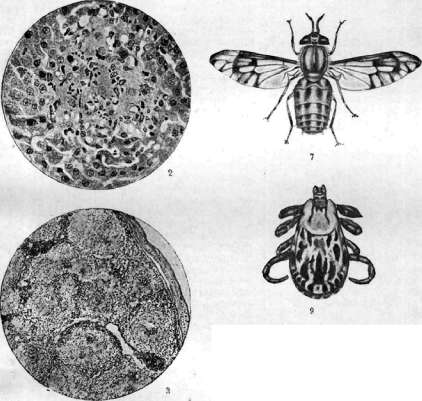
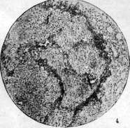
Figure 1. Section from the spleen of a guinea pig that died from acute tularemia two days after infection; a fresh, not yet necrotizing tularemic nodule in the splenic follicle, consisting of polymorphic cells of epithelioid type and polymorphonuclear leukocytes (high magnification). Figure 2. Fresh tularemic nodule in the liver of a guinea pig that died from acute tularemia on the 8th day after infection; among the cells of the granuloma, liver cells in a state of necrobiosis are visible (high magnification). Figure 3. Section from the spleen of a guinea pig that died from acute tularemia on the 1st day after infection; throughout the splenic tissue are scattered numerous non-necrotizing tularemic granulomas, in the centers of which in places there is an accumulation of disintegrating leukocytes; the capsule of the spleen is covered with fibrinous deposits (low magnification). Figure 4. Section from the lymph gland of a guinea pig, regional to the site of introduction of the tularemic strain; death on the 5th day after infection; the tissue of the gland is almost completely replaced by tissue of non-necrotizing tularemic granulomas, in places, especially near the capsule of the gland, with accumulations of nuclear detritus (low magnification). Figure 5. Culture of Bact. tularense. Figure 6. Spleen of a mouse. Figure 7. Fly Hydrophorus discalis (vector). Figure 8. Louse from a water rat. Figure 9. Mite Laelaps echidninus from a water rat. Figure 10. Dermacentor andersoni (vector).
In other organs, mainly in the sinuses of the spleen and in the capillaries of the liver, and even in liver cells, there are numerous accumulations of a huge amount of Bact. tularense. In the spleen, specific tularemic granulomas sometimes develop; there is pronounced congestion of the spleen and a very pronounced karyorrhexis of most follicles. In water rats with T., a predominant lesion of the lungs is noted. In artificially infected domestic rabbits, histological changes in T. are similar to changes in guinea pigs, but granuloma in the lungs is more common. In wild rabbits, the spleen and liver are predominantly affected. The pathological anatomy of T. in humans is little studied. At the site of virus introduction, necrosis and infiltration with polymorphonuclear leukocytes and lymphoid cells develop; soon a purulent ulcer forms. In the tissue of regional lymph glands, small necroses appear, sometimes with fibrin exudation. Along the periphery of the necroses, cells of epithelioid type, sometimes lymphoid type, grow, which in turn necrotize. Later, around the necrotized center, a zone of epithelioid cells with Langhans-type giant cells develops, sometimes with an admixture of lymphoid cells and fibroblasts arranged in a palisade; the entire nodule is surrounded by a connective tissue capsule and very much resembles a solitary tuberculous focus. The healing of necrotized nodules can occur without a leukocytic reaction or from the very beginning polymorphonuclear leukocytes appear in the granulomas. By the end of the month and even earlier, the nodule is surrounded by a connective tissue capsule, on the inner side of which a zone of histiocytes sometimes develops. If the patient quickly died from T. (8th day), the vessels located in the area and near the tularemic granulomas remain unchanged. In a later period of the disease, proliferation of the endothelium, thickening of the walls, and even the development of a histological picture of endarteritis obliterans are observed in them. Due to narrowing and obliteration of the vessel lumen, necroses may form in internal organs. Between the primary ulcer and regional lymph glands under the skin, nodules about 4 millimeters in diameter sometimes develop along the course of the lymph vessels, of the same histological structure as the granulomas in the glands. To the changes at the site of infection entry and in regional lymph glands are added changes in other parts of the body; in the somewhat enlarged and flaccid spleen, in the liver, lungs, adrenal glands, and rarely in other organs, tularemic granulomas of the same histological structure as in the glands develop. In very acute, malignant cases (death on the 4th-8th day), the granulomas are small and numerous, with a predominance of alteration phenomena; in more protracted subacute and chronic cases, the granulomas are few in number and larger, with the size of a pea or more, and with significant development of proliferative processes. In the lungs, in addition to granulomas, tularemic pneumonia can develop. 1 case of tularemic peritonitis and 2 cases of tularemic meningitis have been described; in each of them, infection of guinea pigs with inflammatory exudate (taken from the peritoneum and from the brain) resulted in the development of a typical tularemic picture. Tularemic meningitis in its picture resembles tuberculous meningitis: in the clouded meninges, very delicate necrotizing nodules are visible.
V. Kartasheva. Clinic. The incubation period is 7-14 days; in the overwhelming majority of cases (about 85%) the disease occurs within the first week after infection. Often the incubation period is limited to 2-3 days. According to the clinical course, 3 forms are distinguished: T. glandular, glandulo-ocular, and typhoid-like. At the beginning of the disease, complaints usually consist of pains in all muscles of the body, bones, and joints. The pain is accompanied by stiffness, lethargy, and apathy. In a number of cases, vomiting and nosebleeds are noted at the beginning of the disease. Along with this, mild forms occur, with the onset of the disease in the form of general malaise, weakness, and drowsiness. In such cases, the patient often notes himself the enlargement of lymph glands. The disease is often accompanied by headache and dizziness. 2 clinical symptoms characterize the acute period of T.: remittent fever and enlargement of lymph glands in the glandular form. The enlarged glands are painful during the first week, later becoming painless. In approximately 50% of cases, the enlarged glands suppurate. In natural dissection or puncture of the suppurated glands, it is impossible to find T. microbes either bacteriologically or by infecting laboratory animals. The onset of the disease is usually accompanied by a sharp rise in temperature; the fever lasts 2-3 weeks (Fig. 2). The number of month VI 39 VI VI VI' VI! VII VII 8 UB 10 YN VII 12 VII 14 VII Day of illness b ? 8 38^2 38.0 л л \ 87.0 2 ! J^ f ') -,_1 \ / \ \ \ 36.0 2 \ \ \ 36.0 ! \ ' Figure 2. Temperature curve in tularemia. A remission is often noted after 2-5 days with a subsequent rise in temperature. Daily fluctuations in temperature reach 1-2 degrees. The fever is accompanied by sweating. In severe cases, patients fall into an unconscious state, symptoms of intoxication are sharply expressed, there is febris continua, resembling the curve of acme of abdominal typhus. In cases of subacute course, the initial high rise may give way to subfebrile temperature. In general, the curve in T. is not characteristic for the clinical diagnosis of T.: Glandular form is the main form of T., occurring in 90-100% of cases both in our country and in the USA (in 70% of cases according to Francis). Enlargement of lymph glands is the main symptom of this form of T. Initially, the glands are sharply painful, clearly palpable on superficial palpation; at the end of the 1st week they become tense, then less sensitive, and in the period of convalescence soft, often obviously fluctuating. As a result of painful p. m. e, t. xxxxx. » swelling of the glands, the patient assumes a forced position, e.g. holds the upper extremity in a horizontal position. The lesion of the glands is associated with the place of penetration of the virus. The following table shows the frequency of lesion of T. of various glands. In cases of industrial epidemics, bilateral lesions of glands are often observed with predominant localization on the right side (Hatenever). The enlarged glands reach various sizes, from a bean to a fist of an adult, averaging the size of a chicken and pigeon egg (Hatenever). Sometimes it is possible to observe the formation of papules and pustules at the site of entry gates. When the pustule bursts, an ulcer remains in its place with undermined, raised, sluggantly granulating edges. The size of the ulcer varies from the size of a lentil to a 10-kopek silver coin. T. sometimes gives skin rashes that do not have any specific character. Papules, vesicles, pustules, erythemas or their combinations are observed. The rash appears more often in the second period of the disease, is not painful and does not itch. From the side of the digestive organs, clinical symptoms are not very characteristic. Vomiting occurs in patients with cerebral phenomena. In cases of severe intoxication, the liver is palpable and sensitive on palpation. The spleen is usually unchanged. From the side of the respiratory organs, changes are not frequent, congestion of the mucous membranes, nosebleeds at the beginning of the disease are noted. Organs of circulation are little affected. In the febrile period, tachycardia and muffled heart tones are observed. From the side of the blood, according to Rapoport, in the acute period of the disease, anisocytosis is observed. Hemoglobin gives figures below average. From the side of white blood, leukocytosis is observed, reaching in individual cases 12-24 thousand. Neutrophils give low figures with a moderate regenerative shift; in cases with severe intoxication, phenomena of cell degeneration in the form of round granularity, vacuolization of protoplasm, etc. were noted. Lymphocytosis is noted. An increase in the number of monocytes is observed in approximately 50%. From the side of the nervous system, headache, pain in the lower back, in the extremities, general malaise, drowsiness are noted. In severe cases - meningitis. Francis, describing 24 cases of death from T., notes changes in the meninges in 5 cases. Glandulo-ocular (oculo-glandular) form of T. as a result of penetration of infection through the mucous membrane of the eye occurs extremely rarely. G. reports on 2 cases of this form. According to American authors, the number of cases of this form of T. is 4.7. The mucous membranes of the eyelids and eye are mostly affected. A papule appears, soon turning into an ulcer, acute conjunctivitis develops; from the literature there is 1 case of loss of vision of the affected eye due to perforation of the cornea. Typhoid-like form of T. is usually the result of laboratory infection. The disease begins mostly with chills and high fever, dull headache and pain throughout the body, sweating. From the 3rd day, remission occurs. After some time, the pains intensify, the general condition worsens, the temperature rises again. From the second week, the temperature falls lytically to normal. No local phenomena nor lesions of lymph glands are observed in the typhoid-like form of T. In individual cases, skin rashes are noted. The total duration of the typhoid-like form is 14-21 days, in individual cases it is prolonged to one month and longer. The main clinical symptom of this form is fever. Along with the described picture, clinically mild cases of T. occur, however with a positive agglutination reaction in sufficiently high titers (Hatenever). T. in the USSR has a milder course than in the USA, and almost always ends in recovery without any complications and residual phenomena and without subsequent disability. Lethal outcome occurs very rarely (in our country only 5-6 cases of death have been noted, in the American literature - 40 cases). Repeated cases of T. are observed in rare cases, after 7 and 10 months after the first attack. Bacteriological diagnosis - k. a. Culture of B. tularense directly from the patient cannot be obtained. In the early period of the disease, the diagnosis is made by infecting experimental animals with material in the form of blood, punctate of affected glands, etc. In cases of lethal outcome, the animal is infected with an emulsion of liver and spleen. Dissection of the infected experimental animal is performed with all precautions. During dissection, attention is paid to the condition of the lymph glands near the site of infection. Pieces of liver and spleen are sown on coagulated egg medium. At the same time, smears from different organs, in particular from bone marrow, are prepared. Smears are stained by Giemsa. Passages on subsequent animals are performed either by rubbing the spleen into the skin or infecting it subcutaneously and intraperitoneally. The virus in the organs of dead animals is preserved in glycerine for approximately 40 days. It must be borne in mind that when the corpse cools, ectoparasites leave the host and can cause laboratory infection. Insects are examined after preliminary aseptic treatment. In addition to sowing on egg medium, the contents of the insect require bacterioscopic examination. To infect an animal, an emulsion from crushed insects is rubbed into the skin. Identification of the isolated culture is based on bacterioscopy, study of cultural and pathogenic properties, as well as on the agglutination reaction, which in T. is quite specific. It must be taken into account that serum of T. patients often agglutinates Br. melitensis and B. abortus Bang. In Francis, out of 100 sera of T. patients, 37 had agglutination with Br. melitensis. In most cases, it is noted that the agglutination titer with respect to B. tularense is higher than with Brucella. The same applies to immune rabbit sera. The agglutination reaction with the serum of the patient can be used for diagnosis, starting from the 2nd week of the disease.
Diagnostic titers begin at 1:50, 1:100. Agglutinins reach their maximum height in the 2nd month, after which they begin to decrease. The agglutination reaction can also serve for retrospective diagnosis several years after the disease. Intradermal diagnostic reaction. Rapoport and Birkhovsky prepared a vaccine from cystine agar, killed by heating at 65° for 30 minutes; applying this vaccine intradermally to experimental animals, the authors established a positive reaction in 75% of cases in the form of hyperemia, infiltration, and even necrosis. Experiments by L. A. Levchenko confirmed the positive results of the intradermal reaction in experimental animals. Hatenever, Wolferz, Novikova, and Polumordvinova in 1934 studied the skin test method for early diagnosis under epidemic outbreak conditions. The reaction was performed on 209 people, of whom 131 were patients with T., and the rest were controls with various other diseases and healthy individuals. In all patients with T., the skin test gave a positive result in the form of papule, vesicle, and sometimes pustule formation. In the control group, the skin test gave a negative result, occasionally with slight, quickly passing hyperemia. The intradermal reaction was performed simultaneously with the agglutination reaction. Coincidence of results was established. The skin test gave a positive result at the beginning of the disease (several cases) before the agglutination reaction had become positive. The antigen for the intradermal reaction was prepared by inactivating a culture washed off with physiol. solution with the addition of 3% glycerol, at 60° for 1 hour (Hatenever's method). 0.1 ml of killed emulsion, approximately 25 million microbial bodies, was injected intradermally. In a few cases, the skin test gave a sharply expressed local and mild general reaction. To reduce the local reaction, it is necessary to reduce the dose in further work. The above data allow the use of the intradermal reaction for the diagnosis of T. Prevention and therapy. Measures for personal protection against T. consist of mechanical protection of the skin from the virus (rubber gloves). Francis's vaccine, prepared by heating at 56.5° or by adding 0.1% formalin or 0.5% phenol, did not give positive results in experimental animals. With the help of formalin vaccine and subsequent infection in rabbits, it is possible to obtain a chronically developing infection. The virulent strain used by Francis for vaccination did not give positive results. Hatenever and Levchenko prepared a heated vaccine with 3% glycerol. Infected guinea pigs survived an average of 2-5 months after vaccination. An experiment conducted on a small group of people showed the antigenicity of this vaccine. A weak local post-vaccination reaction is noted. Patients with T. should be advised to remain in bed. Failure to follow the regimen can prolong the period of convalescence. For the treatment of T., salvarsan, iodine preparations, as well as specific serum have been tested. The serum of convalescents in the first experiments did not give positive results. Foshay (1931) published observations on the treatment of T. with goat serum and notes a positive effect. After the application of serum, a decrease in temperature, reduction of lymph glands, and subsidence of general symptoms were observed in patients. The author believes that the therapeutic effect obtained is due to a change in the specific sensitivity of the patient to the antigen. According to Foshay, the therapeutic effect of the serum is even more pronounced when applied during the period of convalescence. l. hatenever.
Related articles
Mentioned in
Cite this page
“TULAREMIA.” Soviet Medical Encyclopedia. English translation of Bolshaya Meditsinskaya Entsiklopediya, 1st ed. (Moscow, 1928–1936), ed. N. A. Semashko. https://sovietmedicalencyclopedia.pages.dev/article/tularemia/