Anthracosis
Historical document, translated for reference. It reflects medical knowledge of the 1920s–30s and is not medical advice.
Summary
Anthracosis is a pneumoconiosis caused by the inhalation of coal dust, leading to its accumulation in the lungs and lymph nodes. The condition is common in urban populations and is particularly severe in coal miners and other workers exposed to high levels of coal dust.
Encyclopedia article (1928–1936)
ANTHRACOSIS, anthracosis (from the Greek anthrax- coal), one of the types of dust diseases, pneumoconioses (see) — the deposition of coal dust in the tissues of the organism. In view of the fact that coal dust is constantly present in the air of living quarters, in the atmosphere of cities and other settlements in a moderate degree not having pathological significance, A. is inherent in all people, beginning in early childhood and gradually increasing with age. In some professions (engineers, stokers, coal miners, blacksmiths, miners) connected with staying in an atmosphere containing a large amount of coal dust, a strong degree of A. can develop, having as a consequence a whole series of pathological changes in the corresponding tissue, and sometimes reflecting on the whole organism. Under such conditions A. must be considered a professional disease. Coal dust in the form of the smallest particles penetrates the organism by inhalation through the respiratory tract (the view of the school of Calmette, assuming the penetration of dust exclusively through the digestive tract, cannot be considered correct). A significant part of the dust particles settles from the inhaled air on the mucous membranes of the nose, pharynx, trachea and bronchi and is removed outward together with the flow of mucus by the movements of the cilia of the ciliated epithelium. Part of the dust penetrates into the lungs and is found here in the form of grains and small rods lying in the alveolar cavities in a free state or, more often, enclosed in phagocytized cells of the alveolar epithelium and macrophages that have emerged from the connective tissue of the lung. These cells with dust particles, designated as "dust cells", can also be found in the sputum. From the alveolar cavities, coal particles enter the lymphatic pathways laid down in the interstitial tissue of the lungs, and here partly are retained, which is the basis of A. of the lungs, partly by the flow of lymph are carried to the regional lymphatic glands (bronchial, bifurcational and tracheal), in which they are deposited, gradually accumulating in an ever-increasing amount (A. of lymphatic glands). A small amount of coal can penetrate further, into larger lymphatic collectors and through them into the blood; moreover, with the accumulation of a large amount of coal in the lungs and lymphatic glands, adhesion of such a mass to the wall of a blood vessel, a violation of the integrity of the latter and the penetration of coal particles directly into the blood can occur. In such cases these particles can be carried by the blood flow and be retained in the spleen, liver, kidneys, bone marrow (A. of these organs). Sometimes the presence of coal is detected in the retroperitoneal lymphatic glands, which is explained by their retrograde penetration here along lymphatic vessels from the bronchial lymphatic glands. - In the initial degree of A. of the lungs, the presence of small black spots, evenly scattered over the pleural surface, is noticeable to the naked eye, which is caused by the deposition of coal in the lymphatic pathways of the pleura at the points of contact of several neighboring lobules of the lung. In the future these spots increase, the uniformity of their arrangement is lost, and the surface of the lungs acquires a spotted gray-black appearance; in addition, the lung tissue on section has a gray tint. Microscopically in these cases one can see the accumulation of coal masses in the form of black lumps, grains and rods in the interstitial tissue of the lung, mainly under the pleura and along the larger bronchi and vessels (see figure). - In places of more significant accumulation of coal usually there is a clear growth and condensation of connective tissue. In the strong degree of professional A. the lungs can be completely black in color and dense in consistency. In such cases a series of very serious sequential changes is observed in the lung tissue; depending on the constant entry of coal dust into the bronchi and alveoli, a state of chronic inflammation (catarrhal bronchitis and desquamative catarrhal pneumonia) develops in the latter; the interstitial tissue of the lung, loaded with coal deposits, grows (cirrhosis of the lungs), as a result of which the alveoli are compressed, decrease in volume, and in places are completely destroyed, giving the picture called "aspid induration of the lungs". Sometimes in the areas of large accumulations of coal their softening and the formation of cavities, cavities, occurs in the lungs, which in old times was called "black tuberculosis" (phthisis atra). - A. of lymphatic glands is expressed in their darkening with an increase in volume and condensation; microscopically the presence of coal particles is found first only in the sinuses, and then in the follicular tissue; in addition, a sequential growth of connective tissue occurs, often capturing the surrounding tissue (periadentitis) and causing adhesion of the glands to neighboring organs. - A. of the liver, spleen, kidneys, bone marrow is often recognized only under a microscope in the form of the presence of coal particles in the elements of the reticuloendothelium and in the interstitial tissue. - If the significance of the moderate, so-called normal degree of A. of the lungs, as well as A. of such organs as the liver, spleen, etc., is negligible, then the pathological role of the strong degree of A. of the lungs is very great. Not to mention the direct significance of chronic bronchitis and pneumonia, it can be noted that the observed cirrhosis of the lungs with a strong degree of A. reduces their respiratory surface, which has as a consequence the oxygen starvation of the organism; in addition, the growth of connective tissue in the lung reflects on the activity of the heart; the closure of the lymphatic pathways of the lungs by coal and their compression by the grown connective tissue promotes the retention in the lungs of various infectious agents, including the tubercle bacillus; moreover, this same state of the lymphatic apparatus of the lungs, with various inflammatory processes in them, makes the resorption of inflammatory exudates extremely difficult and delays the liquidation of inflammation.
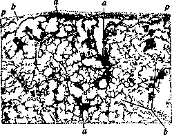
Anthracosis and moderate pulmonary emphysema: a - coal dust accumulations; b - air parenchyma of the lung; p - pleura.
Related articles
Mentioned in
Cite this page
“Anthracosis.” Soviet Medical Encyclopedia. English translation of Bolshaya Meditsinskaya Entsiklopediya, 1st ed. (Moscow, 1928–1936), ed. N. A. Semashko. https://sovietmedicalencyclopedia.pages.dev/article/anthracosis/