Artificial Respiration
Historical document, translated for reference. It reflects medical knowledge of the 1920s–30s and is not medical advice.
Summary
Artificial respiration is a collection of techniques used when natural breathing movements cease, preventing oxygen intake and potentially causing irreversible damage to nerve elements. The article details various causes of respiratory arrest and methods to restore breathing, including reflex stimulation and direct intervention on the respiratory center.
Encyclopedia article (1928–1936)
Artificial Respiration, a collection of techniques applied in cases when natural respiratory movements are absent; in such cases, the intake of oxygen into the organism inevitably ceases, which, due to changes in blood composition, fairly quickly leads to irreversible changes in nerve elements of both the respiratory center and other most important vital centers, and consequently to death. Temporarily replacing natural respiration, artificial respiration at the same time serves to maintain and restore normal excitability of the respiratory center. Relatively frequently, the absence of natural respiratory movements is observed in asphyxia of newborns when, following the cessation of placental blood circulation, adequate excitation of the respiratory center does not occur. In adults, cessation of respiratory movements can be caused by various reasons. 1. Lesion of the respiratory center a) by poisonous substances entering the blood through respiration (e.g., carbon monoxide, illuminating gas, hydrogen sulfide, often chloroform during anesthesia); b) by poisons entering the blood through the digestive tract (most commonly alcohol, opium, morphine, etc.). 2. Sharp decrease in excitability of the higher vital centers due to disturbance of cerebral circulation (e.g., in fainting, traumatic shock, deep exhaustion). 3. Paresis of the muscles of the respiratory apparatus (e.g., in polyneuritis), in particular paresis or paralysis of the diaphragm, and more rarely - spasm of the diaphragm. 4. Difficulty and cessation of air access to the respiratory pathways a) in suffocation, hanging, b) in aspiration of fluid into the respiratory pathways, most commonly in drowning, c) in entry of foreign bodies into the respiratory pathways [see separate table (art. 447-448), fig. 2], d) in narrowing of the glottis by a tumor, coagulated fibrinous exudate, swelling of tissues due to edema, etc. 5. Reflex cessation of functions of the respiratory center (e.g., at the beginning of anesthesia, in cerebral hemorrhage, freezing, electric shock, etc.). In all these cases of respiratory arrest, before proceeding to artificial respiration, it is necessary, if possible, to eliminate the acting cause: 1) to remove the victim from the atmosphere of poisonous gases to fresh air or to a well-ventilated room, to stop anesthesia, to remove the mask; 2) to clear the oral cavity, pharynx and respiratory pathways of foreign substances such as dirt, mucus, silt, water, etc.; 3) to extract foreign bodies from the respiratory pathways; 4) to remove from the stomach (and intestines) introduced poisonous substances by mechanical means, as well as by means of subcutaneous administration of emetics (apomorphine 0.01:10.0, 1 cc3) and laxatives in enema (e.g., 30 g of sodium sulfate in 1/2 glass of warm water). At the same time, it is necessary to free the neck and the entire body from constricting clothing. If foreign bodies cannot be removed or if there is an obstacle in the upper respiratory pathways, tracheotomy must be performed. After all this, and mainly if respiratory arrest occurred due to nervous shock or circulatory disturbance, it is necessary to attempt to reflexively excite the functions of the respiratory center by simpler measures such as spraying with cold water, pouring cold water over the chest, slapping the body and rubbing the skin (in newborn asphyxia, skin rubbing is not applied), tickling the nasal mucosa, bringing irritating substances to the nose (e.g., ammonia, menthol preparations). If this does not help, attempts are made to act directly on the respiratory center by using repeated subcutaneous administration of lobeline (0.01 for adults and 0.003 for children) every 10 minutes, which is a specific excitant of the respiratory center, and other excitants such as camphor oil (in 10- or 20% solution) 1-2 cc3, Cardiazol in 1% solution subcutaneously or in enema, ether (1 cc3), caffeine in 10- or 20% solution (1-2 cc3), adrenaline 1 cc3 of 1:1,000 solution subcutaneously - or as a last measure - directly into the cavity of the left ventricle. In any case, the choice of method for restoring respiration depends on the totality of phenomena observed in the victim. Thus, during anesthesia, if cyanosis appears but pulse filling remains normal, then to restore respiration it is sufficient to remove the mask and vigorously protrude the lower jaw forward by the Esmarch-Heiberg method or to carefully pull the tongue forward. If respiratory arrest is accompanied by a violet tint to facial cyanosis and the pulse becomes small, it is necessary to resort to rhythmic pulling of the tongue by Laborde's method; if this does not help, then proceed to artificial respiration by one of the methods described below. The most dangerous condition requiring immediate application of the most vigorous methods of artificial respiration is one in which, following respiratory arrest, the face becomes very pale and the pulse is not palpable at all. Protrusion of the jaw by Esmarch-Heiberg: standing behind the patient, grasp his head with both hands in such a way that the thumbs press down on the forehead, the index fingers support the upper cheekbones, and the middle fingers, located behind the ascending branches of the lower jaw, protrude it forward, until the lower row of teeth
is positioned in front of the upper row. The head should be bent back as much as possible. This method is used when there is no obstacle in the pharynx and when the tongue does not fall back and block the entrance to the larynx. If the tongue falls back, it should be pulled forward with a handkerchief or by other means. In cases where protrusion of the jaw is impossible or insufficient, the Sylvester method is used: the patient lies on his back, the operator kneels astride the patient's thighs, places his hands on the lower ribs, leans forward, and with straight arms alternately presses on the lower ribs, first on one side, then on the other, with a rhythm corresponding to natural respiration. This method is particularly suitable for cases of drowning. The Schaefer method: the patient lies on his stomach, the operator kneels astride the patient's thighs, places his hands on the lower ribs, leans forward, and with straight arms alternately presses on the lower ribs, first on one side, then on the other, with a rhythm corresponding to natural respiration. This method is particularly suitable for cases of drowning. The method of mouth-to-mouth respiration: after clearing the oral cavity and pharynx, the operator places his mouth on the patient's mouth and blows air into the lungs, then removes his mouth and allows the air to escape. This method is simple and effective but requires practice. The method of using the apparatus for artificial respiration: various apparatuses are used, such as the Dräger apparatus, which allows for controlled ventilation with oxygen or air. The choice of method depends on the specific circumstances and the availability of equipment. In all cases, artificial respiration should be continued until natural breathing is restored or until it is certain that the patient is dead. The duration of artificial respiration may vary from several minutes to several hours, depending on the cause of respiratory arrest and the patient's condition. It is important to monitor the patient's vital signs, such as pulse, breathing, and pupils, during artificial respiration. If the pulse is absent, external cardiac massage should be performed simultaneously with artificial respiration. In some cases, medications such as adrenaline or atropine may be administered to stimulate the heart and respiratory system. The article emphasizes the importance of prompt and correct application of artificial respiration techniques to save lives and prevent brain damage due to oxygen deprivation.
Figure t of the upper (fig. 1). Rhythmic pulling of the tongue according to Laborde as a method of reflex stimulation of the respiratory center is very effective; it is performed as follows: the tip of the tongue is grasped a) with the thumb and index finger wrapped in gauze, or through a towel, handkerchief, etc., or b) with a blunt Esmarch or Kusko tongue depressor in the form of locking forceps with oval or triangular branches, or c) a Mikulicz tongue depressor resembling American, bent at a right angle, bullet forceps, or ordinary bullet forceps, or finally d) a loop of strong sterile thick silk thread passed through the front part of the tongue with the aid of a needle. If the jaws are closed, they must be forcibly opened and held in this position by inserting a hard object between the teeth, such as a cork, a piece of smooth stick, a knife handle, etc. Heister's or Roser's mouth gag spreaders can also be used. While continuously holding the captured tongue, fairly strong pullings are produced every 3-4 seconds in accordance with the normal rhythm of breathing, so that not only the front but also the back part of the tongue is pulled forward. If resistance begins to be felt, this is a sign of the impending resumption of breathing. Then the patient makes several swallowing movements, which are often followed by noisy, gasping breathing. Sometimes rhythmic pulling of the tongue excites respiratory movements very quickly, sometimes it is necessary to continue it for a very long time without losing hope of reviving the victim. Artificial respiration in the proper sense of the word consists of the following methods: mechanical action on the chest; electrical stimulation of the respiratory muscles; introduction of atmospheric air or pure oxygen into the respiratory tract by insufflation; introduction of pure oxygen under the skin. A. Mechanical action on the chest. This method is based on the alternate expansion and compression of the chest, whereby air is rhythmically passively aspirated into the respiratory tract and lungs, and then actively expelled from them, or vice versa. At the same time, since blood circulation still continues, it is possible to a certain extent to maintain respiration and thus prevent irreversible biochemical changes in the nerve cells of the respiratory center, leading to their death. But at the same time, artificially induced respiratory movements, by stimulating the peripheral endings of the respiratory branches of the vagus nerve, reflexly excite the respiratory center, which can lead to the restoration of its functions; as a result of this, the restoration of natural impulses that stimulate in their entirety the movements participating in the act of breathing is obtained. When applying mechanical methods of artificial respiration, it is necessary to take into account the presence of fractures of the ribs or upper extremities and other injuries to the body, since respiratory arrest was caused by attempts at violent death, as occurs in hanging, drowning, suffocation, falling from a height, etc. In all cases, it is necessary to prevent the tongue from falling back, for which it must be pulled out and held in this position by the methods described above. One must always keep in mind that the excitability of the respiratory center can be affected very deeply, and therefore to revive the victim it is necessary to apply one or another method of artificial respiration very persistently, sometimes for many hours and even entire days. Methods of artificial respiration by mechanical action on the chest have been proposed in considerable numbers. 1. Methods of Schultze and Prochovnik (fig. 2) - the most common methods for asphyxia (see) in newborns. The first can hardly be considered completely safe and is therefore now abandoned by many. 2. The method of Silvester (Silvester-Brosch) - one of the most
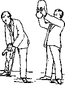
Figure 2.
common methods for performing artificial respiration in adults: the victim lies on his back, and if artificial respiration is performed at the scene of the accident, he may be on the ground, floor, etc., and during anesthesia - on the operating table. The head is tilted back slightly (the designations, as in anatomy, refer to the vertical position of the person), for which a hard pillow or the first thing that comes to hand (e.g. a rolled-up blanket, clothing, etc.) is placed under the arms. The operator stands at the head end of the victim (if he is on the floor - on his knees). The victim's arms are grasped above or below the elbows, whichever is more convenient; with circular movements, they are drawn away from the chest upward so that the shoulders lie next to the head, and held in this position for about 3 seconds to cause expansion of the chest - the phase of inspiration; then the arms are drawn downward and strongly pressed against the lateral wall of the chest for 2 seconds to cause its compression - the phase of expiration. This procedure is methodically, unhurriedly repeated 12-15 times per minute (figure 3). This method, in general not particularly complicated, is very effective, however, it requires considerable effort and is therefore tiring for the operator, especially without practice; the application of excessive force can cause traumatic injury to the brachial plexus in the victim. The supine position prevents the expulsion of mucus, water, etc. from the respiratory tract, is dangerous in vomiting, does not prevent the tongue from falling back, which causes the epiglottis to close. Therefore, it is often necessary to pull out the tongue and press it against the chin with a towel or by other means to keep it pulled out. 3. The method of Pacini. The position of the victim and the operator is the same, but instead of the forearms, the operator grasps the victim's shoulders so that the thumbs are placed in front of the head of the humerus in the axillae, and the other fingers of each hand hold the scapulae. Alternately, first pulling both shoulders upward then lowering them and pressing against the chest, the chest is expanded and narrowed. 4. The method of Schiiller-Roux. The victim lies on his back with the upper part of the torso raised (as in the previous methods), the legs are held bent at the knees and hip joints. The operator
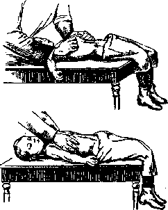
Figure 4. (at the head or on the left side of the victim) grips both of his costal arches with the 4 bent fingers of both hands and alternately pulls them upward and outward as much as possible to mechanically expand the chest—the inhalation phase—and then presses them, pushing with the palms, toward the abdominal cavity—the exhalation phase (Figure 4). 5. Howard's method, most applicable mainly to those who have drowned, choked, etc. 1st act. The victim is laid face down so that the head is bent forward, with the forehead resting on the forearm of one or the other arm, which is brought upward and bent at the elbow at a right angle; thanks to this, the mouth and nose do not come into contact with the surface of the ground, floor, or bed and remain free. A hard pillow or something of the sort is placed under the upper part of the abdomen. The operator, standing over the victim on their knees, so to speak, astride, facing the back of the victim's head, presses strongly but slowly and evenly with their palms on the victim's back, predominantly on the left side at the level of the stomach, with the goal of squeezing out of the stomach and respiratory tract the liquid swallowed and aspirated through the mouth and nose. Then the operator, without losing contact with the chest, leaning back with a sharp movement, temporarily ceases pressure, and after a few seconds resumes it, etc. Both manipulations are performed repeatedly to remove as much of the swallowed liquid as possible. 2nd act. The unconscious person is quickly turned onto their back, with the middle part of the torso being raised, the neck extended, the head tilted backward, and the arms crossed over the head (in case of fractures of the arm bones, they are laid along the chest). To prevent the tongue from falling back, an assistant, positioned at the head of the unconscious person, energetically pushes their lower jaw forward. Respiratory movements are performed in the following manner. The operator again gets on their knees over the unconscious person, so to speak, astride, at the level of their pelvis, and places both hands on the lower part of their chest so that the thumbs are near the xiphoid process, while the other fingers lie on the lower intercostal spaces (costal arches). Resting their elbows against their own sides and leaning forward, they gradually press more and more with the weight of their body onto the chest of the unconscious person, reducing its volume—the exhalation phase. Then, quickly straightening up, they cease this pressure, giving the chest the opportunity to expand by virtue of its natural elasticity—the inhalation phase (Figure 5). Howard's method, as quite expedient, is one of the most common. However, it must be kept in mind that the 1st act—emptying the stomach and respiratory tract—can take up too much precious time, and the 2nd act is not devoid of the negative aspects of the Sylvester method.
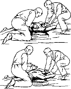
Figure 5.
as in the Howard method. The operator positions themselves over the victim astride, as in the 1st act of the same method, standing on their knees and resting both palms of their outstretched arms on the victim's back outward from the longitudinal spinal muscles, and for 2-3 seconds exerts as much pressure as possible on the lower part of the chest, using the weight of their body for this; thus, the exhalation phase occurs (Figure 6). During the temporary cessation of pressure, for which the operator leans back without removing their palms from the victim's chest, the inhalation phase occurs due to the elasticity of the false ribs. The Schaefer method
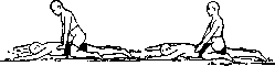
Figure 6.
can be considered one of the best, as it is very simple, easily executable, not very tiring, and demonstrative, thanks to which a completely untrained person can, at first glance, set to work to replace exhausted medical personnel. 7. The Marshall Hall method is also applied mainly to those who have drowned. The victim is laid face down on a hard support, as in the previous method. Then they are turned onto their side to free the chest from pressure and allow it to expand, and again onto their stomach to produce compression of the chest, with the possibility of thereby squeezing abnormal contents out of the respiratory tract. 8. The Flashar method. Two towels folded in width are placed under the chest of the unconscious person lying on their back, at the level of the nipples, with both ends of one towel brought to the right and the other to the left of the torso. Then, gripping them in pairs from one side and the other, two people pull them strongly, as a result of which the lower elastic parts of the chest are compressed and held in such a position for about 2 seconds—the exhalation phase; after that, the tension of both towels is simultaneously relaxed, thanks to which the chest expands by virtue of its natural elasticity—the inhalation phase. 9. The apparatus for Artificial Respiration by R. A. Panis was proposed by the author in 1923 for the attention of the Paris Medical Academy; it is constructed as follows: a metal quadrangular frame about 1/2 meter long, supported in a somewhat inclined position to the horizon by two wedge-shaped stands, serves to accommodate
special movable supports with notches; for supporting the forehead, there is a separate device. By means of the downward movement of a metal lever in the form of a rectangle open on the bottom side, which connects to the aforementioned frame, the chest is gradually pressed against the frame on which it lies, and at the same time, the supports for the shoulders are flattened. Thus, exhalation occurs. The same lever, rising under the influence of 2 springs and the elasticity of the chest, again lifts the shoulders and pulls them to the sides and backward, as a result of which inhalation occurs (Figures 7, 8). The lowering and raising of the lever, alternating with each other, occur very smoothly, and, repeating 15-20 times per minute, cause Artificial Respiration of significant depth. Working with this apparatus is simple and makes it accessible to everyone, even untrained people, while at the same time sparing them from fatigue. Experience with this apparatus has proven its complete suitability and justified the hopes placed in it. B. Electrical stimulation of the respiratory muscles is carried out mainly by means of faradization of the phrenic nerve (n. phrenicus). One of the poles of the induction apparatus is applied to the middle of the outer edge of the sternocleidomastoid muscle, the other to the base of the chest at the level of the attachment of the diaphragm crura. The intermittent current is switched on at equal intervals 15-18 times per minute. It is possible to perform electrical excitation of both phrenic nerves simultaneously, and some authors
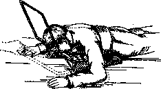
Figure 8.
advise placing both poles of the apparatus on the outer edges of both sternocleidomastoid muscles. After each inhalation movement produced by the current, it is necessary to compress the chest with both hands to induce exhalation. Villette recommends bilateral faradization of the pectoral muscles (mm. pectorales) with electrodes placed at the level of the middle third of the pectoralis major muscle, with inhalation immediately following, ending within 2 seconds. By removing one of the electrodes, exhalation is obtained. C. Insufflation. The simplest method, which was used even in ancient times but has recently been abandoned, is direct mouth-to-mouth air insufflation. This method is unhygienic and rarely achieves its goal. However, in cases of asphyxia of newborns, insufflation is still used, but for this, insufflators with a removable laryngeal tip, having the shape of a cone flattened on the sides, are used. This tip is carefully inserted into the larynx, following the soft tissue of the left index finger, to the posterior surface of the arytenoid cartilages, at the level of which it is lowered slightly, and it easily penetrates into the larynx. Insufflation is performed by squeezing a pear-shaped bulb, the capacity of which corresponds to the average lung capacity of a newborn. Insufflation must be performed unhurriedly, very carefully, to avoid rupturing the pulmonary alveoli. The penetration of the tube by mistake into the esophagus instead of the larynx makes itself known by the gurgling sounds of the returning air. Planchu suggested using for insufflation a thin Nelaton catheter No. 12, which is inserted with the help of a mandrel having the appropriate curvature. The catheter is connected to the longer end of a T-shaped tube; one of the short ends is connected to a bulb, and the other short end to a manometer. Instead of air, especially in cases of poisoning by gases that have a special affinity for hemoglobin, such as carbon monoxide, it is expedient to use the insufflation of pure oxygen. For the introduction of oxygen, several methods have been proposed, including the method of introduction through a tracheostomy fistula (Rosenthal). In hospital conditions, one can use with great success the introduction of oxygen under pressure from a cylinder with the help of apparatus for mixed anesthesia (Roth-Dräger). The best method, in the opinion of Maisonnet, is the method described by Nicloux and Legendre with the help of a hard metal
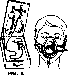
Figure 10.
Nicloux mask: a metallic mask (Nicloux, Legendre) which is placed over the nose and mouth and, thanks to a special pneumatic device, maintains hermetic contact with the skin; O2 is introduced into the respiratory tract from a special inhaler with a balloon (figs. 9 and 10). This apparatus is equipped with the Paris fire brigades. The apparatus gives excellent results in a very short time. It must be taken into account that after the restoration of natural breathing by mechanical means, especially in advanced cases, it is often very useful for quite a long time to maintain the oxidative processes in the body at the proper level by inhaling pure oxygen. G. Subcutaneous introduction of O2 can also be extremely useful for restoring the normal vital activity of the respiratory center, but for this it is necessary to introduce from 1 to 11/2 liters of pure O2 for approximately 20 minutes. This amount is injected in two doses in the area of both buttocks using special devices, for example the Lian apparatus. In the absence of such devices for this purpose, one can use the apparatus for artificial pneumothorax, and the injection needle for introducing O2 under the skin must be significantly thinner. It is also possible to use the Poten apparatus; in view of the presence of hermetic cocks and a pump, with its help it is possible to remove air from the bottle and fill it with O2, and then, by changing the direction of air flow in the pump and opening and closing the corresponding cocks of the inlet and outlet tubes, one can connect the bottle with the oxygen cushion and pump O2 under the skin.
Related articles
Mentioned in
Cite this page
“Artificial Respiration.” Soviet Medical Encyclopedia. English translation of Bolshaya Meditsinskaya Entsiklopediya, 1st ed. (Moscow, 1928–1936), ed. N. A. Semashko. https://sovietmedicalencyclopedia.pages.dev/article/artificial-respiration/