Hyperkinesis
Historical document, translated for reference. It reflects medical knowledge of the 1920s–30s and is not medical advice.
Summary
This article defines hyperkinesis as a broad group of involuntary, excessive movements resulting from disorders of the striatal system. It classifies these movements into pyramidal and extrapyramidal types, detailing specific conditions such as chorea, athetosis, and various forms of epilepsy.
Encyclopedia article (1928–1936)
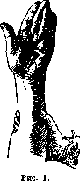
HYPERKINESIS (from Greek hyper-excessive and kinesis-movement), excessive, superfluous movements, according to previous terminology "motor phenomena of irritation," a vast group of involuntary movements, depending for the most part on disease of the striatal system (see Basal ganglia). Muratov (1899) and Bonhoeffer first proved the extrapyramidal origin of choreatic hyperkinesis, linking it with a lesion of the superior cerebellar peduncles (brachia conjunctiva). After the works of Cécile Vogt and Oppenheim on the pathological anatomy of double athetosis (1912), Alzheimer (1911) on the pathological-anatomical substrate of chorea, and Wilson (1912) on lenticular syndrome, the doctrine of striatal hyperkineses began to develop rapidly, and at the present time there is already a large amount of factual material, which has especially grown after the epidemic of encephalitis (see). Organic hyperkineses are divided into pyramidal and extrapyramidal. The former include: 1. A seizure of general epilepsy, depending on diffuse-irritative processes in the cortex. 2. A seizure of cortical, or Jacksonian epilepsy, depending on local, focal cortical irritation; some data suggest that not only cortical but also subcortical apparatuses take part in the genesis of the seizure. 3. Kozhevnikov's epilepsy, or prolonged local clonic convulsions (epilepsia partialis continua), which is a special modification of Jacksonian epilepsy. Tonic spasms of comatose patients and some forms of hypertonia and hyperkineses developing with fresh and massive foci in the hemispheres are also attributed to irritation of the pyramidal tract; however, these spasms must for the most part be interpreted as an expression of increased excitability of spinal centers. From extrapyramidal hyperkineses, the following individual types can be distinguished. Chorea (chorea) represents a motley series of non-rhythmic and highly varied, for the most part rapid movements, each of which is similar to a voluntary contraction, as a result of which patients often produce the first impression of capricious, restless people performing superfluous grimaces and movements. These movements are not limited to any specific muscle group but appear now in one, now in another part, but sometimes are localized in one half of the body (hemichorea). With strong development, choreatic movements hinder speech and phonation and make it impossible to properly hold objects and even stand and walk. Muscle tone is usually lowered, tendon reflexes are preserved (sometimes even lowered); deep atonia of the musculature with the absence of reflexes can simulate true paralysis (chorea mollis). Choreatic movements are observed in chronic progressive sporadic chorea (of arteriosclerotic origin), in chronic progressive hereditary Huntington's chorea, in infectious diseases of the nervous system (Sydenham's chorea minor, or "St. Vitus' dance," as well as in other infections—diphtheria, choreic form of epidemic encephalitis) and often as a focal symptom (usually in the form of hemichorea) with foci in the brain (necrosis, tumors, etc.). Experimentally, chorea has been obtained in mice upon poisoning them with diphtheria toxin (Levy). At the basis of degenerative and toxico-infectious chorea lies a disease, apparently, of the small cells of the neostriatum; focal chorea is also observed in many other localizations (cerebellum, its connections with the red nucleus and through the red nucleus with the thalamus, etc.). Large choreatic movements (so-called hemiballismus) have been observed with foci in the corpus Luysi. Athetosis (athetosis) is the name given to convulsions that are significantly slower, more powerful, and stronger. They are more monotonous and involve predominantly the distal parts, more often of the upper limb. The muscles are usually strongly developed, the joints often allow passive extension of an abnormally large volume, as a result of which the fingers often enter a position of hyperextension (see Figure 1). The spasm is so strong that it is often impossible to overcome it passively. Sometimes athetotic movements of the fingers are replaced for a time by a more persistent convulsion. Often this so-called mobile spasm (spasmus mobilis) is expressed in the proximal parts of the limb, while the phenomena of true athetosis are in the hand and fingers. Athetotic movements in the muscles of the face are not uncommon, especially the platysma myoides and other muscles of the neck. Athetosis has a twofold origin: either it gradually develops after a suffered hemiplegia (post-hemiplegic hemiathetosis), most often in childhood (but also in adults), or from the very beginning, voluntary mobility turns out to be strangely disordered.
Bilateral athetoses, congenital or acquired in early childhood, can subsequently remain stationary (Vogt-Oppenheim type); in other cases, they exhibit a progressive course resulting in "extrapyramidal rigidity." Bilateral idiopathic athetosis, the so-called athetose double, is usually observed as a familial disease. In the majority of cases of athetosis, various diseases of the corpus striatum have been discovered. For stationary congenital double athetosis, C. Vogt and Oppenheim established a specific anatomical substrate in the form of the so-called "marbled state" (status marmoratus, état marbré) of the striatum, clearly visible on preparations stained for myelin. In cases resulting in rigidity, degenerations in the pallidum and substantia nigra have also been observed. At the basis of post-hemiplegic hemiathetosis may lie foci in the corpus striatum, and perhaps in the cerebellum, its dentate nucleus, its superior peduncles, the red nucleus of the tegmentum, and in the thalami. However, it is not possible to distinguish athetosis from chorea in all cases. Convulsions possessing mixed signs of both hyperkineses are encountered. This includes Rossolimo's cerebral amiotaxia, as well as "choreo-athetosis." This term is not particularly successful, and it has been used too widely; thus, choreo-athetoses with foci in the thalamus were often described as one of the components of the so-called thalamic syndrome (see). However, here we are for the most part dealing with a hyperkinesis of a special kind: the so-called "thalamic hand," when left to itself, performs a series of slow involuntary movements in the wrist and finger joints, mainly in the metacarpophalangeal joints, and eventually freezes in a distorted dystonic posture, usually always the same for each given patient. Apparently, the simultaneously observed hemianesthesia, or hemiataxia, is of significance here.
Tremor belongs to the most frequent forms of hyperkinesis. In addition to numerous types of tremor characteristic of various toxic-infectious and autotoxic processes (Basedow's disease, alcoholism, mercury poisoning), various neuroses, senile and hereditary tremor, there are known various types of tremor in more localized organic diseases of the brain. For a long time (Charcot), two clinical forms of tremor have been contrasted: Parkinsonian, characteristic of paralysis agitans, and intention tremor, characteristic of multiple sclerosis. The former continues during complete muscular rest, rhythmically and monotonously (pill-rolling or bread-ball-rolling movements); active movements have the property of inhibiting it for some time. The latter disappears at rest, appears during active movements, is coarser and more sweeping, and is often combined with ataxia, or rather, asynergy. Often both these types are mixed with each other. The question of the localization of different types of tremor cannot yet be considered resolved. It must be thought that tremor occurs with damage to two different systems: on the one hand, the corpus striatum and its connections, and on the other, the cerebellum, its connections with the nucleus ruber, the nucleus ruber itself, and its connections with the thalamus. It is possible that striatal tremor corresponds predominantly to the Parkinsonian type, and cerebellar tremor to the intention type. Within the striatal system itself, it seems that damage to the so-called neostriatum can be expressed by tremor, while damage to the pallidum does not cause it. Paralysis agitans, in which, as the disease progresses, tremor is gradually replaced by rigidity, from this point of view should correspond to the gradual transition of the process from the neostriatum to the palaeostriatum. However, with damage to the substantia nigra, a completely typical Parkinsonian tremor can also be observed. Lightning-fast, single muscle contractions are called myoclonus (myoclonia). This is a collective group uniting several apparently heterogeneous types. In the so-called paramyoclonus multiplex of Friedreich, as well as in the myoclonus epilepsy of Unverricht-Lundborg, the matter concerns rapid, incessant, sometimes fascicular muscle contractions that do not lead to any significant displacement of the given limb segment in space. The spasms affect various muscles. Myoclonus epilepsy is combined with epileptic seizures and can end in extrapyramidal immobilization. In myoclonus, the appearance of peculiar round inclusions, similar to corpora amylacea, has been described, especially in the cells of the thalami, red nuclei, substantia nigra, and even more strongly in the nucleus dentatus cerebelli (Lafora). Hunt found atrophy of the dentatus in the dyssynergia cerebellaris myoclonica described by him. Under the name myoclonus, somewhat different conditions have also been described, where a sudden muscle twitch led to a corresponding movement of the limb or even made standing and walking difficult. Transitions between myoclonic spasms and chorea (chorea electrica) are observed. In other cases, myoclonus is complicated by a syndrome of torsion dystonia. In most cases of myoclonus, the matter concerned pronounced familial diseases. Symptomatological classification within this large group is not yet finished. From myoclonus, one should distinguish the so-called myorrhythmia (myorrhythmia Marinesco). For the first time, this type of hyperkinesis was established in epidemic encephalitis and was described by various authors under different names (localized spasm, myoclonus, hemiclonia, extrapyramidal tic, etc.). This is a sometimes faster, sometimes somewhat slower, but always strictly localized spasm, constantly recurring with a certain rhythm (from 6 to 120 times per minute) in a completely specific muscle or muscle group, regardless of the state of rest or movement, and also regardless of the posture of the limb; the spasm is very persistent and in severe cases continues even during sleep. The most diverse muscles can be affected (most often the facial musculature and the musculature of the anterior abdominal wall, but also the muscles of the limbs, soft palate, and masticatory musculature). Spasmodic hiccups, characteristic of some subspecies of epidemic encephalitis, are obviously akin to this myorrhythmia. Localizing in the muscles of the chest and in the diaphragm, myorrhythmia can cause the so-called "respiratory tic of encephalitics." Often, myorrhythmia is observed simultaneously in different muscle groups, with a special rhythm for each group. The spasm is characteristic of the acute periods of the algio-myoclonic form of epidemic encephalitis and slowly regresses in the further course. Its anatomical substrate is likewise sought in damage to some parts of the extrapyramidal system, and perhaps also the brainstem. Akin to myorrhythmia is the somewhat faster-paced, so-called Klin-Pfeiffer spasm in the muscles of the soft palate, pharynx, larynx, but partly also in the region of the facial nerve, observed several times in processes in the cerebellum related to damage to the nucleus dentatus. This same syndrome was described by French authors under the name "nystagmus of the soft palate." The spasm was observed mainly with pontine foci, perhaps specifically damaging the central tegmental tract, but sometimes also with more distant localizations (dentatus, olives). Torsion spasm (torsion dystonia, dystonia musculorum deformans, dysbasia lordotica progressiva) is a peculiar hyperkinesis consisting of clonic-tonic, very powerful involuntary contractions leading to strong rotational movements of the limbs and especially the spine, predominantly expressed during walking (see Figure 2). The alternation of spasmodic, voluminous, predominantly lordotic curvatures of the spine gives the gait of these patients a peculiar character (camel gait). Torsion spasm is observed as the main symptom of the disease of the same name, described by Ziehen (1908) and Oppenheim (1911). Anatomical studies in this disease, still very scarce, have revealed in several cases the same substrate as in hepatolenticular degeneration. However, both clinically and pathologically-anatomically, it is completely
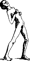
Figure 2.
There are no grounds yet to equate this disease with other forms of hepatolenticular degeneration. The syndrome of torsion spasm can also be observed not infrequently in chronic epidemic encephalitis. More localized spasms of the spinal muscles, which lie at the basis of many cases of spasmodic torticollis and similar conditions that were previously classified in the group of tics, must also be included in the group of torsion dystonia. Alongside these elementary hyperkineses, there exist hyperkineses that are more complexly coordinated, expressed in the contractions of a larger group of synergistic muscles, which brings them closer to voluntary movements. Included here can be the tonic spasm of gaze, not infrequent in epidemic encephalitis, which develops in the form of separate attacks, often combined with an involuntary turning or lifting of the head, as well as attacks of uncontrollable running and uncontrollable laughter. The localization of these complexly coordinated hyperkineses has not yet been clarified. Under the name of striatal or extrapyramidal epilepsy, attacks of general convulsions of extrapyramidal origin are described. They are polymorphic and apparently characterized by a predominance of tonic convulsions. The study of this interesting syndrome is only just beginning. The origin of violent gesticulation and other so-called parakineses remains unclear to us. By this name is understood a peculiar intensification of higher coordinated motor impulses. The affected limb performs a series of violent gestures (e.g., the patient is constantly picking at the blanket with their fingers or rubbing their fingers against each other). Usually, patients of this kind can keep the affected limb motionless; however, involuntary gesticulation appears immediately as soon as the patient diverts their attention from the affected limb. The symptom most often occurs alongside phenomena of mild hemiparesis and certain defects in the intellectual sphere and can hardly be regarded as a true striatal hyperkinesis. Also, the localization of the symptom of involuntary convulsive grasping described by Yanishevsky (frontal lobes? striatum?) remains not entirely clear. Undoubtedly, in the motley mass of hyperkineses, there exist, besides those listed, quite a few types that have been insufficiently studied. Thus, in this area, not everything can be considered fully clear yet. For example, approximately the same territories (mainly the corpus striatum and the efferent system of the cerebellum) turn out to be affected in heterogeneous hyperkineses. This forces one to think that here not only localization is of importance, but also the nature of the pathological process, which is not the same in different cases. Thus, the predominant death of small cells of the striatum apparently causes not athetosis, but chorea. The greater or lesser prevalence of the process (e.g., greater or lesser involvement of the pallidum itself, as well as the varying intensity of the process) can in turn modify the clinical picture. Finally, in striatal lesions, the different age of the patients is apparently also of great importance. Along with the main mass of hyperkineses, which depend not on direct irritation but on the disinhibition of the function of the corresponding centers, the existence of true irritative extrapyramidal hyperkineses (for example, myorhythmia?) is also possible. Finally, a combination of phenomena of irritation and loss in each case is possible. All this makes it very difficult to establish an exact clinico-anatomical parallelism in this area, especially since the number of cases studied in detail by modern histological methods is still very small. Unlike organic hyperkineses, functional hyperkineses possess all the features of suggested or autosuggested hysterical symptoms. Hysterical tremor is encountered most often. Diverse psychogenic tics also belong here. However, the differentiation of organic hyperkineses from functional ones is perhaps far from as simple as is the case, for example, with regard to paralyses: organic hyperkineses can be significantly variable in their course and, for example, in myoclonus, they sharply intensify under the influence of sensory stimuli or the psychic state of the subject (the so-called 'sensory-clonic' and 'psycho-clonic' reactions of Lundborg). Not infrequently, extrapyramidal hyperkineses can even show some improvement under the influence of psychotherapy. Many of the organic hyperkineses currently being studied were previously regarded as psychogenic hysterical symptoms.
Related articles
Mentioned in
Cite this page
“Hyperkinesis.” Soviet Medical Encyclopedia. English translation of Bolshaya Meditsinskaya Entsiklopediya, 1st ed. (Moscow, 1928–1936), ed. N. A. Semashko. https://sovietmedicalencyclopedia.pages.dev/article/hyperkinesis/