Electrical
Historical document, translated for reference. It reflects medical knowledge of the 1920s–30s and is not medical advice.
Summary
This article discusses the electrical properties of the brain's cortex, focusing on bioelectrical currents and their relationship to different cortical fields. It describes how these electrical potentials change in response to stimuli and their significance in understanding brain function.
Encyclopedia article (1928–1936)
Electrical currents, in view of the graphic representation of these currents, have enormous significance for modern knowledge about the structural features of the cerebral cortex, the so-called architectonics of the brain. If until very recently all researchers could only speak about the electrical potentials of the brain when currents were taken from 'two poles of the brain' or two 'areas' of the brain, then modern knowledge about the architectonics of the brain allows one to approach much more closely the understanding of the connections and relationships of these bioelectrical currents with the individual architectonic fields of the cerebral cortex n/v-^vVVA/Vv-' 1»*ЛЛ(/Г^Ал*\M'Ч->^^
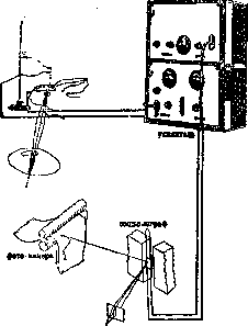
Fig. 7. Diagram of amplifying device for recording bioelectrical phenomena in the cerebral cortex.
with the individual architectonic fields of the cerebral cortex, which opens up new possibilities for penetrating into the dark areas of brain activity. Works in this direction have begun in recent years at the German Brain Institute. At present, the Moscow Institute has not only mastered the appropriate equipment and techniques but has also achieved very important results in this field. Sarkisov and Livanov succeeded in showing that the individual architectonic fields, mentioned above, such as the motor, visual, and auditory areas, have their own characteristic electrical currents for a given area, the so-called intrinsic currents of the cortex (Fig. 8). Furthermore, the research of these authors showed that with appropriate external stimuli, these intrinsic currents produce changes. For example, if intrinsic currents are taken from the visual area nAaM/WVnAn/wWWW"*^--*"i/w"-y-v^^-v/v/ 1 Fig. 9. Action currents of area striata with light stimulation of the eye (1,2); action currents of the auditory area (3). of the cerebral cortex and an external stimulus is immediately applied to the eye with light, then at the moment of applying the stimulus, a change in the curves immediately occurs, which is graphically recorded on an automatically operating camera (Fig. 9). Even more striking is the fact that an external stimulus applied to the visual area of the cortex is not indifferent to the neighboring field, because the intrinsic curves of this neighboring field decrease significantly in amplitude (Fig. 10). This fact has extremely great importance in understanding the mechanisms of cerebral cortex activity, in understanding the relationships of individual areas of the cortex in one or another act. If until now we could, on the basis of the fundamental works of the school of I. P. Pavlov, as well as the works of A. A. Uktomsky and others, speak of a number of important positions about the basic mechanisms of the activity of the cerebral cortex, then undoubtedly the method of capturing and recording bioelectrical currents gives some objective expression of these mechanisms. Along with these data, the study of the nature of the bioelectrical phenomena of the cerebral cortex itself is important - the establishment of a number of regularities about the nature of the curves obtained, their relationships with individual areas of the cerebral cortex, etc. Fig. 10. Intrinsic currents (regio motorica area agranularis) decrease with stimulation of a neighboring field ('); intrinsic currents of regio parietalis change (decrease) with stimulation of a neighboring field area striata (г). Kornmüller noted the position that those areas of the cerebral cortex that are characterized by the presence of small-cellularity (in particular the visual area of the cortex, area striata) give curves with significantly expressed amplitudes, and conversely, areas of the cerebral cortex characterized by the absence of small-cellularity and richness with large cellular elements give curves with small amplitudes. The experiments of Sarkisov and Livanov completely confirm this position. In addition to this, on the question of studying the curves of bioelectrical currents in the sense of establishing a number of regularities, special work has been carried out in the institute's laboratory (M. N. Livanov), which allowed, on the basis of mathematical analysis of these curves by the method of analyzing aperiodic curves of N. A. Bernstein, to arrive at interesting assumptions about the nature of the curves of individual areas, their nature and regularities- «01 Now it is already possible to delve even deeper into the nature of the obtained curves of bioelectrical currents not only in the sense of their connection with individual architectonic fields of the cerebral cortex, but also in the sense of the relationship of individual layers of the cortex in the manifestation of these bioelectrical currents - a fact of enormous importance for clarifying the mechanisms of the work of the cortex as a whole and its various areas individually. Finally, the latest achievements in these research consist in the fact that if in the first period of work bioelectrical currents were obtained directly from the brain, i.e. by opening the skull of the experimental animal, now it is possible to obtain bioelectrical currents without opening the skull, i.e. through bone tissue (fig. 11). Fig. 11. Bioelectrical currents from the motor area through bone tissue (Л; bioelectrical currents of the same area, obtained directly from the cerebral cortex (2); action currents of area striata with visual stimulation of the eye, obtained through bone tissue (3); action currents of area striata, obtained directly from the cerebral cortex (4); action currents of the auditory area, obtained through bone tissue with a clap of the hands (_5). tissue (Fig. 11). True, the currents obtained through bone tissue, unlike the currents obtained directly from the brain, are not so strongly expressed, but here the question can only be about further improvement of the technique and methodology of research. In themselves, these data already open the way to more important research - research on bioelectrical currents in humans. The works of the Viennese scientist Berger (Berger) and similar works begun at the Moscow Brain Institute, as well as the works of Adrian that have appeared recently, show that a new, extremely important path is opening for the study of the human brain.

Related articles
Cite this page
“Electrical.” Soviet Medical Encyclopedia. English translation of Bolshaya Meditsinskaya Entsiklopediya, 1st ed. (Moscow, 1928–1936), ed. N. A. Semashko. https://sovietmedicalencyclopedia.pages.dev/article/electrical/