Hydronephrosis
Historical document, translated for reference. It reflects medical knowledge of the 1920s–30s and is not medical advice.
Summary
This article from the 1928–1936 Soviet Great Medical Encyclopedia defines hydronephrosis as a type of renal retention cyst containing aseptic or diluted urine. It details the historical classification of the condition, its etiology—including mechanical obstructions and congenital anomalies—and the debate surrounding the role of floating kidneys in its development.
Encyclopedia article (1928–1936)
HYDRONEPHROSIS (syn. uronephrosis), a type of renal retention cyst containing aseptic normal or diluted urine. This gave Guyon and Albarran reason to replace the term "hydronephrosis" with "uronephrosis," which was adopted by Schede, Kümmell, Graff, and others, with the caveat that the concepts of hydro- and uronephrosis are entirely identical. Fedorov, distinguishing in the development process of hydronephrosis: 1) an initial stage, characterized by the dilation of only the renal pelvis and its calyces, and 2) a terminal stage, where atrophy of the renal tissue itself due to pressure is added to the aforementioned changes, uses the name hydronephrosis only for this latter stage, while introducing the term "pyeloectasia" for the initial stage. An analogous division, based on the results of X-ray examination (pyelography), was proposed later by Voelker: 1) pyeloectasia (dilation of the "anatomical" renal pelvis proper), 2) nephrectasia (simultaneous dilation of the pelvis and calyces, i.e., the "surgical" pelvis), and 3) hydro- or uronephrosis (with already established atrophy of the renal tissue). Etiology and pathogenesis. Hydronephrosis is encountered at any age. Its greater frequency in women, according to the widespread opinion, is connected with the frequency of floating kidney; the same applies to the greater frequency of right-sided hydronephrosis. However, Israel proved on extensive clinical material that hydronephrosis in women occurs just as often as in men, and that the right and left kidneys are affected with equal frequency. Bilateral hydronephrosis is caused predominantly by conditions that impede the flow of urine from both sides (cancer of the uterus that has spread bilaterally to the parametrium, cancer of the floor of the bladder, far-advanced hypertrophy of the prostate, paralysis of the bladder, etc.) due to compression of the ureters or urine stasis in the bladder. In the majority of cases, hydronephrosis develops as a result of causes that mechanically impede or make the outflow of urine from the kidneys entirely impossible. These causes are sometimes congenital, sometimes acquired, but with all their diversity, they can be reduced, according to Fedorov, to the following 5 groups: 1st group—obstructions lying in the urethra and bladder; 2nd group—obstructions lying along the course of the ureter, but outside its lumen; 3rd group—obstructions caused by deviations in the position and course of the ureter; 4th group—obstructions existing in the lumen of the ureter itself or in the cavity of the pelvis; 5th group—changes in the walls of the ureter or pelvis causing difficulties for the outflow of urine. Of the congenital developmental anomalies, the main significance belongs to atresias, valves, and strictures, which can arise in any section of the urinary tract, from the renal calyces to the external opening of the urethra and the opening of the foreskin.

Figure 1. Crossing between vessels and the ureter. The artery and vein enter the lower pole and cross the ureter at its exit from the pelvis, forming a groove at the site of the crossing; above is the dilation of the pelvis (after Israel).
It is necessary to mention abnormal positioning of the kidney and horseshoe kidney. The etiological role of accessory renal vessels crossing the ureter cannot be accepted unconditionally. The causal link between them and hydronephrosis is more or less clear only in the case where the ureter, compressed or kinked by the vessel, is also dilated along the stretch from the distended pelvis to the site of compression or kinking, if there is no other obvious cause for hydronephrosis (see Figure 1), and if, upon severing the vessel, the pelvis empties and the patient is finally relieved of their suffering (Israel). Among the acquired causes of the development of hydronephrosis, changes in the kidney and ureter must be placed in the first position regarding etiological significance. For a long time, an absolutely exclusive role was attributed to the floating kidney, considering it the primary cause of those kinks, curvatures, and twists of the ureters which, by impeding or delaying the flow of urine entirely, ultimately, after a transient stage of intermittent hydronephrosis, lead to the development of persistent hydronephrosis. Such a point of view finds clinical confirmation in the good therapeutic results from nephropexy surgery in suitable cases of hydronephrosis of the indicated origin. The development of pronounced and persistent hydronephrosis is especially favored by cases of floating kidney complicated by fixed kinks or twists of the ureter. A number of authors, led by Bazy, hold the opposite point of view, considering that the displacement of the kidney is a result of hydronephrosis, not its primary cause. In Bazy's opinion, congenital peculiarities of the structure of the renal pelvis (voluminous, horizontal-type pelves), in the presence of the slightest predisposing factors, lead to urine retention in the pelvis, which in turn leads to the descent of the kidney due to the increase in its weight. Pelvic stones, especially freely mobile ones, lead to the development of hydronephrosis more often than large branched stones that fill the pelvis and calyces. Small stones, neoplasms, strictures, and obliterations of the openings of the renal calyces act in the same direction, causing the development of partial hydronephrosis. There are very many causes for the development of hydronephrosis lying along the course of the ureter. Of these, stones occupy the most important place in practice. The latter disrupt the patency of the ureter to varying degrees. Remaining in the ureter, they are capable of further growth and either sit motionless in a specific place in the ureter or, with significant dilation of the latter, can move along its lumen.
Hydronephrosis can have an intermittent character. Very often, due to an added infection, hydronephrosis on the basis of stones transitions into infected varieties [hydropyonephrosis (see), pyonephrosis]. Ureteral strictures, neoplasms within it, and deviations from its normal course as a result of pressure on it by a pregnant uterus (more often on the right), large tumors of the small pelvis originating from the internal female genital apparatus—predominantly malignant or growing intraligamentously—as well as inflammatory processes of the same localization, act in the same way. Significantly rarer than the purely mechanical causes of hydronephrosis listed above are causes of a dynamic nature. These include those cases of hydronephrosis where one cannot find any obstacles in either the pelvis or the ureter, but the latter turns out to be dilated, sometimes to the size of a small intestine, with a gaping, poorly contracting or non-contracting bladder opening (atony of the ureter) (see Figure 2).
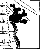
Figure 2. Hydronephrosis with
atony of the ureter. The hydronephrosis sac and the wide ureter are visible.
The hydronephrosis sac and the wide ureter are visible.
The hydronephrosis sac and the wide ureter are visible.
2). For dynamic reasons in the development of hydronephrosis, Israel also attributes those spasms of the muscular apparatus at the boundary of the pelvis and ureter (see Figure 3), which occur with a movable kidney, being a reflex on the irritation of the nerves of the renal pedicle during its stretching and bending, due to the descent of the kidney. Obstacles along the peripheral section of the urinary tract (urinary bladder, urethra) play a significantly smaller role in the development of hydronephrosis. These include urine retention in the bladder due to hypertrophy of the prostate gland, its malignant neoplasms, more rarely stones, paralysis, etc. Long-existing, difficult-to-pass strictures of the urethra lead to hydronephrosis through the intermediary of urine retention in the bladder. It is clear that in the cases listed, the developing hydronephrosis will be bilateral. From the point of view of anatomical changes in hydronephrosis, the pelvis and calyces of the kidney are stretched, and the renal parenchyma is functionally weakened and atrophied, often to its complete disappearance. It is therefore understandable that the contents of the hydronephrotic kidney, which in the initial stages of the disease resemble normal urine, subsequently become increasingly watery, and in old and closed hydronephroses, it does not contain the characteristic components of urine at all. It is also clear that due to the atrophy of the renal parenchyma, functional tests give a negative result not only in closed hydronephroses but also in far-advanced open varieties. Pathological anatomy. Externally, the hydronephrotically altered kidney is more or less enlarged, from barely noticeable increase in its volume to a tumor occupying a large part of the abdominal cavity. In the initial stages of hydronephrosis, especially with an intrarenal type of pelvis and in the absence of obvious visible enlargement of the kidney, the normal density of the kidney is reduced to the point where it can be bent in half. The outer surface of the kidney is either uneven, corresponding to the dilated calyces, or smooth (initial stages of the disease with still well-preserved renal parenchyma and large sacs of old hydronephroses of the unilocular type). From within, as a result of the stretching of the pelvis and calyces, we have a system of communicating cavities separated by partitions (see Figure 4); subsequently, the partitions may completely disappear with the formation of a unilocular cyst (see Figure 5). The boundary between the dilated pelvis and the stretched kidney is sometimes obvious, sometimes blurred. In old large hydronephroses, the vessels are reduced in caliber. In contrast, the ureter in cases of hydronephrosis that developed on the basis of its narrowing participates in the dilation (partially also in the hypertrophy), mainly on the proximal segment. Lengthening, it naturally undergoes bending, however, preserving its mobility in the absence of infection and, consequently, periureteritis. The layer of renal tissue that undergoes gradual atrophy in hydronephrosis, partly from direct pressure by fluid, partly due to the early narrowing of vessels (see below), becomes thinner, with the papillae and medullary substance disappearing first, and then the cortical layer. In severe hydronephrosis, when the kidney turns into a unilocular sac, only remnants of poor-blood and hardened renal tissue can be found in the wall of the latter in a few places; sometimes the latter is no longer found anywhere. In microscopic examination of the kidney, even in the early periods of hydronephrosis, degenerative-atrophic processes can be found in the medullary substance from the side of the tubular epithelium and proliferation of the interstitial connective tissue; later such changes involve the entire kidney. Along with this, a constant thickening of the vessel walls due to the proliferation of the inner membrane and a sharp narrowing of their lumens is noted. In the remnants of renal tissue, found as flattened areas in the wall of the hydronephrotic sac in severe hydronephrosis, the microscope reveals only connective tissue with scattered rare glomeruli (often hyalinized) and collapsed tubules with atrophic epithelium. The pathogenesis of the atrophic changes in the kidney in hydronephrosis can be understood as atrophy from pressure (see Atrophy); on the other hand, the indirect effect of the dilated pelvis on the arteries and, especially, the veins of the kidney in the sense of their mechanical compression or spastic contraction cannot be excluded (Ricker).
Symptomatology and recognition. In its developed and well-known manifestations, hydronephrosis is most characterized by the signs of a tumor. This tumor grows from the hypochondrium downward, into the abdominal cavity, not only approaching the anterior wall but also remaining in contact with the lumbar region. This explains both the good bilateral palpability of the tumor during bimanual examination and the presence of so-called ballottement (see). On palpation, the surface of the tumor is predominantly smooth; the consistency is elastic; sometimes the tumor clearly fluctuates. Fluctuation may be absent both with excessively tight filling of a still relatively thick-walled sac, and in hydronephroses with thin walls, loosely filled with fluid. Very large hydronephrotic sacs, difficult to palpate in the tightly stretched abdomen as a limited fluid-containing tumor, can simulate ascites. With rapid development of hydronephrosis with a significant increase in intrarenal pressure, the consistency of the tumor may appear firm, which may suggest a dense neoplasm of the kidney. In such cases, the painfulness of the tumor on palpation, as well as attacks of spontaneous pain in it, can help in the differential diagnosis of hydronephrosis. Hematuria in hydronephrosis appears at the end of a painful attack; hematuria in kidney tumors precedes pains caused by obstruction of the ureter by blood clots. The tumor has the character of a retroperitoneal one, in front of it usually there is an intestinal sound. Large hydronephrotic sacs, lying on the posterior wall of the abdominal cavity, displace the peritoneal reflection and the large intestine inward, more or less far to the middle line of the abdomen. In these cases, the sound over the tumor, on tapping from the outside and front, is dull; the presence and position of the large intestine connected to the sac can be revealed by inflating the intestine from the anus, which in doubtful cases is an important aid in recognizing the renal origin of the tumor. The excretion of urine is disturbed essentially in quantitative, and partly in qualitative aspects, only during attacks of intermittent hydronephrosis. In this case, the amount of urine is reduced, sometimes to complete anuria, due to retention of urine on the side of the hydronephrosis and the depressing reflex from the diseased kidney to the healthy one; after the attack, polyuria occurs, and on the diseased side, hematuria may occur as a result of venous congestion during obstruction. The recognition of hydronephrosis in the presence of a large tumor is in most cases not difficult, especially in the presence of closed hydronephrosis. One must take into account the constant size of the tumor and the absence of excretion of urine from the corresponding ureter (cystoscopy, ureteral catheterization). With large size, but open hydronephrosis, no significant fluctuations in the size of the sac are noticeable; separate collection of urine will give watery urine of low specific gravity, with a reduced effect of functional tests from the diseased kidney. Thus, fluctuations in the volume of the palpable tumor, accompanied during periods of its increase by sharp painful sensations, and conversely, an increase in the amount of excreted urine with simultaneous subsidence of the tumor and decrease in pain, are characteristic of the intermittent form of hydronephrosis. This variety of hydronephrosis is most often observed with a movable kidney and stones in the pelvis or ureter. Pain does not represent anything characteristic and inevitable in the picture of large hydronephroses, both open and closed. In many cases, hydronephrosis develops without any pain, slowly and gradually; or pains exist to some extent in the early stages of its development, especially when there is still functional renal parenchyma, under conditions of temporarily sharply increased diuresis. In general, the presence of a large hydronephrosis in the patient's abdomen manifests itself not so much in acute pains as in a sensation of heaviness, constriction, and dull pressure in the corresponding side and abdominal cavity. Part of the disorders experienced by patients depends on the pressure of the tumor on the surrounding organs of the abdominal cavity. Of enormous importance in the diagnosis of hydronephrosis are ureteral catheterization and pyelography (see). If from a catheter introduced into the renal pelvis, urine does not flow out in separate drops, periodically, but flows in a continuous stream, this indicates dilation of at least the renal pelvis and the presence of residual urine in it. The degree of dilation of the pelvis is determined

Figure 4. Large hydronephrosis: dilated calyces, separated by partitions, have wide communication with the pelvis. The bottom of the calyces represents the thinned cortical layer (according to IsraeFio).
connective tissue with scattered rare glomeruli (often hyalinized) and collapsed tubules with atrophic epithelium. The pathogenesis of the atrophic changes in the kidney in hydronephrosis can be understood as atrophy from pressure (see Atrophy); on the other hand, the indirect effect of the dilated pelvis on the arteries and, especially, the veins of the kidney in the sense of their mechanical compression or spastic contraction cannot be excluded (Ricker).
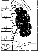
Figure 5. The pelvis and kidney form a continuous sac. The surface of the kidney is lobulated. (From the Urological clinic of 1st Moscow State University).
by the amount of urine found in it, especially compared to the result of a similar investigation of the healthy kidney's pelvis. Everywhere conditions permit, pyelography should currently be used for diagnosis. This method provides a direct view of the size, configuration, and position of the pelvis, as well as important details of the relationship between the pelvis and the ureter (see figures 2, 3, and 5). Despite the undoubted merits of this research method, it is not entirely free from certain sources of erroneous conclusions in this case. The main weakness is that a fairly wide variety of patterns falls within the limits of a normal pyelogram, caused on one hand by the actually existing considerable diversity in the structure of the kidney's cavity system, and on the other hand by the gradual and therefore transitional transition of observed patterns from normal to pathology, i.e., to states of kidney distension; moreover, all this diversity increases and changes depending on the greater or lesser success in filling the pelvis with the injected contrast fluid. Even with great skill in this type of research, there remains sufficient room for subjectivity in interpreting the pyelogram. A certain correction is provided by pyelograms from both sides, since in normal individuals the cavity systems of both kidneys are constructed approximately similarly.-Of the complications of H., one must name the accompanying infection and rupture of H. Infection, by whatever path it occurs, transforms H. into hydropionephrosis (see) or into pyonephrosis, which sharply worsen the patient's condition. A traumatic rupture of H., on the other hand, directly threatens the patient's life with hemorrhage, and in case of spillage of contents into the abdominal cavity, it leads to the development of peritonitis (see), and in cases of spillage into the retroperitoneal tissue, to the development of paranephritis (see). Treatment. Every H. that causes discomfort to the patient should be eliminated. In this respect, moderately sized closed H. that do not cause any disorders do not constitute a fundamental exception, because they cannot be considered completely safe for their carrier, and the possibility of their infection by the hematogenous route always exists. There is no point in arguing against the expediency and attractiveness of surgical methods for treating H., which aim to simultaneously eliminate its cause and preserve the kidney. For this purpose, a whole series of conservative-type operations have been proposed: 1) Reduction of the dilated renal pelvis a) by suturing its wall according to Israel in the manner of gastroplication and b) by partial excision of it (orthopedic resection according to Albarran). 2) Incision of the ureteropelvic spur (Trendelenburg, Fenger); proposed for cases of high origin of the ureter from the pelvis and its fusion with the wall of the pelvis, starting from the orifice and further downward. 3) Formation of an anastomosis between the ureter and the dilated pelvis according to the 'side-to-side' type of anastomosis, according to Albarran, or according to the type of inverting the peripheral end of the ureter, cut below the constriction, 'end-to-side' at the most sloping part of the pelvis (according to Kiister). 4) Ureteroplasty and pyelo-ureteroplasty, according to Fenger, analogous to pyloroplasty; can be applied in cases of short ureteral narrowing, especially of its upper part or at the border between the latter and the renal pelvis. 5) Formation of an anastomosis between the hydronephrotic sac and the urinary bladder (hydro-nephro-cysto-anastomo-sis) as conceived by Firtel, carried out by Reisinger, Snegirev, Schlofter, and others. Applicable in cases of very large sacs with an insurmountable obstacle to urine outflow, inaccessible to excision due to the exceptional density of adhesions, and in disease of the only kidney. This operation has so far been performed by the intraperitoneal route. Kümmel and Graff consider it appropriate to perform it also in cases of unilateral hydronephrosis where the second kidney is functionally inadequate. 6) Division of an accessory vessel to the kidney, according to Ekegård. Permitted only in cases where the anomalous vessel arises directly from the aorta, but not from the renal artery (danger of necrosis of the corresponding part of the kidney). Kummer collected from the literature 56 cases of complete recovery after division of a strangulating ureteral vessel. Mayo also obtained a positive therapeutic result in 13 out of 20 cases. 7) In strictures of the lower end of the ureter at its entrance into a diverticulum of the urinary bladder, etc., the operation of choice will be ureteral transplantation into the urinary bladder, preferably by the transperitoneal route. A priori, one can demand the presence of corresponding conditions that would allow the application of the aforementioned surgical methods, namely, that the hydronephrotic kidney has retained its functional value to a sufficient degree, which is especially important when the paired organ is diseased and absolutely inevitable when H. develops in the only kidney. But if the second kidney is healthy and functionally adequate, the question loses its acuteness, since the healthy kidney will fully replace the diseased one. The functional value of a hydronephrotic kidney is better assessed by the results of a color test rather than by eye based on the thickness of the layer of renal parenchyma: on one hand, renal tissue stretched over a large extent by a thin layer along the wall of the hydronephrotic sac may erroneously seem to be preserved in a negligible amount and functionally worthless, and on the other hand, even a considerable amount of preserved renal parenchyma may be of little value due to degeneration. But even in assessing the color functional test, a considerable amount of subjectivity is inevitably introduced into the decision of the aforementioned question.-The next requirement is the possibility of forming a completely clear picture of the mechanism of origin of H. in each individual case. Not to mention the more difficult cases for proper assessment, the difficulties in distinguishing primary and sequential causes of H., it must be admitted that in a very large, perhaps even overwhelming majority of H. cases, it is not possible for purely technical reasons to identify the mechanism of the given H. (Israel). Conservative operations leave much to be desired and yield to nephrectomies when the second kidney is healthy. Nephrectomy is the method of choice for most surgeons in the treatment of H., except for initial or not very advanced cases of H. with a floating kidney, where clinical cure, in the absence of fixed kinks of the ureter, can sometimes be achieved by nephropexy, or in cases of a stone in the renal pelvis or ureter-by removal of the stone. Of the two methods of nephrectomy-trans- and retroperitoneal-the latter is considered the norm. In very large H., the transperitoneal route has some technical advantages, facilitating the performance of the operation. Due to the loose connection of an aseptic H. with surrounding tissues and the small caliber of the renal vessels in large and old H., their removal often goes more easily than expected. This explains the good results of nephrectomy for H. In bilateral H., treatment is directed toward the primary cause of the disease, with the aim of eliminating chronic urine retention in the urinary bladder. Palliative operations include: 1) catheterization of the renal pelvis, mostly repeated; success can only be expected in the initial stages of H., with a short-lasting stasis and a pelvis wall that has not lost its ability to contract; in view of the ease of introducing infection, Israel is fundamentally against this method in aseptic cases unless removal of H. is planned in the near future;-2) puncture of H., provided strict asepsis is observed; it could find application in cases that cannot be delayed and in conditions that do not allow a more serious operation due to the patient's condition (obstruction with reflex anuria, combination of a large H. with a large abdominal tumor); the therapeutic effect of puncture is short-lived and inferior to puncture with drainage-analogous to the method of Bülow in the treatment of empyema; puncture is always performed retroperitoneally, outward from the colon, along the middle line connecting the end of the XII rib with the iliac crest, 6 cm posterior to the spina ilii ant. sup.; 3) pyelostomy and nephrostomy are indicated even less, as they leave behind persistent urinary fistulas unless the patency of the ureter is restored, and they easily lead to secondary infections from the outside.
Related articles
Mentioned in
Cite this page
“Hydronephrosis.” Soviet Medical Encyclopedia. English translation of Bolshaya Meditsinskaya Entsiklopediya, 1st ed. (Moscow, 1928–1936), ed. N. A. Semashko. https://sovietmedicalencyclopedia.pages.dev/article/hydronephrosis/