Giardia Intestinalis
Historical document, translated for reference. It reflects medical knowledge of the 1920s–30s and is not medical advice.
Summary
This 1930s encyclopedia article describes Giardia intestinalis, a flagellated protozoan parasite inhabiting the intestines of humans and rodents. It details its morphology, life cycle involving cysts, pathogenicity, and historical treatments of associated enterocolitis.
Encyclopedia article (1928–1936)
GIARDIA INTESTINALIS Lambl., 1859 (syn. Cercomonas intestinalis, Lambl., 1859; Hexamitus duodenalis, Davaine, 1875; Megastoma entericum, Grassi, 1881; Lamblia intestinalis, Blanchard, 1888), a flagellate of the family Octomitidae, parasitic in the intestine of humans and rodents (more rarely in dogs and cats). Finding it in the blood of birds has been described. The body shape is pyriform, the structure is bilaterally symmetrical, with 2 nuclei, 4 pairs of flagella, and an axial filament. Size 10–20 x 6–10 µ. The upper side is flattened, forming a surface by which Giardia can attach itself to the epithelium. It multiplies by division. It forms oval cysts 10–13 x 8–9 µ, mostly with 4 nuclei. Nutrition is osmotic. In humans, it is found in the large, small, and duodenum. Vegetative forms are found mostly in liquid stool, while formed stool mostly contains only cysts. With the help of a duodenal sound, vegetative forms can often be found in the contents of the duodenum as well. Cases of finding Giardia in the gallbladder have also been described. Giardia is very frequently found in the intestine of healthy people and occurs everywhere. Infection occurs through water via cysts. For the most part, the presence of Giardia is not associated with any obvious morbid phenomena. According to some data (Brumpt), Giardia may, however, especially in hot zones, be the cause of mucous enterocolitis. It is not excluded, however, that enterocolitis of other origins may favor the enhanced development of Giardia, which then become the cause of secondary complications.
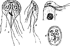
Giardia intestinalis: a—vegetative form from the front; b—vegetative form, from the side; c—vegetative form attached to an epithelial cell; d—cyst.
Giardia infection can proceed in the form of acute enterocolitis with mucous-bloody stools (up to 24 times a day), but for the most part it takes a subacute form (4–6 times a day). Phenomena on the part of the biliary tract are frequent. Cases of ulceration of the large intestine with the finding of Giardia in the thickness of the intestinal wall have been described. Penetrating into capillaries, Giardia can be carried by the blood to other organs as well. Thus, a case of metastatic subdiaphragmatic abscess containing a huge number of Giardia has been described (Turkestan). The treatment of enterocolitis caused by Giardia is carried out with various disinfecting substances (salol, thymol, β-naphthol). There are indications of the favorable effect of bismuth, stovarsol, salvarsan, and emetine.
Related articles
Mentioned in
Cite this page
“Giardia Intestinalis.” Soviet Medical Encyclopedia. English translation of Bolshaya Meditsinskaya Entsiklopediya, 1st ed. (Moscow, 1928–1936), ed. N. A. Semashko. https://sovietmedicalencyclopedia.pages.dev/article/giardia-intestinalis/